

Deciphering the Immune Microenvironment on A Single Archival Formalin-Fixed Paraffin-Embedded Tissue Section by An Immediately Implementable Multiplex Fluorescence Immunostaining Protocol
Open accessThe DOI matches its Crossref record; 38 of the 38 references it cites were found in a registry or an index.
Abstract
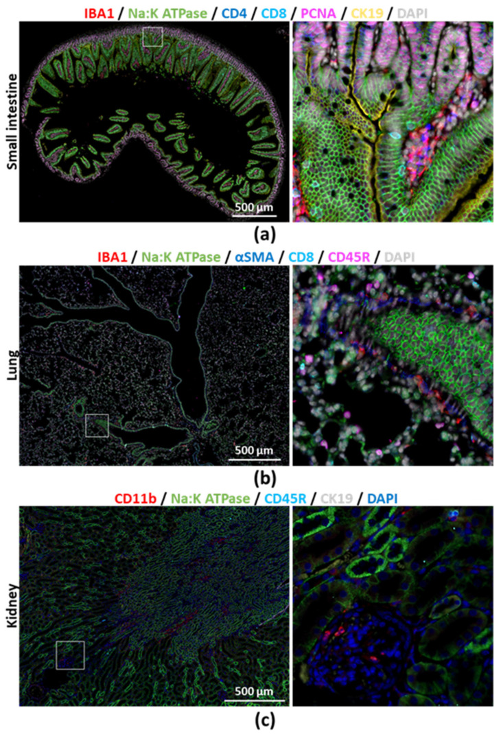
Technological breakthroughs have fundamentally changed our understanding on the complexity of the tumor microenvironment at the single-cell level. Characterizing the immune cell composition in relation to spatial distribution and histological changes may provide important diagnostic and therapeutic information. Immunostaining on formalin-fixed paraffin-embedded (FFPE) tissue samples represents a widespread and simple procedure, allowing the visualization of cellular distribution and processes, on preserved tissue structure. Recent advances in microscopy and molecular biology have made multiplexing accessible, yet technically challenging. We herein describe a novel, simple and cost-effective method for a reproducible and highly flexible multiplex immunostaining on archived FFPE tissue samples, which we optimized for solid organs (e.g., liver, intestine, lung, kidney) from mice and humans. Our protocol requires limited specific equipment and reagents, making multiplexing (>12 antibodies) immediately implementable to any histology laboratory routinely performing immunostaining. Using this method on single sections and combining it with automated whole-slide image analysis, we characterize the hepatic immune microenvironment in preclinical mouse models of liver fibrosis, steatohepatitis and hepatocellular carcinoma (HCC) and on human-patient samples with chronic liver diseases. The data provide useful insights into tissue organization and immune–parenchymal cell-to-cell interactions. It also highlights the profound macrophage heterogeneity in liver across premalignant conditions and HCC.
Overview
The claims and statistics of this paper haven’t been checked yet.
Free: it reads the open-access text and checks the first 5 claims that cite a source we can check. Create a free account to check every claim.
Integrity signals
Registry
Cites
Statistics
Figures




Verification
Claims the paper makes
The claims and statistics of this paper haven’t been checked yet.
Free: it reads the open-access text and checks the first 5 claims that cite a source we can check. Create a free account to check every claim.
Claims other papers make about this one
Each is checked against this paper’s own text: does it say what the citing paper says it does?
The source backs part of the claim, not all of it — or backs it for a different population, condition or comparison.
In the citing paper
“Multiplex immunofluorescence also holds key advantages for molecular pathway exploration in animal models, as this allows for a multidimensional evaluation of cellular phenomena on archival, formalin-fixed and paraffin-embedded tissue sections [343].”
From Inflammation and immunity in liver homeostasis and disease: a nexus of hepatocytes, nonparenchymal cells and immune cells (2025) · doi:10.1038/s41423-025-01313-7 · cites this paper as [343]
Experimental — accuracy not yet measured
Self-check
Percentages, sums and reported differences re-computed from the paper’s own numbers.
The self-check needs the paper’s full text.
Citations
Cites
Fiji: an open-source platform for biological-image analysis
Schindelin, J., Arganda-Carreras, I., Frise, E., Kaynig, V., Longair, M., Pietzsch, T., Preibisch, S., Rueden, C., Saalfeld, S., Schmid, B., et al. (2012) · Nature Methods · doi:10.1038/nmeth.2019 · Cited by 66,890 · cited 4×
“Single channel grayscale pictures were further processed in FIJI [33].”
MethodInfluentialUnderstanding the tumor immune microenvironment (TIME) for effective therapy
Binnewies, M., Roberts, E. W., Kersten, K., Chan, V., Fearon, D. F., Merad, M., Coussens, L. M., Gabrilovich, D. I., Ostrand-Rosenberg, S., Hedrick, C. C., et al. (2018) · Nature Medicine · doi:10.1038/s41591-018-0014-x · Cited by 6,145 · cited 2×
“Dissecting the immune-cell compartment at high resolution in clinical cohorts and model systems is anticipated to pave the way to novel, tailored interventions for inflammatory and malignant diseases [3,4].”
Backgroundilastik: interactive machine learning for (bio)image analysis
Berg, S., Kutra, D., Kroeger, T., Straehle, C. N., Kausler, B. X., Haubold, C., Schiegg, M., Ales, J., Beier, T., Rudy, M., et al. (2019) · Nature Methods · doi:10.1038/s41592-019-0582-9 · Cited by 3,368
Highly multiplexed imaging of tumor tissues with subcellular resolution by mass cytometry
Giesen, C., Wang, H. A. O., Schapiro, D., Zivanovic, N., Jacobs, A., Hattendorf, B., Schüffler, P. J., Grolimund, D., Buhmann, J. M., Brandt, S., et al. (2014) · Nature Methods · doi:10.1038/nmeth.2869 · Cited by 1,951 · cited 1×
“Imaging mass cytometry for instance, would theoretically allow for about 100 markers to be visualized at once at the single cell level, which may provide unprecedented information on cell phenotype [25].”
BackgroundResolving the fibrotic niche of human liver cirrhosis at single-cell level
Ramachandran, P., Dobie, R., Wilson-Kanamori, J. R., Dora, E. F., Henderson, B. E. P., Luu, N. T., Portman, J. R., Matchett, K. P., Brice, M., Marwick, J. A., et al. (2019) · Nature · doi:10.1038/s41586-019-1631-3 · Cited by 1,784 · cited 1×
“It can also be used to validate, in a large number of samples, the presence and localization of distinct cellular subpopulations or phenotypes that have been identified by “omics” technology such as single-cell RNA sequencing [23,24].”
BackgroundSelf-citationLiver Macrophages in Tissue Homeostasis and Disease
Krenkel, O., & Tacke, F. (2017) · Nature Reviews Immunology · doi:10.1038/nri.2017.11 · Cited by 1,424 · cited 1×
“, CK19+ cholangiocytes or αSMA+Desmin+ stellate cells) [10,11].”
Self-citationSternberg (1983) · Computer · doi:10.1109/mc.1983.1654163 · Cited by 930 · cited 1×
“Image preprocessing include tile stitching and background subtraction and autofluorescence subtraction (Figures S13 and S14, respectively) [29].”
BackgroundCellProfiler™: Free, Versatile Software for Automated Biological Image Analysis
Lamprecht, M. R., Sabatini, D. M., & Carpenter, A. E. (2007) · BioTechniques · doi:10.2144/000112257 · Cited by 895
Highly multiplexed single-cell analysis of formalin-fixed, paraffin-embedded cancer tissue
Gerdes, M. J., Sevinsky, C. J., Sood, A., Adak, S., Bello, M. O., Bordwell, A., Can, A., Corwin, A., Dinn, S., Filkins, R. J., et al. (2013) · Proceedings of the National Academy of Sciences · doi:10.1073/pnas.1300136110 · Cited by 724 · cited 1×
“patent 7,741,045) alkaline oxidation-based chemical reaction for irreversible inactivation of antibody-conjugated fluorophores, by which the antibody–fluorophore complex remains on the tissue [20].”
BackgroundLin, J.-R., Izar, B., Wang, S., Yapp, C., Mei, S., Shah, P. M., Santagata, S., & Sorger, P. K. (2018) · eLife · doi:10.7554/elife.31657 · Cited by 692
Tan, W. C. C., Nerurkar, S. N., Cai, H. Y., Ng, H. H. M., Wu, D., Wee, Y. T. F., Lim, J. C. T., Yeong, J., & Lim, T. K. H. (2020) · Cancer Communications · doi:10.1002/cac2.12023 · Cited by 678 · cited 1×
“Alternative methods have been described or are commercially available, allowing for the visualization of multiple cell types within the same sample [6].”
MethodhistoCAT: analysis of cell phenotypes and interactions in multiplex image cytometry data
Schapiro, D., Jackson, H. W., Raghuraman, S., Fischer, J. R., Zanotelli, V. R. T., Schulz, D., Giesen, C., Catena, R., Varga, Z., & Bodenmiller, B. (2017) · Nature Methods · doi:10.1038/nmeth.4391 · Cited by 659 · cited 3×
“The CellProfiler project we used in Figure 5 are provided in Appendix A Cancers 2020, 12, 2449 15 of 17 (Note S9).”
MethodInfluentialTsujikawa, T., Kumar, S., Borkar, R. N., Azimi, V., Thibault, G., Chang, Y. H., Balter, A., Kawashima, R., Choe, G., Sauer, D., et al. (2017) · Cell Reports · doi:10.1016/j.celrep.2017.03.037 · Cited by 568
Biological imaging software tools
Eliceiri, K. W., Berthold, M. R., Goldberg, I. G., Ibáñez, L., Manjunath, B. S., Martone, M. E., Murphy, R. F., Peng, H., Plant, A. L., Roysam, B., et al. (2012) · Nature Methods · doi:10.1038/nmeth.2084 · Cited by 551
CellProfiler Analyst: data exploration and analysis software for complex image-based screens
Jones, T. R., Kang, I. H., Wheeler, D. B., Lindquist, R. A., Papallo, A., Sabatini, D. M., Golland, P., & Carpenter, A. E. (2008) · BMC Bioinformatics · doi:10.1186/1471-2105-9-482 · Cited by 541
Tramu, G., Pillez, A., & Leonardelli, J. (1978) · Journal of Histochemistry & Cytochemistry · doi:10.1177/26.4.207771 · Cited by 505
Spatial heterogeneity in the mammalian liver
Ben-Moshe, S., & Itzkovitz, S. (2019) · Nature Reviews Gastroenterology & Hepatology · doi:10.1038/s41575-019-0134-x · Cited by 485 · cited 1×
“The combination of such large data sets with spatial information (“spatial transcriptomics”) and/or cross-validation with proteomic approaches have helped us gain a highly detailed view of zonation, cellular heterogeneity and key distinctive features of the cellular components in tissues such as the liver [1].”
BackgroundDuctular reaction in liver diseases: pathological mechanisms and translational significances
Sato, K., Marzioni, M., Meng, F., Francis, H., Glaser, S., & Alpini, G. (2019) · Hepatology · doi:10.1002/hep.30150 · Cited by 423 · cited 2×
“One hallm rk of chr nic liver diseases is inflamm tion, notably characterized by macrophage accumulation and biliary epithelial cell-like and liver progenitor cell (or ductul cell) activation [12,13].”
BackgroundAeffner, F., Zarella, M. D., Buchbinder, N., Bui, M. M., Goodman, M. R., Hartman, D. J., Lujan, G. M., Molani, M. A., Parwani, A. V., Lillard, K., et al. (2019) · Journal of Pathology Informatics · doi:10.4103/jpi.jpi_82_18 · Cited by 403
Liver Macrophages: Old Dogmas and New Insights
Guillot, A., & Tacke, F. (2019) · Hepatology Communications · doi:10.1002/hep4.1356 · Cited by 335 · cited 3×
“The balance between these two populations and their respective activation states tremendously regulate the immunological status of the liver, as well as tissue damage or repair [12].”
BackgroundSelf-citationLefere, S., Puengel, T., Hundertmark, J., Penners, C., Frank, A. K., Guillot, A., de Muynck, K., Heymann, F., Adarbes, V., Defrêne, E., et al. (2020) · Journal of Hepatology · doi:10.1016/j.jhep.2020.04.025 · Cited by 310
Self-citationKrenkel, O., Hundertmark, J., Abdallah, A. T., Kohlhepp, M., Puengel, T., Roth, T., Branco, D. P. P., Mossanen, J. C., Luedde, T., Trautwein, C., et al. (2020) · Gut · doi:10.1136/gutjnl-2019-318382 · Cited by 220 · cited 1×
“It can also be used to validate, in a large number of samples, the presence and localization of distinct cellular subpopulations or phenotypes that have been identified by “omics” technology such as single-cell RNA sequencing [23,24].”
BackgroundSelf-citationMultiplex Staining by Sequential Immunostaining and Antibody Removal on Routine Tissue Sections
Bolognesi, M. M., Manzoni, M., Scalia, C. R., Zannella, S., Bosisio, F. M., Faretta, M., & Cattoretti, G. (2017) · Journal of Histochemistry & Cytochemistry · doi:10.1369/0022155417719419 · Cited by 168 · cited 3×
“Thus, the tissue and antigen qualities do not seem to be notably altered along the protocol [18].”
BackgroundMethodRehg, J. E., Bush, D., & Ward, J. M. (2012) · Toxicologic Pathology · doi:10.1177/0192623311430695 · Cited by 117 · cited 2×
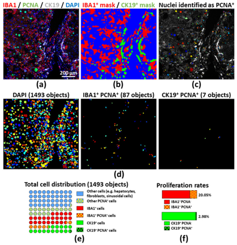
“Liver resident and monocyte-derived macrophages were stained by using an anti-IBA1 antibody, ductular cells were stained for CK19, and proliferation was evaluated with an anti-PCNA antibody (Figures 4a and S9) [15].”
MethodSingle-cell genomic approaches for developing the next generation of immunotherapies
Yofe, I., Dahan, R., & Amit, I. (2020) · Nature Medicine · doi:10.1038/s41591-019-0736-4 · Cited by 114 · cited 1×
“Dissecting the immune-cell compartment at high resolution in clinical cohorts and model systems is anticipated to pave the way to novel, tailored interventions for inflammatory and malignant diseases [3,4].”
BackgroundBartneck, M., Schrammen, P. L., Möckel, D., Govaere, O., Liepelt, A., Krenkel, O., Ergen, C., McCain, M. V., Eulberg, D., Luedde, T., et al. (2019) · Cellular and Molecular Gastroenterology and Hepatology · doi:10.1016/j.jcmgh.2018.10.007 · Cited by 107 · cited 3×
“5 mL/kg body weight) injections from the age of 8 weeks, twice per week for 16 weeks [14].”
BackgroundMethodSelf-citationLin, J., Izar, B., Wang, S., et al. (2017) · bioRxiv (Cold Spring Harbor Laboratory) · doi:10.1101/151738 · Cited by 104
Immunology in the Era of Single-Cell Technologies
Efremova, M., Vento-Tormo, R., Park, J.-E., Teichmann, S. A., & James, K. R. (2020) · Annual Review of Immunology · doi:10.1146/annurev-immunol-090419-020340 · Cited by 79 · cited 1×
“These approaches are particularly valuable to understand the nature and composition of inflammatory foci, as immune cells closely interact with parenchymal cells in homeostasis and disease by dynamically adapting their phenotype [2].”
BackgroundGendusa, R., Scalia, C. R., Buscone, S., & Cattoretti, G. (2014) · Journal of Histochemistry & Cytochemistry · doi:10.1369/0022155414536732 · Cited by 76
Rocha, R., Almeida, C., & Azevedo, N. F. (2018) · PLoS ONE · doi:10.1371/journal.pone.0196522 · Cited by 49
Autofluorescence: A potential pitfall in immunofluorescence-based inflammation grading
Wizenty, J., Ashraf, M., Rohwer, N., Stockmann, M., Weiß, S., Biebl, M., Pratschke, J., Aigner, F., & Wuensch, T. (2018) · Journal of Immunological Methods · doi:10.1016/j.jim.2018.02.007 · Cited by 33
Wizenty, J., Schumann, T., Theil, D., Stockmann, M., Pratschke, J., Tacke, F., Aigner, F., & Wuensch, T. (2020) · Molecules · doi:10.3390/molecules25092095 · Cited by 28 · cited 1×
“This is particularly relevant when performing immunohistochemistry on FFPE samples or when dealing with intrinsic tissue components known to be autofluorescent [21,22].”
BackgroundSelf-citationHistory of Immunohistochemistry
Childs, G. V. (2014) · Elsevier eBooks · doi:10.1016/b978-0-12-386456-7.07401-3 · Cited by 27 · cited 1×
“Immunohistochemistry using primary antibodies directed against the antigen of interest on tissue sections dates back a century [5].”
MethodBraubach, P., Lippmann, T., Raoult, D., Lagier, J., Anagnostopoulos, I., Zender, S., Länger, F. P., Kreipe, H.-H., Kühnel, M., & Jonigk, D. D. (2017) · Frontiers in Medicine · doi:10.3389/fmed.2017.00087 · Cited by 23
Walters, D. K., & Jelinek, D. F. (2019) · Journal of Histochemistry & Cytochemistry · doi:10.1369/0022155419896802 · Cited by 12 · cited 2×
“A similar 2-mercaptoethanol/SDS stripping approach was used to characterize bone marrow immune cell populations [19].”
MethodGuillot, A., Buch, C., & Jourdan, T. (2020) · Methods in molecular biology · doi:10.1007/978-1-0716-0704-6_6 · Cited by 12 · cited 1×
“, CK19+ cholangiocytes or αSMA+Desmin+ stellate cells) [10,11].”
Self-citationRocha, R., Almeida, C., & Azevedo, N. F. (2018) · PLoS ONE · doi:10.1371/journal.pone.0208867 · Cited by 5
Smith‐Togobo, C., Pedersen, M. Ø., Jensen, S. G., Duduyemi, B. M., Gyasi, R. K., Ofori, M. F., Paintsil, V., Renner, L. A., Nørgaard, P. H., & Hviid, L. (2019) · BMC Cancer · doi:10.1186/s12885-019-6488-1 · Cited by 1
Cited by
Alcohol-associated liver disease
Mackowiak, B., Fu, Y., Maccioni, L., & Gao, B. (2024) · Journal of Clinical Investigation · doi:10.1172/jci176345 · Cited by 359
Günsel, G. G., Conlon, T. M., Jeridi, A., Kim, R., Ertüz, Z., Lang, N. J., Ansari, M., Novikova, M., Jiang, D., Strunz, M., et al. (2022) · Nature Communications · doi:10.1038/s41467-022-28809-4 · Cited by 144
Self-citationImmune microenvironment in hepatocellular carcinoma: from pathogenesis to immunotherapy
Seyhan, D., Allaire, M., Fu, Y., Conti, F., Wang, X. W., Gao, B., & Lafdil, F. (2025) · Cellular and Molecular Immunology · doi:10.1038/s41423-025-01308-4 · Cited by 164
“…immunohistochemistry (IHC) or immuno fl uorescence (IF), mIF differentiates itself by allowing the analysis of multiple markers on the same tissue sections with successive staining – stripping protocols, with up to 18 – 20 different markers being able to be visualized on a single section [309, 310].”
Guillot, A., Winkler, M., Silva Afonso, M., Aggarwal, A., Lopez, D., Berger, H., Kohlhepp, M. S., Liu, H., Özdirik, B., Eschrich, J., et al. (2023) · Hepatology · doi:10.1097/hep.0000000000000270 · Cited by 116
“Sequential immunostaining was performed on 2- to 4-μm thick formalin-fixed paraffin-embedded liver sections as previously described.[19] Briefly, heat-mediated Ag retrieval MACROPHAGES DRIVE CHRONIC LIVER DISEASE PROGRESSION | 151”
MethodSelf-citation(2025) · doi:10.1038/s41423-025-01313-7 · Cited by 97
“Multiplex immuno fl uorescence also holds key advantages for molecular pathway exploration in animal models, as this allows for a multidimensional evaluation of cellular phenomena on archival, formalin-fi xed and paraf fi n-embedded tissue sections [343].”
Self-citation
Metadata
- Authors
- Guillot, A., Kohlhepp, M., Bruneau, A., Heymann, F., & Tacke, F.
- Affiliations
- Charité – Universitätsmedizin Berlin (DE)
- Corresponding author
- Tacke, Frank
- Venue
- Cancers
- Publisher
- MDPI AG
- Published
- 2020-08-28
- Volume
- 12
- Issue
- 9
- Pages
- 2449
- ISSN
- 2072-6694 (ISSN-L)
- PMC
- PMC7565194
- OpenAlex
- W3081587935
- Cited by
- 46
- References
- 37
- Citations by year
- 2026: 6 · 2025: 7 · 2024: 10 · 2023: 11 · 2022: 4 · 2021: 5 · 2020: 2
- FWCI
- 1.99
- Type
- journal article
- Language
- en
- Open access
- gold
- doi.org
- Licence
- Locations
- Cancers (publishedVersion, cc-by)
- PubMed Central (publishedVersion, cc-by)
- PubMed (publishedVersion)
- DOAJ (DOAJ: Directory of Open Access Journals) (submittedVersion, cc-by-sa)
- Refubium (Universitätsbibliothek der Freien Universität Berlin) (submittedVersion, cc-by)
- Universitätsbibliothek der FU Berlin Hochschulschriftenstelle u. Dokumentenserver (cc-by)
- Funders
- Deutsche Forschungsgemeinschaft (403224013)
- Topics
- Health Sciences › Medicine › Oncology › Cancer Immunotherapy and Biomarkers
- Life Sciences › Biochemistry, Genetics and Molecular Biology › Molecular Biology › Single-cell and spatial transcriptomics
- Health Sciences › Medicine › Hepatology › Liver physiology and pathology
- Keywords
- formalin-fixed paraffin-embedded tissue, tumor immune microenvironment, hepatic immune microenvironment, liver fibrosis, steatohepatitis, hepatocellular carcinoma, chronic liver disease, macrophage heterogeneity, spatial distribution of immune cells, whole slide image analysis
About these checks
Registry, retraction, Cites and statistics results are computed, not judged, with one exception: a printed reference that no index lists is read once by a model before it counts as a citation error. Claims are extracted by a model and kept only when their quote appears verbatim in the text; each cited source is then checked, with a model’s help, for whether it says what the claim relies on. Those verdicts can be wrong — read the claim’s quote and the cited source before relying on one.
Statistics and arithmetic are recomputed, not judged.
Confidence bands are shown only for fields calibrated on our annotated test set. None is calibrated yet, so no band is shown.
What counts as a claim ›How we measure confidence ›How we check this ›
Are you an author, or do you see a problem with this page? Request a review
Data sources
Bibliographic data: Semantic Scholar, OpenAlex, Crossref.
Contains information from the Semantic Scholar Academic Graph Datasets, made available under the ODC Attribution License (ODC-BY 1.0).
Semantic Scholar’s data is described in: Kinney, R., et al. (2023). The Semantic Scholar Open Data Platform. arXiv:2301.10140.
