

The DOI matches its Crossref record; 5 of the 5 references it cites were found in a registry or an index.
Excitatory Neurons Derived from Human-Induced Pluripotent Stem Cells Show Transcriptomic Differences in Alzheimer’s Patients from Controls
Published version of a preprint
Abstract
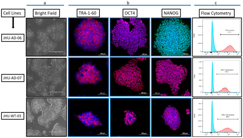
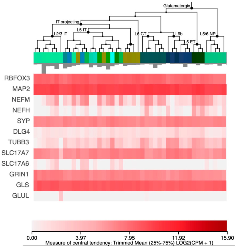
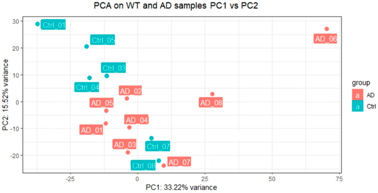
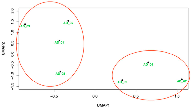
The recent advances in creating pluripotent stem cells from somatic cells and differentiating them into a variety of cell types is allowing us to study them without the caveats associated with disease-related changes. We generated induced Pluripotent Stem Cells (iPSCs) from eight Alzheimer’s disease (AD) patients and six controls and used lentiviral delivery to differentiate them into excitatory glutamatergic neurons. We then performed RNA sequencing on these neurons and compared the Alzheimer’s and control transcriptomes. We found that 621 genes show differences in expression levels at adjusted p < 0.05 between the case and control derived neurons. These genes show significant overlap and directional concordance with genes reported from a single-cell transcriptome study of AD patients; they include five genes implicated in AD from genome-wide association studies and they appear to be part of a larger functional network as indicated by an excess of interactions between them observed in the protein–protein interaction database STRING. Exploratory analysis with Uniform Manifold Approximation and Projection (UMAP) suggests distinct clusters of patients, based on gene expression, who may be clinically different. Our research outcomes will enable the precise identification of distinct biological subtypes among individuals with Alzheimer’s disease, facilitating the implementation of tailored precision medicine strategies.
Overview
The claims and statistics of this paper haven’t been checked yet.
Run the check — free, no account needed. It reads the open-access text and checks the sources it cites.
Integrity signals
Registry
Cites
Statistics
Figures
Verification
Claims the paper makes
The claims and statistics of this paper haven’t been checked yet.
Run the check — free, no account needed. It reads the open-access text and checks the sources it cites.
Claims other papers make about this one
Each is checked against this paper’s own text: does it say what the citing paper says it does?
In the citing paper
“re-analysis of published transcriptomics data sets of iNeurons generated by similar protocols [70, 71] failed to show constitutive receptor expression (data not shown).”
From Neurodegenerative disease-associated microRNAs acting as signaling molecules modulate CNS neuron structure and viability (2025) · cites this paper as [71]
Self-check
Percentages, sums and reported differences re-computed from the paper’s own numbers.
The self-check needs the paper’s full text.
Citations
Cites
Kok, J. S., Berg, I. J., & Scherder, E. J. A. (2013) · Dementia and Geriatric Cognitive Disorders Extra · doi:10.1159/000353441 · Cited by 20,589
Induced pluripotent stem cell technology: a decade of progress.
Shi, Y., Inoue, H., Wu, J. C., & Yamanaka, S. (2017) · Nature Reviews Drug Discovery · doi:10.1038/nrd.2016.245 · Cited by 1,525
Wightman, D. P., Jansen, I. E., Savage, J. E., Shadrin, A. A., Bahrami, S., Holland, D., Rongve, A., Børte, S., Winsvold, B. S., Drange, O. K., et al. (2021) · Nature Genetics · doi:10.1038/s41588-021-00921-z · Cited by 1,011
Lananna, B. V., McKee, C. A., King, M. W., Del-Aguila, J. L., Dimitry, J. M., Farias, F. H. G., Nadarajah, C. J., Xiong, D. D., Guo, C., Cammack, A. J., et al. (2020) · Science Translational Medicine · doi:10.1126/scitranslmed.aax3519 · Cited by 201
Genetics of Gene Expression in the Aging Human Brain Reveal TDP-43 Proteinopathy Pathophysiology
Yang, H.-S., White, C. C., Klein, H.-U., Yu, L., Gaiteri, C., Ma, Y., Felsky, D., Mostafavi, S., Petyuk, V. A., Sperling, R. A., et al. (2020) · Neuron · doi:10.1016/j.neuron.2020.05.010 · Cited by 47
Research works printed but not linked: 52
- Nichols, 2022, Estimation of the global prevalence of dementia in 2019 and forecasted prevalence in 2050: An analysis for the Global Burden of Disease Study 2019, Lancet Public Health, 7, e105, 10.1016/S2468-2667(21)00249-8 · DOI · Paper page
- Goate, 1991, Segregation of a missense mutation in the amyloid precursor protein gene with familial Alzheimer’s disease, Nature, 349, 704, 10.1038/349704a0 · DOI · Paper page
- Sherrington, 1995, Cloning of a gene bearing missense mutations in early-onset familial Alzheimer’s disease, Nature, 375, 754, 10.1038/375754a0 · DOI · Paper page
- Wijsman, 1995, A familial Alzheimer’s disease locus on chromosome 1, Science, 269, 970, 10.1126/science.7638621 · DOI
- Strittmatter, 1993, Apolipoprotein E: High-avidity binding to beta-amyloid and increased frequency of type 4 allele in late-onset familial Alzheimer disease, Proc. Natl. Acad. Sci. USA, 90, 1977, 10.1073/pnas.90.5.1977 · DOI · Paper page
- Reiman, 2007, GAB2 alleles modify Alzheimer’s risk in APOE epsilon4 carriers, Neuron, 54, 713, 10.1016/j.neuron.2007.05.022 · DOI · Paper page
- Myint, 2020, A screen of 1,049 schizophrenia and 30 Alzheimer’s-associated variants for regulatory potential, Am. J. Med Genet. Part B Neuropsychiatr. Genet., 183, 61, 10.1002/ajmg.b.32761 · DOI
- (2023, July 19). A Physician Surplus?|NEJM. Available online: https://www.nejm.org/doi/full/10.1056/NEJM198810063191419. · DOI
- Bellenguez, 2022, New insights into the genetic etiology of Alzheimer’s disease and related dementias, Nat. Genet., 54, 412, 10.1038/s41588-022-01024-z · DOI · Paper page
- Takahashi, 2007, Induction of pluripotent stem cells from adult human fibroblasts by defined factors, Cell, 131, 861, 10.1016/j.cell.2007.11.019 · DOI · Paper page
- Chou, 2011, Efficient human iPS cell derivation by a non-integrating plasmid from blood cells with unique epigenetic and gene expression signatures, Cell Res., 21, 518, 10.1038/cr.2011.12 · DOI
- Liu, 2020, Advances in Pluripotent Stem Cells: History, Mechanisms, Technologies, and Applications, Stem Cell Rev. Rep., 16, 3, 10.1007/s12015-019-09935-x · DOI
- Nishikawa, 2008, The promise of human induced pluripotent stem cells for research and therapy, Nat. Rev. Mol. Cell Biol., 9, 725, 10.1038/nrm2466 · DOI
- Takahashi, 2006, Induction of pluripotent stem cells from mouse embryonic and adult fibroblast cultures by defined factors, Cell, 126, 663, 10.1016/j.cell.2006.07.024 · DOI · Paper page
- Das, 2020, Modeling Psychiatric Disorder Biology with Stem Cells, Curr. Psychiatry Rep., 22, 24, 10.1007/s11920-020-01148-1 · DOI
- Das, 2021, Transcriptome analysis of human induced excitatory neurons supports a strong effect of clozapine on cholesterol biosynthesis, Schizophr. Res., 228, 324, 10.1016/j.schres.2020.12.041 · DOI · Paper page
- Karagiannis, 2019, Induced Pluripotent Stem Cells and Their Use in Human Models of Disease and Development, Physiol. Rev., 99, 79, 10.1152/physrev.00039.2017 · DOI
- Ehrhardt, 2019, Escitalopram for agitation in Alzheimer’s disease (S-CitAD): Methods and design of an investigator-initiated, randomized, controlled, multicenter clinical trial, Alzheimer’s Dement., 15, 1427, 10.1016/j.jalz.2019.06.4946 · DOI
- Mahairaki, 2014, Induced Pluripotent Stem Cells from Familial Alzheimer’s Disease Patients Differentiate into Mature Neurons with Amyloidogenic Properties, Stem Cells Dev., 23, 2996, 10.1089/scd.2013.0511 · DOI
- Das, 2020, Generation and characterization of a novel human iPSC line from a resilient Alzheimer’s disease patient, Stem Cell Res., 48, 101979, 10.1016/j.scr.2020.101979 · DOI
- Chou, 2015, A facile method to establish human induced pluripotent stem cells from adult blood cells under feeder-free and xeno-free culture conditions: A clinically compliant approach, Stem Cells Transl. Med., 4, 320, 10.5966/sctm.2014-0214 · DOI
- Yu, 2009, Human Induced Pluripotent Stem Cells Free of Vector and Transgene Sequences, Science, 324, 797, 10.1126/science.1172482 · DOI · Paper page
- Zhang, 2013, Rapid single-step induction of functional neurons from human pluripotent stem cells, Neuron, 78, 785, 10.1016/j.neuron.2013.05.029 · DOI · Paper page
- Lam, 2021, Transcriptomic data of Clozapine-treated Ngn2-induced Human Excitatory Neurons, Data Brief, 35, 106897, 10.1016/j.dib.2021.106897 · DOI
- Wahbeh, M.H., Peng, X., Bacharaki, S., Hatzimanolis, A., Dimitrakopoulos, S., Wohler, E., Yang, X., Yovo, C., Maher, B.J., and Sobreira, N. (2023). A Missense Variant in CASKIN1′s Proline-Rich Region Segregates with Psychosis in a Three-Generation Family. Genes, 14. · DOI
- Kim, 2019, Salzberg Graph-Based Genome Alignment and Genotyping with HISAT2 and HISAT-genotype, Nat. Biotechnol., 37, 907, 10.1038/s41587-019-0201-4 · DOI · Paper page
- Li, 2009, The Sequence Alignment/Map format and SAMtools, Bioinformatics, 25, 2078, 10.1093/bioinformatics/btp352 · DOI · Paper page
- Pertea, 2015, StringTie enables improved reconstruction of a transcriptome from RNA-seq reads, Nat. Biotechnol., 33, 290, 10.1038/nbt.3122 · DOI
- Pertea, 2016, Transcript-level expression analysis of RNA-seq experiments with HISAT, StringTie and Ballgown, Nat. Protoc., 11, 1650, 10.1038/nprot.2016.095 · DOI
- Soneson, 2015, Robinson. Differential analyses for RNA-seq: Transcript-level estimates improve gene-level inferences, F1000Research, 4, 1521, 10.12688/f1000research.7563.1 · DOI · Paper page
- Love, 2014, Moderated estimation of fold change and dispersion for RNA-seq data with DESeq2, Genome Biol., 15, 550, 10.1186/s13059-014-0550-8 · DOI · Paper page
- Mathys, 2019, Single-cell transcriptomic analysis of Alzheimer’s disease, Nature, 570, 332, 10.1038/s41586-019-1195-2 · DOI · Paper page
- Szklarczyk, 2022, The STRING database in 2023: Protein–protein association networks and functional enrichment analyses for any sequenced genome of interest, Nucleic Acids Res., 51, D638, 10.1093/nar/gkac1000 · DOI · Paper page
- Feuer, K.L., Peng, X., Yovo, C., and Avramopoulos, D. (2023). DPYSL2/CRMP2 isoform B knockout in human iPSC-derived glutamatergic neurons confirms its role in mTOR signaling and neurodevelopmental disorders. Mol. Psychiatry, 1–10. · DOI
- Ho, 2016, Rapid Ngn2-induction of excitatory neurons from hiPSC-derived neural progenitor cells, Methods, 101, 113, 10.1016/j.ymeth.2015.11.019 · DOI · Paper page
- McInnes, L., Healy, J., and Melville, J. (2020). UMAP: Uniform Manifold Approximation and Projection for Dimension Reduction. arXiv.
- Morris, 1993, The Clinical Dementia Rating (CDR): Current version and scoring rules, Neurology, 43, 2412, 10.1212/WNL.43.11.2412-a · DOI · Paper page
- Borkowski, 1967, Word fluency and brain damage, Neuropsychologia, 5, 135, 10.1016/0028-3932(67)90015-2 · DOI
- Folstein, 1975, ‘Mini-mental state’. A practical method for grading the cognitive state of patients for the clinician, J. Psychiatr. Res., 12, 189, 10.1016/0022-3956(75)90026-6 · DOI · Paper page
- Lambert, 2013, Meta-analysis of 74,046 individuals identifies 11 new susceptibility loci for Alzheimer’s disease, Nat. Genet., 45, 1452, 10.1038/ng.2802 · DOI · Paper page
- Tao, 2019, The role of CD2AP in the Pathogenesis of Alzheimer’s Disease, Aging Dis., 10, 901, 10.14336/AD.2018.1025 · DOI · Paper page
- Herold, 2016, Family-based Association Analyses of Imputed Genotypes Reveal Genome-Wide Significant Association of Alzheimer’s disease with OSBPL6, PTPRG and PDCL3, Mol. Psychiatry, 21, 1608, 10.1038/mp.2015.218 · DOI · Paper page
- Gan, 2022, Behavioural and functional evidence revealing the role of RBFOX1 variation in multiple psychiatric disorders and traits, Mol. Psychiatry, 27, 4464, 10.1038/s41380-022-01722-4 · DOI
- Ponthier, 2006, Fox-2 splicing factor binds to a conserved intron motif to promote inclusion of protein 4.1R alternative exon 16, J. Biol. Chem., 281, 12468, 10.1074/jbc.M511556200 · DOI · Paper page
- Alam, 2014, Alternative splicing regulation of APP exon 7 by RBFox proteins, Neurochem. Int., 78, 7, 10.1016/j.neuint.2014.08.001 · DOI
- Chen, Y.-C., Liu, Y.-L., Tsai, S.-J., Kuo, P.-H., Huang, S.-S., and Lee, Y.-S. (2019). LRRTM4 and PCSK5 Genetic Polymorphisms as Markers for Cognitive Impairment in A Hypotensive Aging Population: A Genome-Wide Association Study in Taiwan. J. Clin. Med., 8. · DOI
- Lamont, 2007, The role of circadian clock genes in mental disorders, Dialogues Clin. Neurosci., 9, 333, 10.31887/DCNS.2007.9.3/elamont · DOI
- Bishop, 2012, Genome-wide study links MTMR7 gene to variant Creutzfeldt-Jakob risk, Neurobiol. Aging, 33, 1487.e21, 10.1016/j.neurobiolaging.2011.10.011 · DOI
- Mead, 2013, Genetics of prion diseases, Curr. Opin. Genet. Dev., 23, 345, 10.1016/j.gde.2013.02.012 · DOI
- Huynh, 2015, HS3ST2 expression is critical for the abnormal phosphorylation of tau in Alzheimer’s disease-related tau pathology, Brain, 138, 1339, 10.1093/brain/awv056 · DOI
- Atkin, 2014, F-box only protein 2 (Fbxo2) regulates amyloid precursor protein levels and processing, J. Biol. Chem., 289, 7038, 10.1074/jbc.M113.515056 · DOI
- Williams, 2023, Inhibition of histone methyltransferase Smyd3 rescues NMDAR and cognitive deficits in a tauopathy mouse model, Nat. Commun., 14, 91, 10.1038/s41467-022-35749-6 · DOI
Cited by
Sorted by citations
Kumbol, V., Ivanov, A., McGurran, H., Schüler, J., Zhai, Y., Ludwik, K., Hinkelmann, L., Brehm, M., Krüger, C., Küchler, J., et al. (2025) · Cell Communication and Signaling · doi:10.1186/s12964-025-02199-8 · Cited by 8
Metadata
- Authors
- Sagar, R., Azoidis, I., Zivko, C., Xydia, A., Oh, E. S., Rosenberg, P. B., Lyketsos, C. G., Mahairaki, V., & Avramopoulos, D.
- Affiliations
- Department of Genetic Medicine, Johns Hopkins University School of Medicine, Baltimore, MD 21205, USA
- The Richman Family Precision Medicine Center of Excellence in Alzheimer’s Disease, Johns Hopkins University School of Medicine, Baltimore, MD 21287, USA
- ORCID
- Venue
- Cells
- Publisher
- MDPI AG
- Published
- 2023-08-02
- Volume
- 12
- Issue
- 15
- Pages
- 1990
- ISSN
- 2073-4409
- PMC
- PMC10417412
- Cited by
- 8
- References
- 5
- Language
- en
- Funders
- R21 AG067016 NIH
- Johns Hopkins Richman Family Precision Medicine Center of Excellence in Alzheimer’s Disease
- Richman Family Foundation
- Rick Sharp Alzheimer’s Foundation
- Sharp Family Foundation
- R01AG052510
About these checks
Registry, retraction, Cites and statistics results are computed, not judged, with one exception: a printed reference that no index lists is read once by a model before it counts as a citation error. Claims are extracted by a model and kept only when their quote appears verbatim in the text; each cited source is then checked, with a model’s help, for whether it says what the claim relies on. Those verdicts can be wrong — read the claim’s quote and the cited source before relying on one.
Statistics and arithmetic are recomputed, not judged.
Confidence bands are shown only for fields calibrated on our annotated test set. None is calibrated yet, so no band is shown.
What counts as a claim ›How we measure confidence ›How we check this ›
Are you an author, or do you see a problem with this page? Request a review
