

The DOI matches its Crossref record; 6 of the 6 references it cites were found in a registry or an index.
Important Factors Affecting Induction of Cell Death, Oxidative Stress and DNA Damage by Nano- and Microplastic Particles In Vitro
Abstract
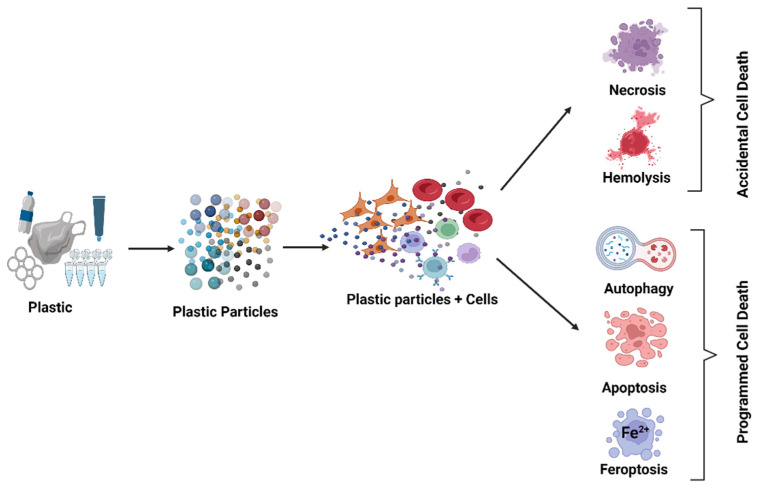
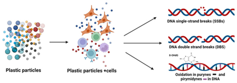
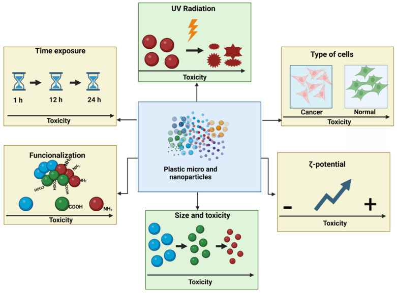
We have described the influence of selected factors that increase the toxicity of nanoplastics (NPs) and microplastics (MPs) with regard to cell viability, various types of cell death, reactive oxygen species (ROS) induction, and genotoxicity. These factors include plastic particle size (NPs/MPs), zeta potential, exposure time, concentration, functionalization, and the influence of environmental factors and cell type. Studies have unequivocally shown that smaller plastic particles are more cytotoxic, penetrate cells more easily, increase ROS formation, and induce oxidative damage to proteins, lipids, and DNA. The toxic effects also increase with concentration and incubation time. NPs with positive zeta potential are also more toxic than those with a negative zeta potential because the cells are negatively charged, inducing stronger interactions. The deleterious effects of NPs and MPs are increased by functionalization with anionic or carboxyl groups, due to greater interaction with cell membrane components. Cationic NPs/MPs are particularly toxic due to their greater cellular uptake and/or their effects on cells and lysosomal membranes. The effects of polystyrene (PS) vary from one cell type to another, and normal cells are more sensitive to NPs than cancerous ones. The toxicity of NPs/MPs can be enhanced by environmental factors, including UV radiation, as they cause the particles to shrink and change their shape, which is a particularly important consideration when working with environmentally-changed NPs/MPs. In summary, the cytotoxicity, oxidative properties, and genotoxicity of plastic particles depends on their concentration, duration of action, and cell type. Also, NPs/MPs with a smaller diameter and positive zeta potential, and those exposed to UV and functionalized with amino groups, demonstrate higher toxicity than larger, non-functionalized and environmentally-unchanged particles with a negative zeta potential.
Overview
The claims and statistics of this paper haven’t been checked yet.
Run the check — free, no account needed. It reads the open-access text and checks the sources it cites.
Integrity signals
Registry
Cites
Statistics
Figures
Verification
Claims the paper makes
The claims and statistics of this paper haven’t been checked yet.
Run the check — free, no account needed. It reads the open-access text and checks the sources it cites.
Claims other papers make about this one
Each is checked against this paper’s own text: does it say what the citing paper says it does?
In the citing paper
“Smaller MP particles, particularly nanosized plastic particles, show a more extensive dispersion, enhanced cellular uptake, and increased toxicity (Płuciennik et al., 2024).”
From Airborne microplastic exposure and urinary oxidative damage biomarkers in populations living in traffic-congested areas (2026) · cites this paper as Płuciennik et al., 2024
Self-check
Percentages, sums and reported differences re-computed from the paper’s own numbers.
The self-check needs the paper’s full text.
Citations
Cites
Apoptosis: A Review of Programmed Cell Death
Elmore, S. (2007) · Toxicologic Pathology · doi:10.1080/01926230701320337 · Cited by 13,853 · cited 1×
“Apoptosis is a crucial process implicated in hormone-dependent atrophy, embryonic development, the cell cycle, normal immune function, and cell death induced by xenobiotics [61].”
BackgroundDiscovery and quantification of plastic particle pollution in human blood
Leslie, H. A., van Velzen, M. J. M., Brandsma, S. H., Vethaak, A. D., Garcia-Vallejo, J. J., & Lamoree, M. H. (2022) · Environment International · doi:10.1016/j.envint.2022.107199 · Cited by 3,906 · cited 1×
“They have also been found in the male reproductive system [13] and in human blood [14].”
BackgroundDetection of microplastics in human lung tissue using μFTIR spectroscopy
Jenner, L. C., Rotchell, J. M., Bennett, R. T., Cowen, M., Tentzeris, V., & Sadofsky, L. R. (2022) · Science of The Total Environment · doi:10.1016/j.scitotenv.2022.154907 · Cited by 1,536
Mechanisms and Consequences of Double‐Strand DNA Break Formation in Chromatin
Cannan, W. J., & Pederson, D. S. (2016) · Journal of Cellular Physiology · doi:10.1002/jcp.25048 · Cited by 499 · cited 1×
“Damage to DNA also proceeds as a result of action of exogenous factors, such as various chemicals [76,77].”
BackgroundFirst Evidence of Microplastics in Human Urine, a Preliminary Study of Intake in the Human Body
Pironti, C., Notarstefano, V., Ricciardi, M., Motta, O., Giorgini, E., & Montano, L. (2022) · Toxics · doi:10.3390/toxics11010040 · Cited by 350
Fan, P., Yu, H., Xi, B., & Tan, W. (2022) · Environment International · doi:10.1016/j.envint.2022.107244 · Cited by 341
Research works printed but not linked: 73
- Gigault, 2018, Current Opinion: What Is a Nanoplastic?, Environ. Pollut., 235, 1030, 10.1016/j.envpol.2018.01.024 · DOI · Paper page
- Lithner, 2011, Environmental and Health Hazard Ranking and Assessment of Plastic Polymers Based on Chemical Composition, Sci. Total Environ., 409, 3309, 10.1016/j.scitotenv.2011.04.038 · DOI · Paper page
- Grote, K., Brüstle, F., and Vlacil, A.-K. (2023). Cellular and Systemic Effects of Micro- and Nanoplastics in Mammals—What We Know So Far. Materials, 16. · DOI
- Mattsson, 2015, Nano-Plastics in the Aquatic Environment, Environ. Sci. Process. Impacts, 17, 1712, 10.1039/C5EM00227C · DOI
- Chae, 2018, Trophic Transfer and Individual Impact of Nano-Sized Polystyrene in a Four-Species Freshwater Food Chain, Sci. Rep., 8, 284, 10.1038/s41598-017-18849-y · DOI
- Costa, 2020, Trophic Transfer of Microplastics From Copepods to Jellyfish in the Marine Environment, Front. Environ. Sci., 8, 571732, 10.3389/fenvs.2020.571732 · DOI · Paper page
- Schwabl, 2019, Detection of Various Microplastics in Human Stool: A Prospective Case Series, Ann. Intern. Med., 171, 453, 10.7326/M19-0618 · DOI
- Huang, 2022, Detection and Analysis of Microplastics in Human Sputum, Environ. Sci. Technol., 56, 2476, 10.1021/acs.est.1c03859 · DOI
- Zhao, 2023, Detection and Characterization of Microplastics in the Human Testis and Semen, Sci. Total Environ., 877, 162713, 10.1016/j.scitotenv.2023.162713 · DOI
- Cetin, 2023, Higher Number of Microplastics in Tumoral Colon Tissues from Patients with Colorectal Adenocarcinoma, Environ. Chem. Lett., 21, 639, 10.1007/s10311-022-01560-4 · DOI
- Salvia, 2023, Fast-Screening Flow Cytometry Method for Detecting Nanoplastics in Human Peripheral Blood, MethodsX, 10, 102057, 10.1016/j.mex.2023.102057 · DOI
- Kik, K., Bukowska, B., Krokosz, A., and Sicińska, P. (2021). Oxidative Properties of Polystyrene Nanoparticles with Different Diameters in Human Peripheral Blood Mononuclear Cells (In Vitro Study). Int. J. Mol. Sci., 22. · DOI
- Guševac Stojanović, I., Drakulić, D., Todorović, A., Martinović, J., Filipović, N., and Stojanović, Z. (2024). Acute Toxicity Assessment of Orally Administered Microplastic Particles in Adult Male Wistar Rats. Toxics, 12. · DOI
- Malinowska, 2022, Polystyrene Nanoparticles: The Mechanism of Their Genotoxicity in Human Peripheral Blood Mononuclear Cells, Nanotoxicology, 16, 791, 10.1080/17435390.2022.2149360 · DOI
- Yin, 2023, Polystyrene Microplastics Promote Liver Inflammation by Inducing the Formation of Macrophages Extracellular Traps, J. Hazard. Mater., 452, 131236, 10.1016/j.jhazmat.2023.131236 · DOI
- Meijer, 2020, The Plastic Brain: Neurotoxicity of Micro- and Nanoplastics, Part. Fibre Toxicol., 17, 24, 10.1186/s12989-020-00358-y · DOI · Paper page
- Zhao, 2024, The Potential Toxicity of Microplastics on Human Health, Sci. Total Environ., 912, 168946, 10.1016/j.scitotenv.2023.168946 · DOI
- Zapata, 2024, Internalization and Toxicity of Polystyrene Nanoplastics on Inmortalized Human Neural Stem Cells, Chemosphere, 355, 141815, 10.1016/j.chemosphere.2024.141815 · DOI
- Domenech, 2020, Nanoplastics as a Potential Environmental Health Factor: Effects of Polystyrene Nanoparticles on Human Intestinal Epithelial Caco-2 Cells, Environ. Sci. Nano, 7, 272, 10.1039/C9EN00523D · DOI
- Schmidt, 2023, Short- and Long-Term Polystyrene Nano- and Microplastic Exposure Promotes Oxidative Stress and Divergently Affects Skin Cell Architecture and Wnt/Beta-Catenin Signaling, Part. Fibre Toxicol., 20, 3, 10.1186/s12989-023-00513-1 · DOI
- Visalli, G., Facciolà, A., Pruiti Ciarello, M., De Marco, G., Maisano, M., and Di Pietro, A. (2021). Acute and Sub-Chronic Effects of Microplastics (3 and 10 Μm) on the Human Intestinal Cells HT-29. Int. J. Environ. Res. Public Health, 18. · DOI
- He, 2020, Cytotoxic Effects of Polystyrene Nanoplastics with Different Surface Functionalization on Human HepG2 Cells, Sci. Total Environ., 723, 138180, 10.1016/j.scitotenv.2020.138180 · DOI
- Annangi, B., Villacorta, A., López-Mesas, M., Fuentes-Cebrian, V., Marcos, R., and Hernández, A. (2023). Hazard Assessment of Polystyrene Nanoplastics in Primary Human Nasal Epithelial Cells, Focusing on the Autophagic Effects. Biomolecules, 13. · DOI
- Duffus, 2007, Glossary of Terms Used in Toxicology, 2nd Edition (IUPAC Recommendations 2007), Pure Appl. Chem., 79, 1153, 10.1351/pac200779071153 · DOI
- Yan, 2023, Polystyrene Nanoplastics Promote the Apoptosis in Caco-2 Cells Induced by Okadaic Acid More than Microplastics, Ecotoxicol. Environ. Saf., 249, 114375, 10.1016/j.ecoenv.2022.114375 · DOI
- Wang, X., Ren, X., Hu, H., Zhang, P., Zhao, F., Zhang, L., He, H., Huang, B., and Pan, X. (2022). Micro- and Nano- Polystyrene Plastic Particles Induce Apoptosis Through the Production of Intracellular ROS on CT26.WT Cells. SSRN J. · DOI
- Mattioda, V., Benedetti, V., Tessarolo, C., Oberto, F., Favole, A., Gallo, M., Martelli, W., Crescio, M.I., Berio, E., and Masoero, L. (2023). Pro-Inflammatory and Cytotoxic Effects of Polystyrene Microplastics on Human and Murine Intestinal Cell Lines. Biomolecules, 13. · DOI
- Steckiewicz, K.P., Adamska, A., Narajczyk, M., Megiel, E., and Inkielewicz—Stepniak, I. (2022). Fluoride Enhances Polystyrene Nanoparticles Cytotoxicity in Colonocytes in Vitro Model. Chem. Biol. Interact., 367. · DOI
- Chen, 2023, Cellular Absorption of Polystyrene Nanoplastics with Different Surface Functionalization and the Toxicity to RAW264.7 Macrophage Cells, Ecotoxicol. Environ. Saf., 252, 114574, 10.1016/j.ecoenv.2023.114574 · DOI
- Jiang, 2015, The Interplay of Size and Surface Functionality on the Cellular Uptake of Sub-10 Nm Gold Nanoparticles, ACS Nano, 9, 9986, 10.1021/acsnano.5b03521 · DOI
- Weiss, M., Fan, J., Claudel, M., Sonntag, T., Didier, P., Ronzani, C., Lebeau, L., and Pons, F. (2021). Density of Surface Charge Is a More Predictive Factor of the Toxicity of Cationic Carbon Nanoparticles than Zeta Potential. J. Nanobiotechnol, 19. · DOI
- Zhang, 2019, The Role of Positively Charged Sites in the Interaction between Model Cell Membranes and γ-Fe2O3 NPs, Sci. Total Environ., 673, 414, 10.1016/j.scitotenv.2019.04.074 · DOI
- Wang, F., Salvati, A., and Boya, P. (2018). Lysosome-Dependent Cell Death and Deregulated Autophagy Induced by Amine-Modified Polystyrene Nanoparticles. Open Biol., 8. · DOI
- Halimu, 2022, Toxic Effects of Nanoplastics with Different Sizes and Surface Charges on Epithelial-to-Mesenchymal Transition in A549 Cells and the Potential Toxicological Mechanism, J. Hazard. Mater., 430, 128485, 10.1016/j.jhazmat.2022.128485 · DOI
- Li, 2022, In Vitro Study on the Toxicity of Nanoplastics with Different Charges to Murine Splenic Lymphocytes, J. Hazard. Mater., 424, 127508, 10.1016/j.jhazmat.2021.127508 · DOI
- Shao, 2015, Independent Effect of Polymeric Nanoparticle Zeta Potential/Surface Charge, on Their Cytotoxicity and Affinity to Cells, Cell Prolif., 48, 465, 10.1111/cpr.12192 · DOI
- Honary, 2013, Effect of Zeta Potential on the Properties of Nano-Drug Delivery Systems—A Review (Part 1), Trop. J. Pharm. Res., 12, 255
- Zhang, 2009, Size-Dependent Endocytosis of Nanoparticles, Adv. Mater., 21, 419, 10.1002/adma.200801393 · DOI
- Schwegmann, 2010, Influence of the Zeta Potential on the Sorption and Toxicity of Iron Oxide Nanoparticles on S. Cerevisiae and E. Coli, J. Colloid Interface Sci., 347, 43, 10.1016/j.jcis.2010.02.028 · DOI
- Xu, 2023, Autophagic Response of Intestinal Epithelial Cells Exposed to Polystyrene Nanoplastics, Environ. Toxicol., 38, 205, 10.1002/tox.23678 · DOI
- Rubio, 2020, Biological Effects, Including Oxidative Stress and Genotoxic Damage, of Polystyrene Nanoparticles in Different Human Hematopoietic Cell Lines, J. Hazard. Mater., 398, 122900, 10.1016/j.jhazmat.2020.122900 · DOI
- Lins, 2022, Toxicity of Nanoplastics to Zooplankton Is Influenced by Temperature, Salinity, and Natural Particulate Matter, Environ. Sci. Nano, 9, 2678, 10.1039/D2EN00123C · DOI
- Shi, 2022, Cytotoxicity and Genotoxicity of Polystyrene Micro- and Nanoplastics with Different Size and Surface Modification in A549 Cells, Int. J. Nanomed., 17, 4509, 10.2147/IJN.S381776 · DOI
- Shi, 2021, Ultraviolet-Induced Photodegradation Elevated the Toxicity of Polystyrene Nanoplastics on Human Lung Epithelial A549 Cells, Environ. Sci. Nano, 8, 2660, 10.1039/D1EN00465D · DOI
- Sun, 2023, Combined Toxicity of Micro/Nanoplastics Loaded with Environmental Pollutants to Organisms and Cells: Role, Effects, and Mechanism, Environ. Int., 171, 107711, 10.1016/j.envint.2022.107711 · DOI
- Tang, 2019, The Molecular Machinery of Regulated Cell Death, Cell Res., 29, 347, 10.1038/s41422-019-0164-5 · DOI
- Phuc, 2019, Polystyrene Nanoparticles Induce Apoptosis or Necrosis With or Without Epidermal Growth Factor, J. Nanosci. Nanotechnol., 19, 4812, 10.1166/jnn.2019.16347 · DOI
- Xia, 2008, Cationic Polystyrene Nanosphere Toxicity Depends on Cell-Specific Endocytic and Mitochondrial Injury Pathways, ACS Nano, 2, 85, 10.1021/nn700256c · DOI · Paper page
- Duchnowicz, 2023, The Effects of Non-Functionalized Polystyrene Nanoparticles with Different Diameters on Human Erythrocyte Membrane and Morphology, Toxicol. Vitr., 91, 105634, 10.1016/j.tiv.2023.105634 · DOI
- Sarma, D.K., Dubey, R., Samarth, R.M., Shubham, S., Chowdhury, P., Kumawat, M., Verma, V., Tiwari, R.R., and Kumar, M. (2022). The Biological Effects of Polystyrene Nanoplastics on Human Peripheral Blood Lymphocytes. Nanomaterials, 12. · DOI
- Gopinath, 2019, Assessment on Interactive Prospectives of Nanoplastics with Plasma Proteins and the Toxicological Impacts of Virgin, Coronated and Environmentally Released-Nanoplastics, Sci. Rep., 9, 8860, 10.1038/s41598-019-45139-6 · DOI
- Rudnicka, 2011, Apoptosis and autophagy—Mechanisms and detection methods, Postep. Biol. Komorki, 38, 247
- Jeon, 2023, Polystyrene Microplastic Particles Induce Autophagic Cell Death in BEAS-2B Human Bronchial Epithelial Cells, Environ. Toxicol., 38, 359, 10.1002/tox.23705 · DOI
- Lu, 2022, Size-Dependent Effects of Polystyrene Nanoplastics on Autophagy Response in Human Umbilical Vein Endothelial Cells, J. Hazard. Mater., 421, 126770, 10.1016/j.jhazmat.2021.126770 · DOI
- Seca, 2019, Autophagy-Dependent Toxicity of Amino-Functionalized Nanoparticles in Ovarian Cancer Cells, J. Mater. Chem. B, 7, 5376, 10.1039/C9TB00935C · DOI
- Pinsino, 2017, Amino-Modified Polystyrene Nanoparticles Affect Signalling Pathways of the Sea Urchin (Paracentrotus lividus) Embryos, Nanotoxicology, 11, 201, 10.1080/17435390.2017.1279360 · DOI
- Malinowska, 2023, The Effects of Non-Functionalized Polystyrene Nanoparticles of Different Diameters on the Induction of Apoptosis and MTOR Level in Human Peripheral Blood Mononuclear Cells, Chemosphere, 335, 139137, 10.1016/j.chemosphere.2023.139137 · DOI
- Wu, H. (2014). Cell Death, Springer. · DOI
- Han, 2020, Ferroptosis and Its Potential Role in Human Diseases, Front. Pharmacol., 11, 239, 10.3389/fphar.2020.00239 · DOI
- Cao, 2016, Mechanisms of Ferroptosis, Cell. Mol. Life Sci., 73, 2195, 10.1007/s00018-016-2194-1 · DOI · Paper page
- Wu, 2024, Polystyrene Nanoplastics Lead to Ferroptosis in the Lungs, J. Adv. Res., 56, 31, 10.1016/j.jare.2023.03.003 · DOI
- Sun, 2023, Involvement of the JNK/HO-1/FTH1 Signaling Pathway in Nanoplastic-induced Inflammation and Ferroptosis of BV2 Microglia Cells, Int. J. Mol. Med., 52, 61, 10.3892/ijmm.2023.5264 · DOI
- Chen, 2024, Understanding the Mechanistic Roles of Microplastics Combined with Heavy Metals in Regulating Ferroptosis: Adding New Paradigms Regarding the Links with Diseases, Environ. Res., 242, 117732, 10.1016/j.envres.2023.117732 · DOI
- Poma, A., Vecchiotti, G., Colafarina, S., Zarivi, O., Aloisi, M., Arrizza, L., Chichiriccò, G., and Di Carlo, P. (2019). In Vitro Genotoxicity of Polystyrene Nanoparticles on the Human Fibroblast Hs27 Cell Line. Nanomaterials, 9. · DOI · Paper page
- Domenech, J., De Britto, M., Velázquez, A., Pastor, S., Hernández, A., Marcos, R., and Cortés, C. (2021). Long-Term Effects of Polystyrene Nanoplastics in Human Intestinal Caco-2 Cells. Biomolecules, 11. · DOI
- Vecchiotti, G., Colafarina, S., Aloisi, M., Zarivi, O., Di Carlo, P., and Poma, A. (2021). Genotoxicity and Oxidative Stress Induction by Polystyrene Nanoparticles in the Colorectal Cancer Cell Line HCT116. PLoS ONE, 16. · DOI
- Kim, 2018, Imaging DNA Single-Strand Breaks Generated by Reactive Oxygen Species Using a Liquid Crystal-Based Sensor, Anal. Biochem., 556, 1, 10.1016/j.ab.2018.06.009 · DOI
- Barnes, 2018, Carcinogens and DNA Damage, Biochem. Soc. Trans., 46, 1213, 10.1042/BST20180519 · DOI
- Hossain, M.A., Lin, Y., and Yan, S. (2018). Single-Strand Break End Resection in Genome Integrity: Mechanism and Regulation by APE2. Int. J. Mol. Sci., 19. · DOI
- Da Silva, M.S. (2021). DNA Double-Strand Breaks: A Double-Edged Sword for Trypanosomatids. Front. Cell Dev. Biol., 9. · DOI
- Ballesteros, 2020, Genotoxic and Immunomodulatory Effects in Human White Blood Cells after Ex Vivo Exposure to Polystyrene Nanoplastics, Environ. Sci. Nano, 7, 3431, 10.1039/D0EN00748J · DOI
- Tejeda, 2023, Detrimental Effects of Individual versus Combined Exposure to Tetrabromobisphenol A and Polystyrene Nanoplastics in Fish Cell Lines, Environ. Toxicol. Pharmacol., 98, 104072, 10.1016/j.etap.2023.104072 · DOI
- Sulukan, 2020, Polystyrene Nanoplastics (20 Nm) Are Able to Bioaccumulate and Cause Oxidative DNA Damages in the Brain Tissue of Zebrafish Embryo (Danio Rerio), NeuroToxicology, 77, 51, 10.1016/j.neuro.2019.12.010 · DOI
Cited by
Sorted by citations
Navasumrit, P., Chompoobut, C., Rattanayut, P., Kuntinuguntanon, A., Waeya, S., Kanitwithayanun, J., & Ruchirawat, M. (2026) · Journal of Hazardous Materials Advances · doi:10.1016/j.hazadv.2026.101571
Metadata
- Authors
- Płuciennik, K., Sicińska, P., Misztal, W., & Bukowska, B.
- Affiliations
- University of Lodz, Faculty of Biology and Environmental Protection, Department of Biophysics of Environmental Pollution, Pomorska 141/143, 90-236 Lodz, Poland
- Venue
- Cells
- Publisher
- MDPI AG
- Published
- 2024-04-30
- Volume
- 13
- Issue
- 9
- Pages
- 768
- ISSN
- 2073-4409
- PMC
- PMC11083305
- Cited by
- 132
- References
- 6
- Language
- en
- Funders
- Department of Biophysics of Environmental Pollution, Faculty of Biology and Environmental Protection, University of Lodz (B2011000000191.01)
About these checks
Registry, retraction, Cites and statistics results are computed, not judged, with one exception: a printed reference that no index lists is read once by a model before it counts as a citation error. Claims are extracted by a model and kept only when their quote appears verbatim in the text; each cited source is then checked, with a model’s help, for whether it says what the claim relies on. Those verdicts can be wrong — read the claim’s quote and the cited source before relying on one.
Statistics and arithmetic are recomputed, not judged.
Confidence bands are shown only for fields calibrated on our annotated test set. None is calibrated yet, so no band is shown.
What counts as a claim ›How we measure confidence ›How we check this ›
Are you an author, or do you see a problem with this page? Request a review
How to cite
Cite CoChat for the checks on this page, and the paper for its own findings.
- This page
- CoChat. (2026). Citation check: Important Factors Affecting Induction of Cell Death, Oxidative Stress and DNA Damage by Nano- and Microplastic Particles In Vitro. https://app.cochat.ai/papers/10.3390/cells13090768
- The paper
- Płuciennik, K., Sicińska, P., Misztal, W., & Bukowska, B. (2024). Important Factors Affecting Induction of Cell Death, Oxidative Stress and DNA Damage by Nano- and Microplastic Particles In Vitro. Cells. https://doi.org/10.3390/cells13090768
