

The DOI matches its Crossref record; 4 of the 4 references it cites were found in a registry or an index.
Microfluidic Technology for the Isolation and Analysis of Exosomes
Abstract
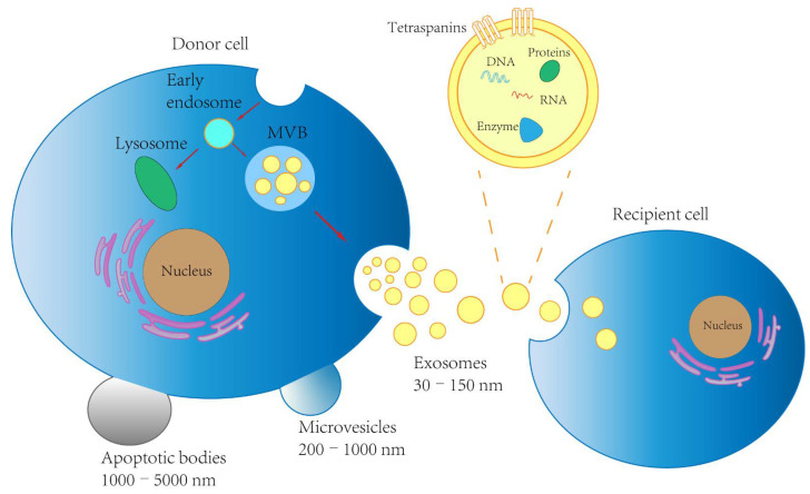
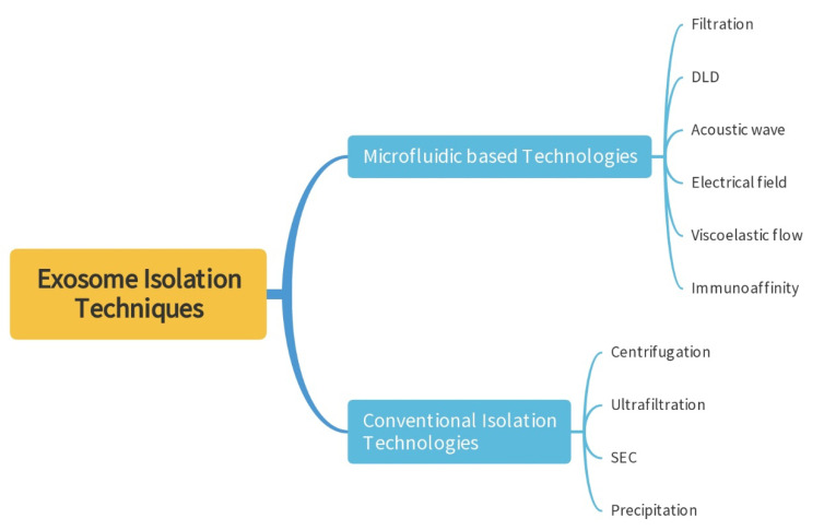
Exosomes are lipid-bilayer enclosed vesicles with diameters of 30–150 nm, which play a pivotal role in cell communication by transporting their cargoes such as proteins, lipids, and genetic materials. In recent years, exosomes have been under intense investigation, as they show great promise in numerous areas, especially as bio-markers in liquid biopsies. However, due to the high heterogeneity and the nano size of exosomes, the separation of exosomes is not easy. This review will deliver an outline of the conventional methods and the microfluidic-based technologies for exosome separation. Particular attention is devoted to microfluidic devices, highlighting the efficiency of exosome isolation by these methods. Additionally, this review will introduce advances made in the integrated microfluidics technologies that enable the separation and analysis of exosomes.
Overview
The claims and statistics of this paper haven’t been checked yet.
Run the check — free, no account needed. It reads the open-access text and checks the sources it cites.
Integrity signals
Registry
Cites
Statistics
Figures
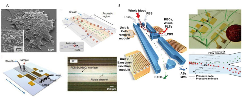
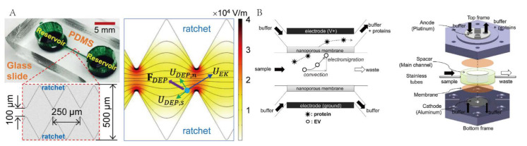
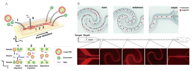
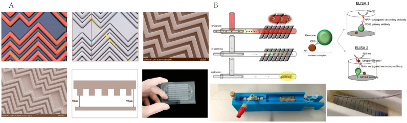
Verification
Claims the paper makes
The claims and statistics of this paper haven’t been checked yet.
Run the check — free, no account needed. It reads the open-access text and checks the sources it cites.
Claims other papers make about this one
Each is checked against this paper’s own text: does it say what the citing paper says it does?
In the citing paper
“Moreover, automation of exosome isolation using systems like microfluidics or centrifugal filtration could increase efficiency and reduce labor costs. Technologies that minimize handling time and reduce manual interventions would also help mitigate batch-to-batch variability [67].”
From Emerging role of exosomes in cancer therapy: progress and challenges (2025) · cites this paper as [67]
Self-check
Percentages, sums and reported differences re-computed from the paper’s own numbers.
The self-check needs the paper’s full text.
Citations
Cites
The biology , function , and biomedical applications of exosomes
Kalluri, R., & LeBleu, V. S. (2020) · Science · doi:10.1126/science.aau6977 · Cited by 10,770
The origins and the future of microfluidics
Whitesides, G. M. (2006) · Nature · doi:10.1038/nature05058 · Cited by 9,687
Johnstone, R. M., Adam, M., Hammond, J. R., Orr, L., & Turbide, C. (1987) · Journal of Biological Chemistry · doi:10.1016/s0021-9258(18)48095-7 · Cited by 2,931 · cited 2×
“For differential ultracentrifugation, the samples were first centrifuged at low speed (300× g) to remove cellular debris, then the large vesicles, such as apoptotic bodies and microvesicles, were removed under a higher speed centrifuge (<20,000× g), and finally the exosomes were precipitated under the action of high c…”
BackgroundMethodGlypican-1 identifies cancer exosomes and detects early pancreatic cancer
Melo, S. A., Luecke, L. B., Kahlert, C., Fernandez, A. F., Gammon, S. T., Kaye, J., LeBleu, V. S., Mittendorf, E. A., Weitz, J., Rahbari, N., et al. (2015) · Nature · doi:10.1038/nature14581 · Cited by 2,880
Self-citation
Research works printed but not linked: 97
- Farooqi, 2018, Exosome biogenesis, bioactivities and functions as new delivery systems of natural compounds, Biotechnol. Adv., 36, 328, 10.1016/j.biotechadv.2017.12.010 · DOI · Paper page
- Raposo, 2018, Shedding light on the cell biology of extracellular vesicles, Nat. Rev. Mol. Cell Biol., 19, 213, 10.1038/nrm.2017.125 · DOI · Paper page
- Raposo, 2013, Extracellular vesicles: Exosomes, microvesicles, and friends, J. Cell Biol., 200, 373, 10.1083/jcb.201211138 · DOI · Paper page
- Shao, 2018, New Technologies for Analysis of Extracellular Vesicles, Chem. Rev., 118, 1917, 10.1021/acs.chemrev.7b00534 · DOI
- Mathieu, 2019, Specificities of secretion and uptake of exosomes and other extracellular vesicles for cell-to-cell communication, Nat. Cell Biol., 21, 9, 10.1038/s41556-018-0250-9 · DOI
- Wortzel, 2019, Exosome-Mediated Metastasis: Communication from a Distance, Dev. Cell, 49, 347, 10.1016/j.devcel.2019.04.011 · DOI
- Jakobsen, 2015, Exosomal proteins as potential diagnostic markers in advanced non-small cell lung carcinoma, J. Extracell Vesicles, 4, 26659, 10.3402/jev.v4.26659 · DOI
- Doyle, L.M., and Wang, M.Z. (2019). Overview of Extracellular Vesicles, Their Origin, Composition, Purpose, and Methods for Exosome Isolation and Analysis. Cells, 8. · DOI
- Cai, 2015, Accessing Genetic Information with Liquid Biopsies, Trends Genet., 31, 564, 10.1016/j.tig.2015.06.001 · DOI
- Yu, 2022, Exosomes as a new frontier of cancer liquid biopsy, Mol. Cancer, 21, 56, 10.1186/s12943-022-01509-9 · DOI · Paper page
- Thery, 2002, Exosomes: Composition, biogenesis and function, Nat. Rev. Immunol., 2, 569, 10.1038/nri855 · DOI
- Yang, 2020, Progress, opportunity, and perspective on exosome isolation-efforts for efficient exosome-based theranostics, Theranostics, 10, 3684, 10.7150/thno.41580 · DOI
- Le, 2021, Exosome isolation using nanostructures and microfluidic devices, Biomed. Mater., 16, 022005
- Royo, F., Thery, C., Falcon-Perez, J.M., Nieuwland, R., and Witwer, K.W. (2020). Methods for Separation and Characterization of Extracellular Vesicles: Results of a Worldwide Survey Performed by the ISEV Rigor and Standardization Subcommittee. Cells, 9. · DOI
- Witwer, 2013, Standardization of sample collection, isolation and analysis methods in extracellular vesicle research, J. Extracell Vesicles, 2, 20360, 10.3402/jev.v2i0.20360 · DOI
- Vidal, 1997, Aggregation reroutes molecules from a recycling to a vesicle-mediated secretion pathway during reticulocyte maturation, J. Cell Sci., 110, 1867, 10.1242/jcs.110.16.1867 · DOI
- Poliakov, 2009, Structural heterogeneity and protein composition of exosome-like vesicles (prostasomes) in human semen, Prostate, 69, 159, 10.1002/pros.20860 · DOI
- Kalra, 2013, Comparative proteomics evaluation of plasma exosome isolation techniques and assessment of the stability of exosomes in normal human blood plasma, Proteomics, 13, 3354, 10.1002/pmic.201300282 · DOI
- 2017, Isolation of Extracellular Vesicles by Ultracentrifugation, Methods Mol. Biol., 1660, 25, 10.1007/978-1-4939-7253-1_3 · DOI
- Zhu, 2019, Exosomes from nicotine-stimulated macrophages accelerate atherosclerosis through miR-21-3p/PTEN-mediated VSMC migration and proliferation, Theranostics, 9, 6901, 10.7150/thno.37357 · DOI
- Li, 2017, Progress in Exosome Isolation Techniques, Theranostics, 7, 789, 10.7150/thno.18133 · DOI
- Alvarez, 2012, Comparison of protein, microRNA, and mRNA yields using different methods of urinary exosome isolation for the discovery of kidney disease biomarkers, Kidney Int., 82, 1024, 10.1038/ki.2012.256 · DOI
- Busatto, S., Vilanilam, G., Ticer, T., Lin, W.L., Dickson, D.W., Shapiro, S., Bergese, P., and Wolfram, J. (2018). Tangential Flow Filtration for Highly Efficient Concentration of Extracellular Vesicles from Large Volumes of Fluid. Cells, 7. · DOI
- Dehghani, 2019, Tangential Flow Microfluidics for the Capture and Release of Nanoparticles and Extracellular Vesicles on Conventional and Ultrathin Membranes, Adv. Mater. Technol., 4, 1900539, 10.1002/admt.201900539 · DOI
- Heinemann, 2017, Sequential Filtration: A Gentle Method for the Isolation of Functional Extracellular Vesicles, Methods Mol. Biol., 1660, 33, 10.1007/978-1-4939-7253-1_4 · DOI
- Haraszti, 2018, Exosomes Produced from 3D Cultures of MSCs by Tangential Flow Filtration Show Higher Yield and Improved Activity, Mol. Ther., 26, 2838, 10.1016/j.ymthe.2018.09.015 · DOI · Paper page
- Nordin, 2015, Ultrafiltration with size-exclusion liquid chromatography for high yield isolation of extracellular vesicles preserving intact biophysical and functional properties, Nanomedicine, 11, 879, 10.1016/j.nano.2015.01.003 · DOI
- Roura, 2019, Extracellular vesicle isolation methods: Rising impact of size-exclusion chromatography, Cell. Mol. Life Sci., 76, 2369, 10.1007/s00018-019-03071-y · DOI
- Sidhom, K., Obi, P.O., and Saleem, A. (2020). A Review of Exosomal Isolation Methods: Is Size Exclusion Chromatography the Best Option?. Int. J. Mol. Sci., 21. · DOI
- Franquesa, 2016, Size-Exclusion Chromatography-based isolation minimally alters Extracellular Vesicles’ characteristics compared to precipitating agents, Sci. Rep., 6, 33641, 10.1038/srep33641 · DOI
- Stranska, 2018, Comparison of membrane affinity-based method with size-exclusion chromatography for isolation of exosome-like vesicles from human plasma, J. Transl. Med., 16, 1, 10.1186/s12967-017-1374-6 · DOI
- Navajas, 2019, Serum Exosome Isolation by Size-Exclusion Chromatography for the Discovery and Validation of Preeclampsia-Associated Biomarkers, Methods Mol. Biol., 1959, 39, 10.1007/978-1-4939-9164-8_3 · DOI
- Weng, 2016, Effective isolation of exosomes with polyethylene glycol from cell culture supernatant for in-depth proteome profiling, Analyst, 141, 4640, 10.1039/C6AN00892E · DOI
- Arbelaiz, 2015, Methods for extracellular vesicles isolation in a hospital setting, Front. Immunol., 6, 50
- Niu, Z., Pang, R.T.K., Liu, W., Li, Q., Cheng, R., and Yeung, W.S.B. (2017). Polymer-based precipitation preserves biological activities of extracellular vesicles from an endometrial cell line. PLoS ONE, 12. · DOI
- Patel, 2019, Comparative analysis of exosome isolation methods using culture supernatant for optimum yield, purity and downstream applications, Sci. Rep., 9, 5335, 10.1038/s41598-019-41800-2 · DOI
- Tian, 2020, Quality and efficiency assessment of six extracellular vesicle isolation methods by nano-flow cytometry, J. Extracell Vesicles, 9, 1697028, 10.1080/20013078.2019.1697028 · DOI · Paper page
- Culbertson, 2014, Micro total analysis systems: Fundamental advances and biological applications, Anal. Chem., 86, 95, 10.1021/ac403688g · DOI
- Liga, 2015, Exosome isolation: A microfluidic road-map, Lab A Chip, 15, 2388, 10.1039/C5LC00240K · DOI
- Singh, P.K., Patel, A., Kaffenes, A., Hord, C., Kesterson, D., and Prakash, S. (2022). Microfluidic Approaches and Methods Enabling Extracellular Vesicle Isolation for Cancer Diagnostics. Micromachines, 13. · DOI
- Liu, 2017, The Exosome Total Isolation Chip, ACS Nano, 11, 10712, 10.1021/acsnano.7b04878 · DOI
- Wang, 2013, Ciliated micropillars for the microfluidic-based isolation of nanoscale lipid vesicles, Lab A Chip, 13, 2879, 10.1039/c3lc41343h · DOI
- Wunsch, 2016, Nanoscale lateral displacement arrays for the separation of exosomes and colloids down to 20 nm, Nat. Nanotechnol., 11, 936, 10.1038/nnano.2016.134 · DOI
- Wu, 2017, Isolation of exosomes from whole blood by integrating acoustics and microfluidics, Proc. Natl. Acad. Sci. USA, 114, 10584, 10.1073/pnas.1709210114 · DOI
- Ibsen, 2017, Rapid Isolation and Detection of Exosomes and Associated Biomarkers from Plasma, ACS Nano, 11, 6641, 10.1021/acsnano.7b00549 · DOI
- Zhou, 2019, Submicron Particle Focusing and Exosome Sorting by Wavy Microchannel Structures within Viscoelastic Fluids, Anal. Chem., 91, 4577, 10.1021/acs.analchem.8b05749 · DOI
- Zhang, 2021, A herringbone mixer based microfluidic device HBEXO-chip for purifying tumor-derived exosomes and establishing miRNA signature in pancreatic cancer, Sens. Actuators B Chem., 332, 129511, 10.1016/j.snb.2021.129511 · DOI
- Sebastian, 2020, Isolation of exosomes from whole blood by a new microfluidic device: Proof of concept application in the diagnosis and monitoring of pancreatic cancer, J. Nanobiotechnol., 18, 150, 10.1186/s12951-020-00701-7 · DOI
- Song, Z., Mao, J., Barrero, R.A., Wang, P., Zhang, F., and Wang, T. (2020). Development of a CD63 Aptamer for Efficient Cancer Immunochemistry and Immunoaffinity-Based Exosome Isolation. Molecules, 25. · DOI
- Davies, 2012, Microfluidic filtration system to isolate extracellular vesicles from blood, Lab A Chip, 12, 5202, 10.1039/c2lc41006k · DOI
- Cho, 2016, Isolation of extracellular vesicle from blood plasma using electrophoretic migration through porous membrane, Sens. Actuators B Chem., 233, 289, 10.1016/j.snb.2016.04.091 · DOI
- Chen, 2021, Exosome detection via the ultrafast-isolation system: EXODUS, Nat. Methods, 18, 212, 10.1038/s41592-020-01034-x · DOI
- Chen, 2018, Detection of exosomes by ZnO nanowires coated three-dimensional scaffold chip device, Biosens. Bioelectron., 122, 211, 10.1016/j.bios.2018.09.033 · DOI
- Salafi, 2019, A Review on Deterministic Lateral Displacement for Particle Separation and Detection, Nanomicro Lett., 11, 77
- Huang, 2004, Continuous particle separation through deterministic lateral displacement, Science, 304, 987, 10.1126/science.1094567 · DOI
- Hattori, 2019, Micro- and Nanopillar Chips for Continuous Separation of Extracellular Vesicles, Anal. Chem., 91, 6514, 10.1021/acs.analchem.8b05538 · DOI
- Ku, 2018, Acoustic Enrichment of Extracellular Vesicles from Biological Fluids, Anal. Chem., 90, 8011, 10.1021/acs.analchem.8b00914 · DOI
- Ding, 2014, Cell separation using tilted-angle standing surface acoustic waves, Proc. Natl. Acad. Sci. USA, 111, 12992, 10.1073/pnas.1413325111 · DOI
- Wu, 2017, Acoustic Separation of Nanoparticles in Continuous Flow, Adv. Funct. Mater., 27, 1606039, 10.1002/adfm.201606039 · DOI
- Chen, 2021, Review on Strategies and Technologies for Exosome Isolation and Purification, Front. Bioeng. Biotechnol., 9, 811971, 10.3389/fbioe.2021.811971 · DOI · Paper page
- Lee, 2015, Acoustic purification of extracellular microvesicles, ACS Nano, 9, 2321, 10.1021/nn506538f · DOI
- Shi, 2019, Rapid and label-free isolation of small extracellular vesicles from biofluids utilizing a novel insulator based dielectrophoretic device, Lab A Chip, 19, 3726, 10.1039/C9LC00902G · DOI
- Malekanfard, 2021, AC Insulator-Based Dielectrophoretic Focusing of Particles and Cells in an “Infinite” Microchannel, Anal. Chem., 93, 5947, 10.1021/acs.analchem.1c00697 · DOI
- Sen, 2021, Label-Free Isolation of Exosomes Using Microfluidic Technologies, ACS Nano, 15, 17047, 10.1021/acsnano.1c03469 · DOI
- Asghari, 2020, Oscillatory Viscoelastic Microfluidics for Efficient Focusing and Separation of Nanoscale Species, ACS Nano, 14, 422, 10.1021/acsnano.9b06123 · DOI
- Lim, 2014, Inertio-elastic focusing of bioparticles in microchannels at high throughput, Nat. Commun., 5, 4120, 10.1038/ncomms5120 · DOI
- Liu, 2017, Field-Free Isolation of Exosomes from Extracellular Vesicles by Microfluidic Viscoelastic Flows, ACS Nano, 11, 6968, 10.1021/acsnano.7b02277 · DOI
- Yang, 2017, Exosome separation using microfluidic systems: Size-based, immunoaffinity-based and dynamic methodologies, Biotechnol. J., 12, 1600699, 10.1002/biot.201600699 · DOI
- Sharma, 2018, Immunoaffinity-based isolation of melanoma cell-derived exosomes from plasma of patients with melanoma, J. Extracell Vesicles, 7, 1435138, 10.1080/20013078.2018.1435138 · DOI
- Yu, 2021, ExoSD chips for high-purity immunomagnetic separation and high-sensitivity detection of gastric cancer cell-derived exosomes, Biosens. Bioelectron., 194, 113594, 10.1016/j.bios.2021.113594 · DOI
- Lo, 2020, Microfluidic device for high-throughput affinity-based isolation of extracellular vesicles, Lab A Chip, 20, 1762, 10.1039/C9LC01190K · DOI
- Yi, 2021, Aptamer-Exosomes for Tumor Theranostics, ACS Sens., 6, 1418, 10.1021/acssensors.0c02237 · DOI
- Dong, 2018, Highly Sensitive Electrochemical Detection of Tumor Exosomes Based on Aptamer Recognition-Induced Multi-DNA Release and Cyclic Enzymatic Amplification, Anal. Chem., 90, 4507, 10.1021/acs.analchem.7b04863 · DOI
- Bai, 2019, Rapid Isolation and Multiplexed Detection of Exosome Tumor Markers Via Queued Beads Combined with Quantum Dots in a Microarray, Nanomicro Lett., 11, 59
- Yu, 2021, Exosome-based liquid biopsies in cancer: Opportunities and challenges, Ann. Oncol., 32, 466, 10.1016/j.annonc.2021.01.074 · DOI
- Li, 2017, Exosomal proteins as potential markers of tumor diagnosis, J. Hematol. Oncol., 10, 175, 10.1186/s13045-017-0542-8 · DOI
- Kanwar, 2014, Microfluidic device (ExoChip) for on-chip isolation, quantification and characterization of circulating exosomes, Lab A Chip, 14, 1891, 10.1039/C4LC00136B · DOI
- Lu, 2017, Elevated glypican-1 expression is associated with an unfavorable prognosis in pancreatic ductal adenocarcinoma, Cancer Med., 6, 1181, 10.1002/cam4.1064 · DOI
- Kleeff, 1998, The cell-surface heparan sulfate proteoglycan glypican-1 regulates growth factor action in pancreatic carcinoma cells and is overexpressed in human pancreatic cancer, J. Clin. Investig., 102, 1662, 10.1172/JCI4105 · DOI
- Tayebi, 2020, Exosome Purification and Analysis Using a Facile Microfluidic Hydrodynamic Trapping Device, Anal. Chem., 92, 10733, 10.1021/acs.analchem.0c02006 · DOI
- Panagopoulou, 2020, Phenotypic analysis of extracellular vesicles: A review on the applications of fluorescence, J. Extracell Vesicles, 9, 1710020, 10.1080/20013078.2019.1710020 · DOI
- Lu, 2022, Integrated microfluidic system for isolating exosome and analyzing protein marker PD-L1, Biosens. Bioelectron., 204, 113879, 10.1016/j.bios.2021.113879 · DOI
- Zhou, 2020, Integrated Microfluidic Device for Accurate Extracellular Vesicle Quantification and Protein Markers Analysis Directly from Human Whole Blood, Anal. Chem., 92, 1574, 10.1021/acs.analchem.9b04852 · DOI
- Zhang, 2016, Ultrasensitive microfluidic analysis of circulating exosomes using a nanostructured graphene oxide/polydopamine coating, Lab A Chip, 16, 3033, 10.1039/C6LC00279J · DOI
- Chen, 2019, Microfluidic device for on-chip isolation and detection of circulating exosomes in blood of breast cancer patients, Biomicrofluidics, 13, 054113, 10.1063/1.5110973 · DOI
- Gholizadeh, 2017, Microfluidic approaches for isolation, detection, and characterization of extracellular vesicles: Current status and future directions, Biosens. Bioelectron., 91, 588, 10.1016/j.bios.2016.12.062 · DOI
- Liang, 2021, Emerging methods in biomarker identification for extracellular vesicle-based liquid biopsy, J. Extracell Vesicles, 10, e12090, 10.1002/jev2.12090 · DOI
- Chen, 2019, An integrated microfluidic system for on-chip enrichment and quantification of circulating extracellular vesicles from whole blood, Lab A Chip, 19, 3305, 10.1039/C9LC00624A · DOI
- Im, 2014, Label-free detection and molecular profiling of exosomes with a nano-plasmonic sensor, Nat. Biotechnol., 32, 490, 10.1038/nbt.2886 · DOI
- Zhu, 2014, Label-free quantitative detection of tumor-derived exosomes through surface plasmon resonance imaging, Anal. Chem., 86, 8857, 10.1021/ac5023056 · DOI
- Lv, 2019, Label-Free Exosome Detection Based on a Low-Cost Plasmonic Biosensor Array Integrated with Microfluidics, Langmuir, 35, 9816, 10.1021/acs.langmuir.9b01237 · DOI
- Wang, 2019, Direct quantification of cancerous exosomes via surface plasmon resonance with dual gold nanoparticle-assisted signal amplification, Biosens. Bioelectron., 135, 129, 10.1016/j.bios.2019.04.013 · DOI
- Yu, 2022, Recent Advances in Bio-Sensing Methods for the Detection of Tumor Exosomes, Crit. Rev. Anal. Chem., 52, 356, 10.1080/10408347.2020.1802220 · DOI
- Huang, 2019, A Sensitive Aptasensor Based on a Hemin/G-Quadruplex-Assisted Signal Amplification Strategy for Electrochemical Detection of Gastric Cancer Exosomes, Small, 15, e1900735, 10.1002/smll.201900735 · DOI
- Zhou, 2016, Development of an aptasensor for electrochemical detection of exosomes, Methods, 97, 88, 10.1016/j.ymeth.2015.10.012 · DOI
- Jeong, 2016, Integrated Magneto-Electrochemical Sensor for Exosome Analysis, ACS Nano, 10, 1802, 10.1021/acsnano.5b07584 · DOI
- Kim, 2020, Detachable microfluidic device implemented with electrochemical aptasensor (DeMEA) for sequential analysis of cancerous exosomes, Biosens. Bioelectron., 169, 112622, 10.1016/j.bios.2020.112622 · DOI
Cited by
Sorted by citations
Emerging role of exosomes in cancer therapy: progress and challenges
Li, J., Wang, J., & Chen, Z. (2025) · Molecular Cancer · doi:10.1186/s12943-024-02215-4 · Cited by 297
Metadata
- Authors
- Wu, Y., Wang, Y., Lu, Y., Luo, X., Huang, Y., Xie, T., Pilarsky, C., Dang, Y., & Zhang, J.
- Venue
- Micromachines
- Publisher
- MDPI AG
- Published
- 2022-09-22
- Volume
- 13
- Issue
- 10
- Pages
- 1571
- ISSN
- 2072-666X
- PMC
- PMC9607600
- Cited by
- 91
- References
- 4
- Language
- en
- Funders
- National Key R&D Program of China (2021YFE0202000, 2021ZDZX2006, PX-1020420, pdjh2022b0425, 2021A080, 02-408-2203-2019, 202002030410)
- Fund of Guangdong Education Department (2021YFE0202000, 2021ZDZX2006, PX-1020420, pdjh2022b0425, 2021A080, 02-408-2203-2019, 202002030410)
- College Student Laboratory Opening Project of Guangzhou Medical University (2021YFE0202000, 2021ZDZX2006, PX-1020420, pdjh2022b0425, 2021A080, 02-408-2203-2019, 202002030410)
- Undergraduates Research Project (2021YFE0202000, 2021ZDZX2006, PX-1020420, pdjh2022b0425, 2021A080, 02-408-2203-2019, 202002030410)
- Fund of Guangzhou Science and Technology Program (2021YFE0202000, 2021ZDZX2006, PX-1020420, pdjh2022b0425, 2021A080, 02-408-2203-2019, 202002030410)
About these checks
Registry, retraction, Cites and statistics results are computed, not judged, with one exception: a printed reference that no index lists is read once by a model before it counts as a citation error. Claims are extracted by a model and kept only when their quote appears verbatim in the text; each cited source is then checked, with a model’s help, for whether it says what the claim relies on. Those verdicts can be wrong — read the claim’s quote and the cited source before relying on one.
Statistics and arithmetic are recomputed, not judged.
Confidence bands are shown only for fields calibrated on our annotated test set. None is calibrated yet, so no band is shown.
What counts as a claim ›How we measure confidence ›How we check this ›
Are you an author, or do you see a problem with this page? Request a review
How to cite
Cite CoChat for the checks on this page, and the paper for its own findings.
- This page
- CoChat. (2026). Citation check: Microfluidic Technology for the Isolation and Analysis of Exosomes. https://app.cochat.ai/papers/10.3390/mi13101571
- The paper
- Wu, Y., Wang, Y., Lu, Y., Luo, X., Huang, Y., Xie, T., Pilarsky, C., Dang, Y., & Zhang, J. (2022). Microfluidic Technology for the Isolation and Analysis of Exosomes. Micromachines. https://doi.org/10.3390/mi13101571
