

The DOI matches its Crossref record; 369 of the 389 references it cites were found in a registry or an index; 75 of the 234 sources its claims cite were checked for support.
Vitamin A Update: Forms, Sources, Kinetics, Detection, Function, Deficiency, Therapeutic Use and Toxicity
Open accessAbstract
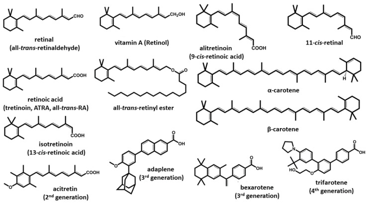
Vitamin A is a group of vital micronutrients widely present in the human diet. Animal-based products are a rich source of the retinyl ester form of the vitamin, while vegetables and fruits contain carotenoids, most of which are provitamin A. Vitamin A plays a key role in the correct functioning of multiple physiological functions. The human organism can metabolize natural forms of vitamin A and provitamin A into biologically active forms (retinol, retinal, retinoic acid), which interact with multiple molecular targets, including nuclear receptors, opsin in the retina.
Overview
CoChat Summary
## TL;DR
This is a narrative review of vitamin A covering its chemical forms, dietary sources, absorption and transport, biological functions, deficiency, therapeutic uses, detection methods, and toxicity. It emphasizes that vitamin A is essential but can be harmful in excess, and that analytical methods must be carefully chosen because endogenous retinoids are present at very low levels.
## Key findings
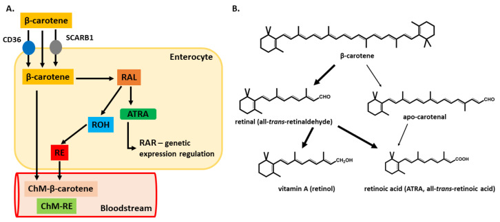
- Vitamin A comes from animal foods as preformed retinol/retinyl esters and from plants as provitamin A carotenoids; only a few carotenoids are important dietary sources, especially β-carotene, α-carotene, and β-cryptoxanthin.
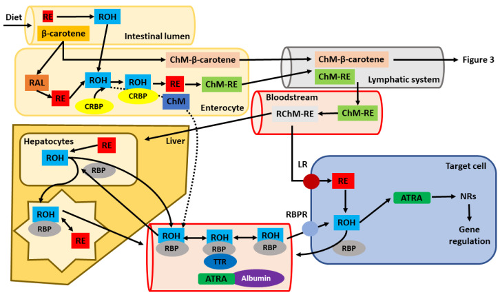
- In the body, retinol is transported mainly by retinol-binding protein (RBP) and transthyretin (TTR); vitamin A is stored largely as retinyl esters in the liver and adipose tissue, and deficiency may take months to appear after intake stops.
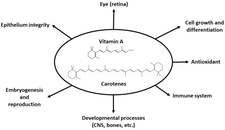
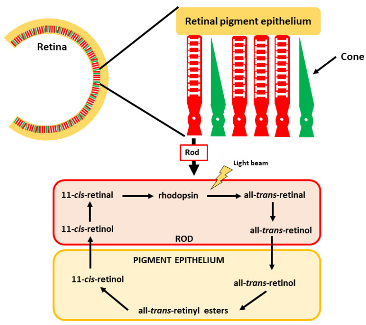
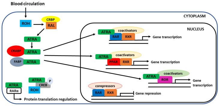
- The review states that vitamin A is involved in vision, gene regulation through nuclear receptors, immune function, epithelial integrity, embryogenesis, bone homeostasis, and other processes; it also notes that ATRA is the major active form of vitamin A.
- Deficiency is linked to night blindness, xerophthalmia, impaired immunity, infections, anemia, developmental problems, and blindness; the paper states that about one-third of the world’s infant population suffers from hypovitaminosis A, around 15% of pregnant women in developing countries are deficient, and vitamin A deficiency causes more than 650,000 child deaths per year in developing countries.
## Methods
- Narrative review article synthesizing previously published literature on vitamin A across sources, pharmacokinetics, function, deficiency, therapy, toxicity, and detection.
- The review draws on reported analytical and biological studies and summarizes findings from dietary, clinical, preclinical, and methodological papers; no original sample was collected.
- For detection methods, it compares instrumental approaches such as (U)HPLC with UV-Vis/DAD, FLD, ECD, MS/MS, SFC, GC-MS, and immunoassays, focusing on sensitivity, specificity, and suitability for endogenous retinoids.
## Limitations
- Not stated in the paper.
- Evident from the text: this is a review, so it does not present original experimental data or a new systematic synthesis with explicit search strategy, inclusion criteria, or risk-of-bias assessment.
- Evident from the text: many conclusions are based on heterogeneous prior studies, and the paper itself notes contradictory findings in areas such as cancer-related effects of retinoids and β-carotene.
Model-written summary. Check it against the paper before relying on it.
Integrity signals
Registry
Cites
Statistics
Figures
Verification
Claims the paper makes
46 Valid · 10 Not valid · 49 Not checked · 36 Own results and interpretations
The cited sources support only part of this claim — read each source’s verdict below.
In the paper
“The vitamin can be provided in the diet either via products of an animal origin in the form of vitamin A (retinol and its close derivatives) or as provitamin A (carotenoids) from vegetables [1].”
§ Vitamin A Update: Forms, Sources, Kinetics, Detection, Function, Deficiency, Therapeutic Use and Toxicity > 1. Introduction and Forms of Vitamin A
What the sources say
- Partly supportedcites [1] Clugston, 2011, Vitamin A metabolism: An update, Nutrients sourceExperimental — accuracy not yet measured
Section 2.1 (page not specified) says dietary retinoids arrive either as preformed retinol and retinyl ester or as proretinoid carotenoids. The passages support the two dietary forms but do not specify animal products as the source of the former or vegetables as the source of the latter. (LLM, conf=0.84)
plain- Asserted: passes — The authors put it forward as true.
- Checkable: passes — It can be checked against evidence.
- Load-bearing: unclear — It is unclear whether the paper’s conclusions depend on it.
- Based: passes — The paper gives a reason why it holds.
- Partly supported
Every source the paper cites for this claim was checked and supports it.
In the paper
“Although the term vitamin A is mostly associated with retinol, and retinol is, in fact, the predominant form of retinoids in the human body, the main biologically active molecules are the oxidized derivates 11-cis-retinal and all-trans-retinoid acid (ATRA) [1,2].”
§ Vitamin A Update: Forms, Sources, Kinetics, Detection, Function, Deficiency, Therapeutic Use and Toxicity > 1. Introduction and Forms of Vitamin A
What the sources say
- Supportedcites [1] Clugston, 2011, Vitamin A metabolism: An update, Nutrients source
Section 5.3 describes 11-cis-retinoid as required for vision, while Section 2.2 reports that retinoic acid acts through its receptors to regulate gene expression. The paper’s retinoid-binding-protein table identifies both 11-cis-retinal and all-trans-retinoic acid. Together, these passages support the sentence’s characterization of them as major biologically active vitamin A derivatives (page numbers unavailable in our copy). (LLM, conf=0.85)
- Supportedcites [2] Kam, 2012, Retinoic acid synthesis and functions in early embryonic development, Cell. Biosci. source
Section 2, “Metabolism of vitamin A and the production of all-trans retinoic acid,” identifies atRA and 11-cis retinal as the key mediators of vitamin A function. Section 2.1 describes the formation of 11-cis retinal by oxidation of 11-cis retinol; section 2.2 describes oxidation in the pathway to atRA. (LLM, conf=0.95)
plain- Asserted: passes — The authors put it forward as true.
- Checkable: passes — It can be checked against evidence.
- Load-bearing: unclear — It is unclear whether the paper’s conclusions depend on it.
- Based: passes — The paper gives a reason why it holds.
- Supported
The cited sources support only part of this claim — read each source’s verdict below.
In the paper
“The values found in individual studies vary considerably, but the highest vitamin A content in the liver is usually reported in pigs [56,57,58,59,60].”
§ Vitamin A Update: Forms, Sources, Kinetics, Detection, Function, Deficiency, Therapeutic Use and Toxicity > 2. Sources of Vitamin A
What the sources say
- Not checked — no full text of the sourcecites [56] Majchrzak, 2006, Vitamin A content (retinol and retinyl esters) in livers of different animals, Food Chem. source
- Not checked — no full text of the sourcecites [57] Vicario, 2015, Carotenoid and Vitamin A Contents in Biological Fluids and Tissues of Animals as an Effect of the Diet: A Review, Food Rev. Int. source
- Not checked — no full text of the sourcecites [58] Ollilainen, 1988, Carotenoids and retinoids in finnish foods: Meat and meat products, J. Food Compos. Anal. source
- Could not verifycites [59] Darwish, 2016, β-carotene and retinol contents in the meat of herbivorous ungulates with a special reference to their public health importance, J. Vet. Med. Sci. sourceExperimental — accuracy not yet measured
The retinol-results passage (indexed under Abstract; page unavailable) compares rats, sheep and other ungulates, but not pigs. A search for pig, pork, swine and porcine found no passage assessing whether pig liver usually has the highest vitamin A content. (LLM, conf=0.78)
- Partly supportedcites [60] Schweigert, 2001, Effect of dietary β-carotene on the accumulation of β-carotene and vitamin A in plasma and tissues of gilts, Reprod. Nutr. Dev. Reprod. Nutr. Dev. sourceExperimental — accuracy not yet measured
Section 3.2 reports vitamin A concentrations in gilt livers and says they were higher than in the other tissues examined. It does not compare liver concentrations across species, so it cannot establish that pigs usually have the highest liver vitamin A content. (LLM, conf=0.79)
hedged- Asserted: passes — The authors put it forward as true.
- Checkable: passes — It can be checked against evidence.
- Load-bearing: unclear — It is unclear whether the paper’s conclusions depend on it.
- Based: passes — The paper gives a reason why it holds.
The cited sources support only part of this claim — read each source’s verdict below.
In the paper
“Drugs such as estrogens and oral contraceptives have been reported to elevate plasma concentrations of RBP, thereby elevating retinoid blood levels [111].”
§ Vitamin A Update: Forms, Sources, Kinetics, Detection, Function, Deficiency, Therapeutic Use and Toxicity > 3. Vitamin A Pharmacokinetics > 3.2. Other Factors Influencing Vitamin A Pharmacokinetics
What the sources say
- Partly supportedcites [111] Jung, 2013, Effects of estrogen receptor alpha and beta on the expression of visfatin and retinol-binding protein 4 in 3T3-L1 adipocytes, Int. J. Mol. Med. sourceExperimental — accuracy not yet measured
The Discussion (page not available) reports that estradiol increased RBP4 secretion in cultured 3T3-L1 adipocytes and describes a correlation between plasma RBP and estradiol levels. It does not report that estrogens or oral contraceptives elevate plasma RBP, or that either raises retinoid blood levels; its proposed effect on retinoic acid metabolism is qualified as an expectation. (LLM, conf=0.86)
hedgedstates a cause- Asserted: passes — The authors put it forward as true.
- Checkable: passes — It can be checked against evidence.
- Load-bearing: unclear — It is unclear whether the paper’s conclusions depend on it.
- Based: passes — The paper gives a reason why it holds.
- Partly supported
The cited sources support only part of this claim — read each source’s verdict below.
In the paper
“Although the placenta has been believed to protect the fetus against high-vitamin A intake from pregnant women being over-supplemented with vitamin A, several studies have reported that high vitamin A intake by the mother can lead to teratogenic effects in the developing fetus [117,118,119].”
§ Vitamin A Update: Forms, Sources, Kinetics, Detection, Function, Deficiency, Therapeutic Use and Toxicity > 3. Vitamin A Pharmacokinetics > 3.3. Pharmacokinetics of Vitamin A during Pregnancy
What the sources say
- Supportedcites [117] Bastos Maia, S., Rolland Souza, A., Costa Caminha, M., Lins Da Silva, S., Callou Cruz, R., Carvalho Dos Santos, C., and Batista Filho, M. (2019). Vitamin A and Pregnancy: A Narrative Review. Nutrients, 11. source
Section 4 reports that high maternal intake of preformed vitamin A during pregnancy may pose a teratogenic risk and describes malformations in children whose mothers consumed high doses. The review distinguishes preformed vitamin A from beta-carotene; it does not suggest that high beta-carotene intake has the same effect. (LLM, conf=0.94)
- Supportedcites [118] Rothman, 1995, Teratogenicity of high vitamin A intake, N. Engl. J. Med. source
The Abstract reports a higher prevalence of birth defects among babies whose mothers consumed high amounts of preformed vitamin A and concludes that high intake appears teratogenic. The Discussion likewise describes a potential teratogenic effect, particularly with intake during early pregnancy; it distinguishes preformed vitamin A from beta carotene. (LLM, conf=0.94)
- Not matchedcites [119] Hunt, 1996, Teratogenicity of high vitamin A intake, N. Engl. J. Med.
Couldn’t be matched to a paper in Crossref, so it can’t be checked.
plainstates a cause- Asserted: passes — The authors put it forward as true.
- Checkable: passes — It can be checked against evidence.
- Load-bearing: passes — The paper’s conclusions depend on it.
- Based: passes — The paper gives a reason why it holds.
- Supported
68 more claims · 25 with their cited sources checked
Create a free account to see all 141 claims and every source verdict.
- It may state two separate claims as one. 52
- Probably not a substantial claim. 23
Claims other papers make about this one
Each is checked against this paper’s own text: does it say what the citing paper says it does?
The source backs part of the claim, not all of it — or backs it for a different population, condition or comparison.
In the citing paper
“Excessive and prolonged beta-carotene consumption causes yellow-orange discolouration of skin thick areas, such as palms and soles, however this is a benign, reversible condition [1,19].”
From The Role of Vitamin A in Retinal Diseases (2022) · cites this paper as [1]
Experimental — accuracy not yet measuredThe source backs part of the claim, not all of it — or backs it for a different population, condition or comparison.
In the citing paper
“In adults, stores of vitamin A in the body can last around 1 to 2 years while in children stores last much shorter and signs of deficiency develop more rapidly [1].”
From The Role of Vitamin A in Retinal Diseases (2022) · cites this paper as [1]
Experimental — accuracy not yet measuredThe source backs part of the claim, not all of it — or backs it for a different population, condition or comparison.
In the citing paper
“It is crucial for reproduction, growth and development of the embryo, normal brain function, immune system functioning, skeletal development, growth of hair and nails, and epithelial cell RNA synthesis including the conjunctiva and cornea.”
From The Role of Vitamin A in Retinal Diseases (2022) · cites this paper as [1]
Experimental — accuracy not yet measuredThe cited source says what the claim attributes to it.
In the citing paper
“One of the most important vitamin A functions is its involvement in visual phototransduction, where it serves as the crucial part of photopigment, the first molecule in the process of transforming photons of light into electrical signals.”
From The Role of Vitamin A in Retinal Diseases (2022) · cites this paper as [1]
The cited source says what the claim attributes to it.
In the citing paper
“It is essential for several physiological processes.”
From The Role of Vitamin A in Retinal Diseases (2022) · cites this paper as [1]
8 more claims about this paper
Create a free account to see every claim other papers make about this one.
Self-check
Percentages, sums and reported differences re-computed from the paper’s own numbers.
Each contradiction between two passages listed here was confirmed by an independent check.
Rated major: it would touch a result — a value a result rests on, reported and recomputed p-values on opposite sides of the significance level, two passages that conflict with no sign which is right, or a cited work retracted before this paper was published.
In the paper
§ Vitamin A Update: Forms, Sources, Kinetics, Detection, Function, Deficiency, Therapeutic Use and Toxicity > 7. Therapeutic Use of Vitamin A
“Whereas most retinoids are usually available in oral preparations, acitretin is indicated for topical use.”
§ Vitamin A Update: Forms, Sources, Kinetics, Detection, Function, Deficiency, Therapeutic Use and Toxicity > 3. Vitamin A Pharmacokinetics > 3.1. Absorption, Distribution, Metabolism and Elimination
“Etretinate and acitretin, which are second-generation retinoids, are also used in oral form in therapy.”
Citations
Cites
Trumbo (2001) · doi:10.1016/s0002-8223(01)00078-5 · Cited by 4,852
Effects of a combination of beta carotene and vitamin A on lung cancer and cardiovascular disease
Omenn, G. S., Goodman, G. E., Thornquist, M. D., Balmes, J., Cullen, M. R., Glass, A., Keogh, J. P., Meyskens, F. L., Valanis, B., Williams, J. H., et al. (1996) · New England Journal of Medicine · doi:10.1056/nejm199605023341802 · Cited by 3,638
A human retinoic acid receptor which belongs to the family of nuclear receptors
Petkovich, M., Brand, N. J., Krust, A., & Chambon, P. (1987) · Nature · doi:10.1038/330444a0 · Cited by 2,214
Serum retinol binding protein 4 contributes to insulin resistance in obesity and type 2 diabetes
Yang, Q., Graham, T. E., Mody, N., Preitner, F., Peroni, O. D., Zabolotny, J. M., Kotani, K., Quadro, L., & Kahn, B. B. (2005) · Nature · doi:10.1038/nature03711 · Cited by 2,086
The epidemiology of global micronutrient deficiencies
Bailey, R. L., West Jr., K. P., & Black, R. E. (2015) · Annals of Nutrition and Metabolism · doi:10.1159/000371618 · Cited by 2,035
Induction of differentiation of the human promyelocytic leukemia cell line (HL-60) by retinoic acid
Breitman, T. R., Selonick, S. E., & Collins, S. J. (1980) · Proceedings of the National Academy of Sciences · doi:10.1073/pnas.77.5.2936 · Cited by 1,966
Identification of a receptor for the morphogen retinoic acid
Giguere, V., Ong, E. S., Segui, P., & Evans, R. M. (1987) · Nature · doi:10.1038/330624a0 · Cited by 1,965
Lammer, E. J., Chen, D. T., Hoar, R. M., Agnish, N. D., Benke, P. J., Braun, J. T., Curry, C. J., Fernhoff, P. M., Grix, A. W., Lott, I. T., et al. (1985) · New England Journal of Medicine · doi:10.1056/nejm198510033131401 · Cited by 1,942
9-cis retinoic acid is a high affinity ligand for the retinoid X receptor
Heyman, R. A., Mangelsdorf, D. J., Dyck, J. A., Stein, R. B., Eichele, G., Evans, R. M., & Thaller, C. (1992) · Cell · doi:10.1016/0092-8674(92)90479-v · Cited by 1,780
TISSUE CHANGES FOLLOWING DEPRIVATION OF FAT-SOLUBLE A VITAMIN
Wolbach, S. B., & Howe, P. R. (1925) · Journal of Experimental Medicine · doi:10.1084/jem.42.6.753 · Cited by 1,647
Research works printed but not linked: 2
- Seroczynska, 2006, Relationship between carotenoids content and flower or fruit flesh colour of winter squash (Cucurbita maxima Duch.), Folia Hortic., 18, 51
- Fedele, 2004, Seasonal variation in retinol concentration of goat milk associated with grazing compared to indoor feeding, S. Afr. J. Anim. Sci., 34, 148
Cited by
A review on the influence of nutraceuticals and functional foods on health
Vignesh, A., Amal, T. C., Sarvalingam, A., & Vasanth, K. (2024) · Food Chemistry Advances · doi:10.1016/j.focha.2024.100749 · Cited by 196
Human Skin Aging and the Anti-Aging Properties of Retinol
Quan, T. (2023) · Biomolecules · doi:10.3390/biom13111614 · Cited by 162
The Role of Vitamin A in Retinal Diseases
Sajovic, J., Meglič, A., Glavač, D., Markelj, Š., Hawlina, M., & Fakin, A. (2022) · International Journal of Molecular Sciences · doi:10.3390/ijms23031014 · Cited by 109
“They are also present in spinach, peas, parsley, pistachios, kale, broccoli and Swiss chard, where green chlorophyll masks the carotenoid pigment [1,10].”
BackgroundInfluentialAshraf, M. V., Khan, S., Misri, S., Gaira, K. S., Rawat, S., Rawat, B., Khan, M. A. H., Shah, A. A., Asgher, M., & Ahmad, S. (2024) · Pharmaceuticals · doi:10.3390/ph17080975 · Cited by 102
The Role of Nutrition on Thyroid Function
Shulhai, A.-M., Rotondo, R., Petraroli, M., Patianna, V. D., Predieri, B., Iughetti, L., Esposito, S., & Street, M. E. (2024) · Nutrients · doi:10.3390/nu16152496 · Cited by 133
“Additionally, vitamin A status appears to modulate the peripheral metabolism of thyroid hormones, including the conversion of T4 to the biologically active T3 hormone [119,125].”
Background
Metadata
- Authors
- Carazo, A., Macáková, K., Matoušová, K., Krčmová, L. K., Protti, M., & Mladěnka, P.
- Affiliations
- Charles University (CZ)
- University Hospital Hradec Králové (CZ)
- University of Bologna (IT)
- Corresponding author
- Carazo, Alejandro
- ORCID
- Venue
- Nutrients
- Publisher
- MDPI AG
- Published
- 2021-05-18
- Volume
- 13
- Issue
- 5
- Pages
- 1703
- ISSN
- 2072-6643 (ISSN-L)
- PMC
- PMC8157347
- OpenAlex
- W3160109639
- Cited by
- 461
- References
- 389
- Citations by year
- 2026: 75 · 2025: 152 · 2024: 120 · 2023: 68 · 2022: 39 · 2021: 7
- FWCI
- 5.96
- Type
- journal article
- Language
- en
- Open access
- gold
- doi.org
- Licence
- Locations
- Nutrients (publishedVersion, cc-by)
- PubMed Central (publishedVersion, cc-by)
- PubMed (publishedVersion)
- Archivio istituzionale della ricerca (Alma Mater Studiorum Università di Bologna) (submittedVersion, cc-by)
- DOAJ (DOAJ: Directory of Open Access Journals) (submittedVersion, cc-by-sa)
- Funders
- ERASMUS+ project strategic partnership (2020-1-CZ01-KA203-078218)
- EFSA-CDN project co-funded by the ERDF (CZ.02.1.01/0.0/0.0/16_019/0000841)
- Topics
- Life Sciences › Biochemistry, Genetics and Molecular Biology › Molecular Biology › Retinoids in leukemia and cellular processes
- Health Sciences › Medicine › Biochemistry (medical) › Antioxidant Activity and Oxidative Stress
- Health Sciences › Medicine › Public Health, Environmental and Occupational Health › Nutrition and Health Studies
- Keywords
- vitamin A, retinol, retinal, retinoic acid, provitamin A, carotenoids, retinyl esters, vitamin A metabolism, vitamin A deficiency, vitamin A toxicity, vitamin A supplementation, nuclear receptors, opsin
- MeSH terms
- Humans, Nutritional Physiological Phenomena, Vitamin A, Vitamin A Deficiency
About these checks
Registry, retraction, Cites and statistics results are computed, not judged, with one exception: a printed reference that no index lists is read once by a model before it counts as a citation error. Claims are extracted by a model and kept only when their quote appears verbatim in the text; each cited source is then checked, with a model’s help, for whether it says what the claim relies on. Those verdicts can be wrong — read the claim’s quote and the cited source before relying on one.
The claim verdicts this report counts are experimental: their accuracy is not yet measured.
A language model drew the claims from the paper on 2026-10-01, under version 3 of the claim definition (model deepseek-v4.1-flash, prompt extract-v3).
A second model labelled each claim on 2026-10-01 (model jev-1.13-20260917, definition revision gold-guideline-v0.2).
Cited sources are checked with a language model’s help (gpt-6-sol).
Statistics and arithmetic are recomputed, not judged.
Confidence bands are shown only for fields calibrated on our annotated test set. None is calibrated yet, so no band is shown.
What counts as a claim ›How we measure confidence ›How we check this ›
Are you an author, or do you see a problem with this page? Request a review
How to cite
Cite CoChat for the checks on this page, and the paper for its own findings.
- This page
- CoChat. (2026). Citation check: Vitamin A Update: Forms, Sources, Kinetics, Detection, Function, Deficiency, Therapeutic Use and Toxicity. https://app.cochat.ai/papers/10.3390/nu13051703
- The paper
- Carazo, A., Macáková, K., Matoušová, K., Krčmová, L. K., Protti, M., & Mladěnka, P. (2021). Vitamin A Update: Forms, Sources, Kinetics, Detection, Function, Deficiency, Therapeutic Use and Toxicity. Nutrients. https://doi.org/10.3390/nu13051703
Data sources
Bibliographic data: Semantic Scholar, OpenAlex, Crossref.
Contains information from the Semantic Scholar Academic Graph Datasets, made available under the ODC Attribution License (ODC-BY 1.0).
Semantic Scholar’s data is described in: Kinney, R., et al. (2023). The Semantic Scholar Open Data Platform. arXiv:2301.10140.
