

The DOI matches its Crossref record; 258 of the 309 references it cites were found in a registry or an index.
Vitamin A in Reproduction and Development
Open accessAbstract
The requirement for vitamin A in reproduction was first recognized in the early 1900’s, and its importance in the eyes of developing embryos was realized shortly after. A greater understanding of the large number of developmental processes that require vitamin A emerged first from nutritional deficiency studies in rat embryos, and later from genetic studies in mice. It is now generally believed that all-trans retinoic acid (RA) is the form of vitamin A that supports both male and female reproduction as well as embryonic development. This conclusion is based on the ability to reverse most reproductive and developmental blocks found in vitamin A deficiency induced either by nutritional or genetic means with RA, and the ability to recapitulate the majority of embryonic defects in retinoic acid receptor compound null mutants. The activity of the catabolic CYP26 enzymes in determining what tissues have access to RA has emerged as a key regulatory mechanism, and helps to explain why exogenous RA can rescue many vitamin A deficiency defects. In severely vitamin A-deficient (VAD) female rats, reproduction fails prior to implantation, whereas in VAD pregnant rats given small amounts of carotene or supported on limiting quantities of RA early in organogenesis, embryos form but show a collection of defects called the vitamin A deficiency syndrome or late vitamin A deficiency. Vitamin A is also essential for the maintenance of the male genital tract and spermatogenesis. Recent studies show that vitamin A participates in a signaling mechanism to initiate meiosis in the female gonad during embryogenesis, and in the male gonad postnatally. Both nutritional and genetic approaches are being used to elucidate the vitamin A-dependent pathways upon which these processes depend.
Overview
The claims and statistics of this paper haven’t been checked yet.
Run the check — free, no account needed. It reads the open-access text and checks the sources it cites.
Integrity signals
Registry
Cites
Statistics
Figures
Verification
Claims the paper makes
The claims and statistics of this paper haven’t been checked yet.
Run the check — free, no account needed. It reads the open-access text and checks the sources it cites.
Claims other papers make about this one
Each is checked against this paper’s own text: does it say what the citing paper says it does?
The source backs part of the claim, not all of it — or backs it for a different population, condition or comparison.
In the citing paper
“Vitamin A or retinol is essential for human survival at every point, from embryogenesis to adulthood ( 2,3 ).”
From Vitamin A and retinoid signaling: genomic and nongenomic effects (2013) · cites this paper as [3]
Experimental — accuracy not yet measuredThe source backs part of the claim, not all of it — or backs it for a different population, condition or comparison.
In the citing paper
“Moreover, several studies show a connection between maternal vitamin A deficiency and diabetes mellitus in adulthood and gestational diabetes. During pregnancy, as shown by several animal studies, vitamin A deficiency affects fetal development negatively, particularly in endocrine pancreas development, suggesting a dominant impact of this micronutrient in the diabetic pathogenesis [128,129].”
From Maternal Overnutrition and Fetal Programming: Long-Term Metabolic, Cognitive, and Epigenetic Consequences (2026) · cites this paper as [129]
Experimental — accuracy not yet measuredThe source backs part of the claim, not all of it — or backs it for a different population, condition or comparison.
In the citing paper
“It is widely known that, conversely, excess vitamin A has teratogenic effects. A vitamin A intake exceeding 10,000 UI at the beginning of the first trimester can lead to serious congenital malformations involving the central nervous system and cardiovascular system, causing miscarriage in the most serious cases.”
From Maternal Overnutrition and Fetal Programming: Long-Term Metabolic, Cognitive, and Epigenetic Consequences (2026) · cites this paper as [129]
Experimental — accuracy not yet measuredThe source backs part of the claim, not all of it — or backs it for a different population, condition or comparison.
In the citing paper
“Vitamin A is a vital micronutrient during pregnancy for both the mother and the baby. It plays a key role in vision, as it is a precursor of rhodopsin, a crucial pigment in retinal photoreceptor cells. It is also involved in cell differentiation, helps preserve eye health, and prevents xerophthalmia; its deficiency remains the leading cause of avoidable blindness worldwide.”
From Maternal Overnutrition and Fetal Programming: Long-Term Metabolic, Cognitive, and Epigenetic Consequences (2026) · cites this paper as [129]
Experimental — accuracy not yet measuredThe check wasn’t confident either way, so it gives no verdict — never disputed.
In the citing paper
“Vitamin A deficiency is a major cause of anemia. Recent studies have shown that vitamin A supplementation during pregnancy increases hemoglobin levels. Although the exact mechanism linking serum retinol levels and anemia is not yet fully understood, it is clear that vitamin A influences hematopoiesis, iron metabolism, and immune function.”
From Maternal Overnutrition and Fetal Programming: Long-Term Metabolic, Cognitive, and Epigenetic Consequences (2026) · cites this paper as [129]
Experimental — accuracy not yet measured
Self-check
Percentages, sums and reported differences re-computed from the paper’s own numbers.
The self-check needs the paper’s full text.
Citations
Cites
A decade of molecular biology of retinoic acid receptors
Chambon, P. (1996) · The FASEB Journal · doi:10.1096/fasebj.10.9.8801176 · Cited by 2,889 · cited 1×
“There are three major subtypes of RAR protein (α, β and γ) with additional isoforms resulting from alternate promoter usage and splicing [29].”
BackgroundThe coregulator exchange in transcriptional functions of nuclear receptors
Glass, C. K., & Rosenfeld, M. G. (2000) · Genes & Development · doi:10.1101/gad.14.2.121 · Cited by 2,233 · cited 1×
“RAREs serve as enhancer elements and when occupied by the RA/RAR/RXR complex, facilitate chromatin opening and changes in RA target gene transcriptional activity [34,35].”
BackgroundInsulin-promoter-factor 1 is required for pancreas development in mice
Jonsson, J., Carlsson, L., Edlund, T., & Edlund, H. (1994) · Nature · doi:10.1038/371606a0 · Cited by 1,879 · cited 1×
“pancreatic buds to grow and differentiate [279–281].”
TISSUE CHANGES FOLLOWING DEPRIVATION OF FAT-SOLUBLE A VITAMIN
Wolbach, S., & Howe, P. R. (1925) · Journal of Experimental Medicine · doi:10.1084/jem.42.6.753 · Cited by 1,674 · cited 2×
“Using dietary manipulation to induce deficiency in rats, the importance of vitamin A in both male and female reproduction was soon discovered [2,3].”
ResultDefects in the kidney and enteric nervous system of mice lacking the tyrosine kinase receptor Ret
Schuchardt, A., D'Agati, V., Larsson-Blomberg, L. B., Costantini, F., & Pachnis, V. (1994) · Nature · doi:10.1038/367380a0 · Cited by 1,658
Graham, A. (2003) · Current Biology · doi:10.1016/s0960-9822(03)00315-4 · Cited by 1,485 · cited 1×
“Much of the craniofacial skeleton originates from neural crest cells [226].”
BackgroundEmbryonic retinoic acid synthesis is essential for early mouse post-implantation development
Niederreither, K., Subbarayan, V., Dollé, P., & Chambon, P. (1999) · Nature Genetics · doi:10.1038/7788 · Cited by 1,111 · cited 3×
“RALDH2 null mutant mice lack nephric ducts [123] and in RARα/β2 double receptor mutants, nephron progenitors, stromal cells and ureteric bud tips are all greatly reduced or completely absent at birth [98,236].”
BackgroundRetinoic Acid Synthesis and Signaling during Early Organogenesis
Duester, G. (2008) · Cell · doi:10.1016/j.cell.2008.09.002 · Cited by 1,098 · cited 1×
“5 in the mouse [136].”
Kessel, M., & Gruss, P. (1991) · Cell · doi:10.1016/0092-8674(91)90574-i · Cited by 1,030 · cited 1×
“5 rat), produces anterior transformations starting at vertebrae 15 (thoracic vertebra 8, T8) whereas excess RA at even later times, yields both rostral posteriorizing and caudal anteriorizations [223,224].”
BackgroundRetinoid signaling determines germ cell fate in mice
Bowles, J., Knight, D., Smith, C., Wilhelm, D., Richman, J., Mamiya, S., Yashiro, K., Chawengsaksophak, K., Wilson, M. J., Rossant, J., et al. (2006) · Science · doi:10.1126/science.1125691 · Cited by 1,011 · cited 3×
“Raldh2 is largely responsible for RA synthesis in the mesonephros, with some contribution from Raldh3 in the region of the mesonephric duct [71,80].”
Background
Research works printed but not linked: 48
- Evans, 1928, The effects of inadequate vitamin A on the sexual physiology of the female, J. Biol. Chem., 77, 651, 10.1016/S0021-9258(20)74018-4 · DOI
- Carpenter, 1997, Experiments That Changed Nutritional Thinking, J. Nutr., 127, 1017S, 10.1093/jn/127.5.1017S · DOI
- Karrer, P. (1966). Nobel Lectures, Chemistry (1922-1941), Elsevier.
- Chithalen, 2002, HPLC-MS/MS analysis of the products generated from all-trans-retinoic acid using recombinant human CYP26A, J. Lipid Res., 43, 1133, 10.1194/jlr.M100343-JLR200 · DOI
- Sporn, M.B., Roberts, A.B., and Goodman, D.S. (1994). The Retinoids: Biology, Chemistry, and Medicine, Raven Press. [2nd].
- Sporn, M.B., Roberts, A.B., and Goodman, D.S. (1994). The Retinoids: Biology, Chemistry and Medicine, Raven Press. [2nd].
- Balmer, 2002, Gene expression regulation by retinoic acid, J. Lipid Res., 43, 1773, 10.1194/jlr.R100015-JLR200 · DOI · Paper page
- Unni, 1983, Histological & ultrastructural studies on the effect of vitamin A depletion & subsequent repletion with vitamin A on germ cells & Sertoli cells in rat testis, Indian J. Exp. Biol., 21, 180
- 1990, Synchronization of the seminiferous epithelium after vitamin A replacement in vitamin A-deficient mice, Biol. Reprod., 43, 363, 10.1095/biolreprod43.3.363 · DOI
- 1990, The origin of the synchronization of the seminiferous epithelium in vitamin A-deficient rats after vitamin A replacement, Biol. Reprod., 42, 677, 10.1095/biolreprod42.4.677 · DOI
- 1991, Retinoic acid is able to reinitiate spermatogenesis in vitamin A-deficient rats and high replicate doses support the full development of spermatogenic cells, Endocrinology, 128, 697, 10.1210/endo-128-2-697 · DOI
- Evans, 1922, On an invariable and characteristic disturbance of reproductive function in animals reared on a diet poor in fat soluble vitamine A, Anat. Rec., 23, 17
- Koubova, 2006, Retinoic acid regulates sex-specific timing of meiotic initiation in mice, Proc. Natl. Acad. Sci. USA, 103, 2474, 10.1073/pnas.0510813103 · DOI · Paper page
- Dickman, 1997, Temporally-regulated retinoic acid depletion produces specific neural crest, ocular and nervous system defects, Development, 124, 3111, 10.1242/dev.124.16.3111 · DOI
- Lohnes, 1994, Function of the retinoic acid receptors (RARs) during development (I). Craniofacial and skeletal abnormalities in RAR double mutants, Development, 120, 2723, 10.1242/dev.120.10.2723 · DOI
- Mendelsohn, 1994, Function of the retinoic acid receptors (RARs) during development (II). Multiple abnormalities at various stages of organogenesis in RAR double mutants, Development, 120, 2749, 10.1242/dev.120.10.2749 · DOI
- Mascrez, 1998, The RXRalpha ligand-dependent activation function 2 (AF-2) is important for mouse development, Development, 125, 4691, 10.1242/dev.125.23.4691 · DOI
- Farese, 1996, A novel function for apolipoprotein B: lipoprotein synthesis in the yolk sac is critical for maternal-fetal lipid transport in mice, J. Lipid Res., 37, 347, 10.1016/S0022-2275(20)37621-5 · DOI
- Mic, 2002, Novel retinoic acid generating activities in the neural tube and heart identified by conditional rescue of Raldh2 null mutant mice, Development, 129, 2271, 10.1242/dev.129.9.2271 · DOI
- Niederreither, 2000, Retinoic acid synthesis and hindbrain patterning in the mouse embryo, Development, 127, 75, 10.1242/dev.127.1.75 · DOI
- Begemann, 2001, The zebrafish neckless mutation reveals a requirement for raldh2 in mesodermal signals that pattern the hindbrain, Development, 128, 3081, 10.1242/dev.128.16.3081 · DOI
- Grandel, 2002, Retinoic acid signalling in the zebrafish embryo is necessary during pre-segmentation stages to pattern the anterior-posterior axis of the CNS and to induce a pectoral fin bud, Development, 129, 2851, 10.1242/dev.129.12.2851 · DOI
- Wendling, 2001, Roles of retinoic acid receptors in early embryonic morphogenesis and hindbrain patterning, Development, 128, 2031, 10.1242/dev.128.11.2031 · DOI
- Dupe, 1999, Key roles of retinoic acid receptors alpha and beta in the patterning of the caudal hindbrain, pharyngeal arches and otocyst in the mouse, Development, 126, 5051, 10.1242/dev.126.22.5051 · DOI
- Dupe, 2001, Hindbrain patterning involves graded responses to retinoic acid signalling, Development, 128, 2199, 10.1242/dev.128.12.2199 · DOI
- MacLean, 2001, Cloning of a novel retinoic-acid metabolizing cytochrome P450, Cyp26B1, and comparative expression analysis with Cyp26A1 during early murine developmen, Mech. Dev., 107, 195, 10.1016/S0925-4773(01)00463-4 · DOI
- Schneider, 2001, Local retinoid signaling coordinates forebrain and facial morphogenesis by maintaining FGF8 and SHH, Development, 128, 2755, 10.1242/dev.128.14.2755 · DOI
- Rohrer, 1991, Retinoic acid induces NGF-dependent survival response and high-affinity NGF receptors in immature chick sympathetic neurons, Development, 112, 813, 10.1242/dev.112.3.813 · DOI
- Plum, 1996, All-trans retinoic acid stimulates and maintains neurite outgrowth in nerve growth factor-supported developing chick embryonic sympathetic neurons, Dev. Biol., 205, 52
- McCaffery, 1991, Aldehyde dehydrogenase is a positional marker in the retina, Development, 112, 693, 10.1242/dev.112.3.693 · DOI
- Mori, 2001, Systematic immunolocalization of retinoid receptors in developing and adult mouse eyes, Invest. Ophthalmol. Vis. Sci., 42, 1312
- Kastner, 1994, Genetic analysis of RXR alpha developmental function: convergence of RXR and RAR signaling pathways in heart and eye morphogenesis, Cell, 78, 987, 10.1016/0092-8674(94)90274-7 · DOI
- Kastner, 1997, Genetic evidence that the retinoid signal is transduced by heterodimeric RXR/RAR functional units during mouse development, Development, 124, 313, 10.1242/dev.124.2.313 · DOI
- Gage, 1999, Dosage requirement of Pitx2 for development of multiple organs, Development, 126, 4643, 10.1242/dev.126.20.4643 · DOI
- Sawada, 2001, Fgf/MAPK signalling is a crucial positional cue in somite boundary formation, Development, 128, 4873, 10.1242/dev.128.23.4873 · DOI
- Kessel, 1992, Respecification of vertebral identities by retinoic acid, Development, 115, 487, 10.1242/dev.115.2.487 · DOI
- Niederreither, 2001, Embryonic retinoic acid synthesis is essential for heart morphogenesis in the mouse, Development, 128, 1019, 10.1242/dev.128.7.1019 · DOI
- Mendelsohn, 1999, Stromal cells mediate retinoid-dependent functions essential for renal development, Development, 126, 1139, 10.1242/dev.126.6.1139 · DOI
- Schuchardt, 1996, Renal agenesis and hypodysplasia in ret-k- mutant mice result from defects in ureteric bud development, Development, 122, 1919, 10.1242/dev.122.6.1919 · DOI
- Brenner, 1997, Nephron mass as a risk factor for progression of renal disease, Kidney Int. Suppl., 63, S124
- Keller, 2003, Nephron number in patients with primary hypertension, New Engl. J. Med., 348, 101, 10.1056/NEJMoa020549 · DOI
- Hind, 2002, Temporal/spatial expression of retinoid binding proteins and RAR isoforms in the postnatal lung, Am. J. Physiol. Lung Cell. Mol. Physiol., 282, L468, 10.1152/ajplung.00196.2001 · DOI
- Malpel, 2000, Regulation of retinoic acid signaling during lung morphogenesis, Development, 127, 3057, 10.1242/dev.127.14.3057 · DOI
- Mollard, 2000, Tissue-specific expression of retinoic acid receptor isoform transcripts in the mouse embryo, Mech. Dev., 94, 223, 10.1016/S0925-4773(00)00303-8 · DOI
- Massaro, 2003, Retinoic acid receptor-alpha regulates pulmonary alveolus formation in mice after, but not during, perinatal perio, Am. J. Physiol. Lung Cell. Mol. Physiol., 284, L431, 10.1152/ajplung.00245.2002 · DOI
- Ahlgren, 1996, The morphogenesis of the pancreatic mesenchyme is uncoupled from that of the pancreatic epithelium in IPF1/PDX1-deficient mice, Development, 122, 1409, 10.1242/dev.122.5.1409 · DOI · Paper page
- Offield, 1996, PDX-1 is required for pancreatic outgrowth and differentiation of the rostral duodenum, Development, 122, 983, 10.1242/dev.122.3.983 · DOI
- Niederreither, 2002, Embryonic retinoic acid synthesis is required for forelimb growth and anteroposterior patterning in the mouse, Development, 129, 3563, 10.1242/dev.129.15.3563 · DOI
Cited by
Sorted by citations
Micronutrient deficiencies in pregnancy worldwide: health effects and prevention
Gernand, A. D., Schulze, K. J., Stewart, C. P., West, K. P., & Christian, P. S. (2016) · Nature Reviews Endocrinology · doi:10.1038/nrendo.2016.37 · Cited by 760
Carazo, A., Macáková, K., Matoušová, K., Krčmová, L. K., Protti, M., & Mladěnka, P. (2021) · Nutrients · doi:10.3390/nu13051703 · Cited by 461
Vitamin A and retinoid signaling: genomic and nongenomic effects
Al Tanoury, Z., Piskunov, A., & Rochette-Egly, C. (2013) · Journal of Lipid Research · doi:10.1194/jlr.r030833 · Cited by 408
Scientific Opinion on Dietary Reference Values for vitamin A
EFSA Panel on Dietetic Products, N. (2015) · EFSA Journal · doi:10.2903/j.efsa.2015.4028 · Cited by 247
“The main symptoms observed in case of deficiency of vitamin A are intrauterine and post-natal growth retardation and a large array of congenital malformations collectively referred to as the fetal “vitamin A deficiency syndrome”, which is well documented in animals (Clagett-Dame and Knutson, 2011).”
BackgroundRetinoic acid signaling pathways in development and diseases
Das, B. C., Thapa, P., Karki, R., Das, S., Mahapatra, S., Liu, T.-C., Torregroza, I., Wallace, D. P., Kambhampati, S., Van Veldhuizen, P., et al. (2014) · Bioorganic & Medicinal Chemistry · doi:10.1016/j.bmc.2013.11.025 · Cited by 276
Functions, Therapeutic Applications, and Synthesis of Retinoids and Carotenoids
Álvarez, R., Vaz, B., Gronemeyer, H., & de Lera, Á. R. (2014) · Chemical Reviews · doi:10.1021/cr400126u · Cited by 260
The vicious cycle of vitamin a deficiency: A review
Wiseman, E. M., Dadon, S. B.-E., & Reifen, R. M. (2016) · Critical Reviews in Food Science and Nutrition · doi:10.1080/10408398.2016.1160362 · Cited by 261
“Vitamin A is not only essential for both male and female reproduction processes (Clagett-Dame and Knutson, 2011), it is also crucial for the prevention of malformations, the maintenance of vertebral identity, and for the development of skeletal elements during embryogenesis (Baume et al.”
BackgroundInfluentialSchweiggert, R., Mezger, D., Schimpf, F., Steingass, C., & Carle, R. (2012) · Food Chemistry · doi:10.1016/j.foodchem.2012.07.035 · Cited by 208
Role of the steroidogenic acute regulatory protein in health and disease
Manna, P. R., Stetson, C. L., Slominski, A. T., & Pruitt, K. (2015) · Endocrine · doi:10.1007/s12020-015-0715-6 · Cited by 218
“The systemic administration of RAs has been shown to reverse most reproductive and developmental blocks in vitamin A deficient (VAD) rats and mice, demonstrating that retinoid signaling rescues reproductive defects as well as steroidogenesis in VAD animals [201, 207, 208].”
BackgroundEnzymology of retinoic acid biosynthesis and degradation
Kedishvili, N. Y. (2013) · Journal of Lipid Research · doi:10.1194/jlr.r037028 · Cited by 196
Vitamin A Deficiency and the Lung
Timoneda, J., Rodríguez‐Fernández, L., Zaragozá, R., Marín, M., Cabezuelo, M. A., Torres, L., Vinã, J. R., & Barber, T. (2018) · Nutrients · doi:10.3390/nu10091132 · Cited by 220
On the role of skin in the regulation of local and systemic steroidogenic activities
Slominski, A. T., Manna, P. R., & Tuckey, R. C. (2015) · Steroids · doi:10.1016/j.steroids.2015.04.006 · Cited by 188
Microencapsulation of vitamin A: A review
Gonçalves, A., Estevinho, B., & Rocha, F. (2016) · Trends in Food Science & Technology · doi:10.1016/j.tifs.2016.03.001 · Cited by 154
Ghiaur, G., Yegnasubramanian, S., Perkins, B. N., Gucwa, J. L., Gerber, J. M., & Jones, R. J. (2013) · Proceedings of the National Academy of Sciences · doi:10.1073/pnas.1305937110 · Cited by 136
“Although RA signaling is essential for the homeostasis of diverse biological systems (5), its role in hematopoiesis, and HSC biology in particular, is still unclear.”
BackgroundImpact of oxidative stress on oocyte competence for in vitro embryo production programs.
Soto-Heras, S., & Paramio, M. (2020) · Research in Veterinary Science · doi:10.1016/j.rvsc.2020.07.013 · Cited by 134
Billings, S. E., Pierzchalski, K., Tjaden, N. E. B., Pang, X., Trainor, P. A., Kane, M. A., & Moise, A. R. (2013) · The FASEB Journal · doi:10.1096/fj.13-227967 · Cited by 128
“Thus, DHRS3 is found in many embryonic tissues where retinoid metabolism and retinoid signaling play important roles (1, 2, 64).”
BackgroundMechanistic aspects of carotenoid health benefits – where are we now?
Bohn, T., Bonet, M. L., Borel, P., Keijer, J., Landrier, J., Milisav, I., Ribot, J., Riso, P., Winklhofer‐Roob, B. M., Sharoni, Y., et al. (2021) · Nutrition Research Reviews · doi:10.1017/s0954422421000147 · Cited by 155
“importance for processes of embryonic development(362,363), reproduction(364,365), epidermal homeostasis and regeneration(366,367), immune responses(368,369) and maintenance of bone, brain, nervous and cardiovascular functions(370–372).”
BackgroundRole of Vitamin A/Retinoic Acid in Regulation of Embryonic and Adult Hematopoiesis
Cañete, A., Cano, E., Muñoz‐Chápuli, R., & Carmona, R. (2017) · Nutrients · doi:10.3390/nu9020159 · Cited by 124
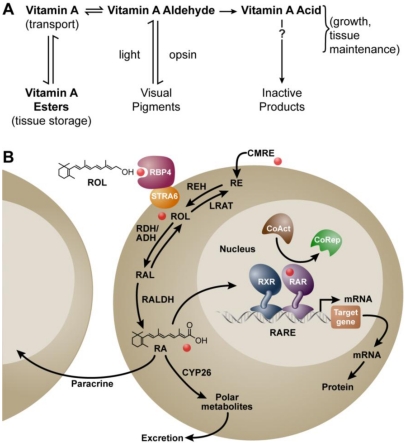
“In the nucleus, the RAR/RXR complex is bound to a specific sequence of DNA (RARE: retinoic acid response element) and usually performs repressor roles in the absence of ligands, activating transcription of target genes when bound to them ([16,17]) (Figure 1).”
BackgroundSandell, L. J., Lynn, M. L., Inman, K. E., McDowell, W., & Trainor, P. A. (2012) · PLoS ONE · doi:10.1371/journal.pone.0030698 · Cited by 127
“Retinoic acid (RA) is a derivative of Vitamin A (retinol) that plays an essential role in many vertebrate biological processes including energy metabolism, brain function, immune response, reproduction and embryonic development (reviewed in [1,2,3, 4,5,6]).”
BackgroundAdams, M. K., Belyaeva, O., Wu, L., & Kedishvili, N. (2014) · Journal of Biological Chemistry · doi:10.1074/jbc.m114.552257 · Cited by 98
Rochette‐Egly, C. (2014) · Biochimica et Biophysica Acta (BBA) - Molecular and Cell Biology of Lipids · doi:10.1016/j.bbalip.2014.04.003 · Cited by 102
Retinol dehydrogenase 10 is indispensible for spermatogenesis in juvenile males
Tong, M.-H., Yang, Q.-E., Davis, J. C., & Griswold, M. (2012) · Proceedings of the National Academy of Sciences of the United States of America · doi:10.1073/pnas.1214883110 · Cited by 95
Piskunov, A., & Rochette-Egly, C. (2012) · Oncogene · doi:10.1038/onc.2011.499 · Cited by 95
“Retinoic acid (RA), a pleiotropic signaling molecule derived from vitamin A, regulates critical genetic programs that control development and homeostasis, cell proliferation and differentiation, as well as cell death or survival (Clagett-Dame and Knutson, 2011).”
BackgroundMolecular regulation of the mitosis/meiosis decision in multicellular organisms.
Kimble, J. (2011) · Cold Spring Harbor Perspectives in Biology · doi:10.1101/cshperspect.a002683 · Cited by 93
“is that VAD blocks meiotic entry (Bowles and Koopman 2007; Clagett-Dame and Knutson 2011).”
Amimo, J. O., Michael, H., Chepngeno, J., Raev, S. A., Saif, L. J., & Vlasova, A. N. (2022) · Nutrients · doi:10.3390/nu14235038 · Cited by 109
“VA importance in reproduction and embryo development was demonstrated in studies showing that VAD female rats had difficulty becoming pregnant and that those that became pregnant produced pups with birth defects [50].”
BackgroundLutein, zeaxanthin and mammalian development: metabolism, functions and implications for health
Giordano, E., & Quadro, L. (2018) · Archives of Biochemistry and Biophysics · doi:10.1016/j.abb.2018.04.008 · Cited by 90
“The essential nutrient vitamin A indeed supports proper mammalian development by exerting critical transcriptional regulatory activities mediated by its active form retinoic acid (18, 19).”
BackgroundPaik, J., Haenisch, M., Muller, C., Goldstein, A., Arnold, S. L. M., Isoherranen, N., Brabb, T., Treuting, P., & Amory, J. (2014) · Journal of Biological Chemistry · doi:10.1074/jbc.m113.540211 · Cited by 84
“In males, either vitamin A deficiency (3) or mutations in genes involved in retinoid signaling pathways leads to perturbations in meiosis and spermatogenesis (1, 2).”
BackgroundSun, L., Kwak, S., & Jin, Y.-S. (2019) · ACS Synthetic Biology · doi:10.1021/acssynbio.9b00217 · Cited by 81
Molecular and Cellular Mechanisms of Intramuscular Fat Development and Growth in Cattle
Tan, Z., & Jiang, H. (2024) · International Journal of Molecular Sciences · doi:10.3390/ijms25052520 · Cited by 92
“Vitamin A is a fat-soluble vitamin crucial for maintaining normal growth and development, immune system function, vision, skin health, and reproduction [114].”
BackgroundFu, X.-M., Feng, C., Wang, C.-Y., Yin, X.-R., Lu, P., Grierson, D., Xu, C.-J., & Chen, K.-S. (2014) · Journal of Experimental Botany · doi:10.1093/jxb/eru257 · Cited by 80
Youness, R. A., Dawoud, A., Eltahtawy, O., & Farag, M. A. (2022) · Nutrition & Metabolism · doi:10.1186/s12986-022-00696-y · Cited by 99
Naturally Occurring Antioxidant Therapy in Alzheimer’s Disease
Collins, A. E., Saleh, T. M., & Kalisch, B. E. (2022) · Antioxidants · doi:10.3390/antiox11020213 · Cited by 93
“Vitamin A maintains essential roles in vision and synaptic function [58], bone growth and development [59], gene expression [60], cell division [61], reproduction [62], the maintenance of epithelial cells in respiratory, intestinal and urinary tracts, and a healthy immune system [63,64].”
BackgroundRetinoids in health and disease: A role for hepatic stellate cells in affecting retinoid levels
Haaker, M. W., Vaandrager, A. B., & Helms, J. B. (2020) · Biochimica et Biophysica Acta (BBA) - Molecular and Cell Biology of Lipids · doi:10.1016/j.bbalip.2020.158674 · Cited by 86
Importance of ALDH1A enzymes in determining human testicular retinoic acid concentrations
Arnold, S. L. M., Kent, T., Hogarth, C., Schlatt, S., Prasad, B., Haenisch, M., Walsh, T., Muller, C., Griswold, M., Amory, J., et al. (2015) · Journal of Lipid Research · doi:10.1194/jlr.m054718 · Cited by 72
McClements, D. J., & Öztürk, B. (2021) · Foods · doi:10.3390/foods10020365 · Cited by 109
“Vitamin A is known to improve visual health, help reproduction, boost the immune system, and promote growth [21].”
BackgroundMolecular Basis for Vitamin A Uptake and Storage in Vertebrates
Chelstowska, S., Widjaja-Adhi, M., Silvaroli, J., & Golczak, M. (2016) · Nutrients · doi:10.3390/nu8110676 · Cited by 72
Metabolic and functional interplay between gut microbiota and fat-soluble vitamins
Stacchiotti, V., Rezzi, S., Eggersdorfer, M. L., & Galli, F. (2020) · Critical Reviews in Food Science and Nutrition · doi:10.1080/10408398.2020.1793728 · Cited by 84
“These bind specific DNA sequences, e.g., the RA response elements, thereby activating transcription of target genes (i.e., genes of immune response and reproduction) (Clagett-Dame and Knutson 2011; Erkelens and Mebius 2017).”
BackgroundWijekoon, M. M. J. O., Mahmood, K., Ariffin, F., Nafchi, A. M., & Zulkurnain, M. (2023) · International Journal of Biological Macromolecules · doi:10.1016/j.ijbiomac.2023.124539 · Cited by 65
Gut microbiota supports male reproduction via nutrition, immunity, and signaling
Cai, H., Cao, X., Qin, D., Liu, Y., Liu, Y., Hua, J., & Peng, S. (2022) · Frontiers in Microbiology · doi:10.3389/fmicb.2022.977574 · Cited by 75
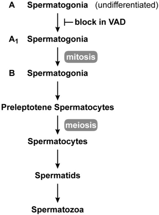
“Vitamin A deficiency leads to the failure of type A spermatogonial stem cells differentiation into type A1; the stratified squamous keratinizing epithelium replaces epithelia of the prostate, epididymis, and seminal vesicle, slowing sperm production (Clagett-Dame and Knutson, 2011).”
BackgroundGlover‐Amengor, M., Aryeetey, R., Afari, E., & Nyarko, A. (2016) · Food Science & Nutrition · doi:10.1002/fsn3.395 · Cited by 64
A Clinician’s Guide to Topical Retinoids
Motamedi, M., Chehade, A., Sanghera, R., & Grewal, P. S. (2021) · Journal of Cutaneous Medicine and Surgery · doi:10.1177/12034754211035091 · Cited by 86
Effects of micronutrients on placental function: evidence from clinical studies to animal models.
Baker, B., Hayes, D., & Jones, R. L. (2018) · Reproduction · doi:10.1530/rep-18-0130 · Cited by 60
“Hypervitaminosis A results in congenital malformations, due to wide-ranging developmental actions, including axial polarisation and cell morphogenesis (Clagett-Dame & Knutson 2011).”
BackgroundTransport of vitamin A across blood‐tissue barriers is facilitated by STRA6
Kelly, M., Widjaja‐Adhi, M. A. K., Palczewski, G., & von Lintig, J. (2016) · The FASEB Journal · doi:10.1096/fj.201600446r · Cited by 60
The Biological Role of Vitamins in Athletes’ Muscle, Heart and Microbiota
Brancaccio, M., Mennitti, C., Cesaro, A., Fimiani, F., Vano, M., Gargiulo, B., Caiazza, M., Amodio, F., Coto, I., D’Alicandro, G., et al. (2022) · International Journal of Environmental Research and Public Health · doi:10.3390/ijerph19031249 · Cited by 66
Kim, J. A., Jang, J., & Lee, S. (2021) · Nutrients · doi:10.3390/nu13093162 · Cited by 68
“Recently, there have been study results indicating that vitamin A is required for the formation of the reproduction system in both males and females [61].”
BackgroundRetinoic acid signaling in ovarian folliculogenesis and steroidogenesis
Damdimopoulou, P., Chiang, C., & Flaws, J. (2019) · Reproductive Toxicology · doi:10.1016/j.reprotox.2019.04.007 · Cited by 56
Li, R., Kang, C., Song, X., Yu, L., Liu, D., He, S., Zhai, H., & Liu, Q. (2017) · Plant Science · doi:10.1016/j.plantsci.2017.05.014 · Cited by 65
Candelier, J. (2015) · Cell Adhesion & Migration · doi:10.1080/19336918.2015.1093275 · Cited by 89
Retinoic acid promotes Sertoli cell differentiation and antagonises activin-induced proliferation.
Nicholls, P. K., Harrison, C., Rainczuk, K., Vogl, A., & Stanton, P. G. (2013) · Molecular and Cellular Endocrinology · doi:10.1016/j.mce.2013.06.034 · Cited by 55
Obesity and male fertility disorders
Cannarella, R., Crafa, A., Curto, R., Condorelli, R. A., Vignera, S. L., & Calogero, A. E. (2024) · Molecular Aspects of Medicine · doi:10.1016/j.mam.2024.101273 · Cited by 63
The molecular aspects of absorption and metabolism of carotenoids and retinoids in vertebrates
Widjaja‐Adhi, M. A. K., & Golczak, M. (2019) · Biochimica et Biophysica Acta (BBA) - Molecular and Cell Biology of Lipids · doi:10.1016/j.bbalip.2019.158571 · Cited by 67
“It is impossible to overestimate the significance of this regulatory molecule for the physiology and pathophysiology of vertebrates as it is involved in the transcriptional regulation of over 500 different genes [5, 156-159].”
BackgroundVitamin A and the eye: an old tale for modern times.
Faustino, J., Ribeiro-Silva, A., Dalto, R. F., de Souza, M. M., Furtado, J. M., Rocha, G. D. M., Alves, M., & Rocha, E. (2016) · Arquivos Brasileiros de Oftalmologia · doi:10.5935/0004-2749.20160018 · Cited by 51
Role of Vitamin A in Mammary Gland Development and Lactation
Cabezuelo, M. T., Zaragozá, R., Barber, T., & Vinã, J. R. (2019) · Nutrients · doi:10.3390/nu12010080 · Cited by 64
Role of Milk Micronutrients in Human Health
Cimmino, F., Catapano, A., Petrella, L., Villano, I., Tudisco, R., & Cavaliere, G. (2023) · Frontiers in Bioscience-Landmark · doi:10.31083/j.fbl2802041 · Cited by 59
“It also sustains fetal development and embryonic growth [95].”
BackgroundAll-trans Retinoic Acid as a Versatile Cytosolic Signal Modulator Mediated by CRABP1
Nagpal, I., & Wei, L. (2019) · International Journal of Molecular Sciences · doi:10.3390/ijms20153610 · Cited by 52
“For cells that do not generate RA, they can obtain RA from RA-producing neighboring cells as a paracrine or from circulation as an endocrine factor [54].”
BackgroundLai, X., Wu, X., Hou, N., Liu, S., Li, Q., Yang, T., Miao, J., Dong, Z., Chen, J., & Li, T. (2017) · Molecular Nutrition & Food Research · doi:10.1002/mnfr.201700754 · Cited by 51
“As one of the important micronutrients for growth and development, VA has multiple physiological functions during embryonic development [39] .”
BackgroundPoly (ADP-ribose) glycohydrolase regulates retinoic acid receptor-mediated gene expression.
May, N. L., Iltis, I., Amé, J., Zhovmer, A., Biard, D., Egly, J., Schreiber, V., & Coin, F. (2012) · Molecules and Cells · doi:10.1016/j.molcel.2012.09.021 · Cited by 48
“The all-trans retinoic acid (t-RA), the ligand of RAR, is a pleiotropic signaling molecule derived from vitamin A that regulates critical genetic programs, thereby controlling cell proliferation and differentiation, as well as cell death or survival (Clagett-Dame and Knutson, 2011).”
BackgroundFrom in planta Function to Vitamin-Rich Food Crops: The ACE of Biofortification
Strobbe, S., Lepeleire, J. D., & Straeten, D. V. D. (2018) · Frontiers in Plant Science · doi:10.3389/fpls.2018.01862 · Cited by 54
Dumont, L., Oblette, A., Rondanino, C., Jumeau, F., Bironneau, A., Liot, D., Duchesne, V., Wils, J., & Rives, N. (2016) · Molecular Human Reproduction · doi:10.1093/molehr/gaw063 · Cited by 54
Vitamin A, endocrine tissues and hormones: interplay and interactions
Brossaud, J., Pallet, V., & Corcuff, J. (2017) · Endocrine Connections · doi:10.1530/ec-17-0101 · Cited by 49
“RA is a critical factor for the formation of the gonads in man and one of the major consequences of vitamin A deficiency apart from blindness is infertility (35, 76).”
BackgroundManna, P., Słominski, A., King, S., Stetson, C., & Stocco, D. (2014) · Endocrinology · doi:10.1210/en.2013-1694 · Cited by 45
Ghasemzadeh-Hasankolaei, M., Eslaminejad, M., & Sedighi-Gilani, M. (2015) · In Vitro Cellular & Developmental Biology - Animal · doi:10.1007/s11626-015-9945-4 · Cited by 44
“Their undeniable functions in cell proliferation, differentiation and apoptosis, reproductive competence, GC production, and triggering GC meiosis and gametogenesis have been described (Bowles and Koopman 2007; Clagett-Dame and Knutson 2011; Gudas and Wagner 2011).”
BackgroundPetrelli, B., Bendelac-Kapon, L., Hicks, G. G., & Fainsod, A. (2019) · genesis · doi:10.1002/dvg.23278 · Cited by 54
“Deviations from the nor- mal physiological condition can result in severe developmental malfor- mations (Adams, 2010; Campo-Paysaa, Marlétaz, Laudet, & Schubert, 2008; Clagett-Dame & Knutson, 2011; Kedishvili, 2013; Mark, Ghyse- linck, & Chambon, 2009).”
D’Aniello, E., & Waxman, J. S. (2014) · Developmental Dynamics · doi:10.1002/dvdy.24232 · Cited by 44
“…et al., 2006; Pan and Baker, 2007; Duester, 2008; Niederreither and Dolle, 2008; Pares et al., 2008; Mark et al., 2009; Pennimpede et al., 2010; Clagett-Dame and Knutson, 2011; Rhinn and Dolle, 2012; Samarut and RochetteEgly, 2012; Schilling et al., 2012; Duester, 2013; Kedishvili, 2013; Das…”
BackgroundKashyap, V., Laursen, K. B., Brenet, F. A., Viale, A. J., Scandura, J. M., & Gudas, L. J. (2012) · Journal of Cell Science · doi:10.1242/jcs.119701 · Cited by 68
Nutrition, genetic variation and male fertility
Vanderhout, S. M., Panah, M. R., García‐Bailo, B., Grace-Farfaglia, P., Samsel, K., Dockray, J., Jarvi, K. A., & El‐Sohemy, A. (2021) · Translational Andrology and Urology · doi:10.21037/tau-20-592 · Cited by 54
“Vitamin A deficiency damages the seminiferous epithelium of the epididymis, prostate, and the seminal vesicle, which results in the termination of spermatogenesis (71).”
BackgroundAlterations in retinoic acid signaling affect the development of the mouse coronary vasculature
Wang, S., Huang, W., Castillo, H. A., Kane, M. A., Xavier‐Neto, J., Trainor, P. A., & Moise, A. R. (2018) · Developmental Dynamics · doi:10.1002/dvdy.24639 · Cited by 43
“Retinoic acid (RA), an active metabolite of vitamin A, carries out regulatory roles in many essential processes during both embryonic and postembryonic life (Clagett-Dame and Knutson, 2011).”
BackgroundAbashev, T. M., Metzler, M. A., Wright, D., & Sandell, L. L. (2017) · Developmental Dynamics · doi:10.1002/dvdy.24476 · Cited by 42
“RA, the active metabolite of vitamin A (all-trans-retinol), is a small lipid soluble molecule that regulates many aspects of embryogenesis and adult health (reviewed in Clagett-Dame and Knutson, 2011).”
BackgroundMarzinke, M. A., & Clagett-Dame, M. (2012) · Experimental Cell Research · doi:10.1016/j.yexcr.2011.10.002 · Cited by 42
Self-citationThe Expanding Role of Cancer Stem Cell Marker ALDH1A3 in Cancer and Beyond
McLean, M. E., MacLean, M. R., Cahill, H. F., Arun, R., Walker, O. L., Wasson, M., Fernando, W., Venkatesh, J., & Marcato, P. (2023) · Cancers · doi:10.3390/cancers15020492 · Cited by 41
“RA treatment can reduce the severity of asthma [18,19], while in contrast, retinoid and vitamin A deficiency exacerbate the condition [20,21].”
BackgroundUp-regulation of steroid biosynthesis by retinoid signaling: Implications for aging
Manna, P. R., Stetson, C. L., Daugherty, C., Shimizu, I., Syapin, P. J., Garrel, G., Cohen-Tannoudji, J., Huhtaniemi, I. T., Slominski, A. T., Pruitt, K., et al. (2015) · Mechanisms of Ageing and Development · doi:10.1016/j.mad.2015.08.007 · Cited by 41
“Retinoids, the metabolically active structural derivatives of vitamin A, especially atRA and 9-cis RA, exert a wide range of effects on development, differentiation, reproduction and epidermal homeostasis (Clagett-Dame and Knutson, 2011; Inoue et al., 2012; Jacobs et al., 2006; Vernet et al., 2006).”
InfluentialChen, W., Li, J.-Z., Ma, Y., Shi, R.-J., Yu, H.-Y., Gantumur, M.-A., Bilawal, A., & Jiang, Z.-M. (2022) · Food Hydrocolloids · doi:10.1016/j.foodhyd.2022.108140 · Cited by 38
Modulation of retinoid signaling: therapeutic opportunities in organ fibrosis and repair
Wang, S., Yu, J., Kane, M. A., & Moise, A. R. (2019) · Pharmacology & Therapeutics · doi:10.1016/j.pharmthera.2019.107415 · Cited by 44
Mambwe, B., Mellody, K. T., Kiss, O., O’Connor, C., Bell, M., Watson, R. E. B., & Langton, A. K. (2024) · International Journal of Cosmetic Science · doi:10.1111/ics.13013 · Cited by 45
Vitamin A Deficiency Induces Congenital Spinal Deformities in Rats
Li, Z., Shen, J.-X., Wu, W., Wang, X., Liang, J.-Q., Qiu, G., & Liu, J.-M. (2012) · PLoS ONE · doi:10.1371/journal.pone.0046565 · Cited by 37
“Nervous system, cardiovascular, and axial patterning defects may be caused by early VAD, whereas a less well-developed nasal region, salivary gland hypoplasia, agenesis of the Harderian glands, hypoplasia of the intestinal villi and a number of skeleton abnormalities arise if VAD occurs at later times [22].”
BackgroundResultEffects of nutrition and gestational alcohol consumption on fetal growth and development
Naik, V. D., Lee, J., Wu, G.-Y., Washburn, S. E., & Ramadoss, J. (2022) · Nutrition reviews · doi:10.1093/nutrit/nuab119 · Cited by 36
Danio rerio embryos on Prozac Effects on the detoxification mechanism and embryo development
Cunha, V. F. P. M., Rodrigues, P., Santos, M. M., Moradas‐Ferreira, P., & Ferreira, M. (2016) · Aquatic Toxicology · doi:10.1016/j.aquatox.2016.08.003 · Cited by 50
“…RXR), play an important role in embryonic development, since both an excess and a deficiency of vitamin A can lead to a wide range of abnormalities in the developing embryos (Klymkowsky et al., 2010; Clagett-Dame and Knutson, 2011; André et al., 2014; Xavier-Neto et al., 2015; Samarut et al., 2015).”
BackgroundThe lycopene β-cyclase plays a significant role in provitamin A biosynthesis in wheat endosperm
Zeng, J., Wang, C., Chen, X., Zang, M., Yuan, C., Wang, X., Wang, Q., Li, M., Li, X., Chen, L., et al. (2015) · BMC Plant Biology · doi:10.1186/s12870-015-0514-5 · Cited by 34
“Given the huge daily consumption of wheat-based products in populations worldwide, increasing the β-carotene content in wheat grains could significantly impact VAD.”
BackgroundInfluentialChen, X., Dodge, J., Komorowski, R., & Drobyski, W. (2012) · Blood · doi:10.1182/blood-2012-08-445130 · Cited by 35
“RA is an active metabolite of vitamin A that is involved in many important biological processes in vivo.(12,13) Within the immune system, RA influences many immune cell lineages and regulates an array of immune responses.”
BackgroundManna, P., Sennoune, S., Martínez‐Zaguilán, R., Słominski, A., & Pruitt, K. (2015) · Biochemical and Biophysical Research Communications - BBRC · doi:10.1016/j.bbrc.2015.06.150 · Cited by 34
Pu, Y., Wang, Z.-F., Bian, Y.-N., Zhang, F., Yang, P., Li, Y.-S., Zhang, Y.-H., Liu, Y., Fang, F., Cao, H.-G., et al. (2014) · Animal science journal = Nihon chikusan Gakkaiho · doi:10.1111/asj.12216 · Cited by 34
Regulation of germ cell meiosis in the fetal ovary.
Spiller, C. M., Bowles, J., & Koopman, P. (2012) · International Journal of Developmental Biology · doi:10.1387/ijdb.120142pk · Cited by 34
Vitamin A Requirements in Pregnancy and Lactation
Gannon, B. M., Jones, C., & Mehta, S. (2020) · Current Developments in Nutrition · doi:10.1093/cdn/nzaa142 · Cited by 33
“Hypervitaminosis A has also led to altered placental morphology and agenesis (154), as well as fetal congenital malformations (15, 17).”
BackgroundBiological Roles of Lipocalins in Chemical Communication, Reproduction, and Regulation of Microbiota
Stopková, R., Otcenásková, T., Matějková, T., Kuntová, B., & Stopka, P. (2021) · Frontiers in Physiology · doi:10.3389/fphys.2021.740006 · Cited by 33
Chen, W., Yu, H.-Y., Shi, R.-J., Ma, C., Gantumur, M.-A., Qayum, A., Bilawal, A., Liang, G.-Z., Oh, K.-C., Jiang, Z.-M., et al. (2021) · Food Hydrocolloids · doi:10.1016/j.foodhyd.2021.106662 · Cited by 33
Zhong, G. G., Hogarth, C. A., Snyder, J. M., Palau, L., Topping, T., Huang, W., Czuba, L. C., LaFrance, J., Ghiaur, G., & Isoherranen, N. (2019) · Journal of Biological Chemistry · doi:10.1074/jbc.ra119.009023 · Cited by 33
“5 (4) Cyp26a1 / 440 153 (4) 267 110 (4) 182 92 (4) 113 61 (3) 8.”
Metabolic disturbances of the vitamin A pathway in human diaphragmatic hernia.
Coste, K., Beurskens, L., Blanc, P., Gallot, D., Delabaere, A., Blanchon, L., Tibboel, D., Labbé, A., Rottier, R., & Sapin, V. (2015) · American Journal of Physiology - Lung cellular and Molecular Physiology · doi:10.1152/ajplung.00108.2014 · Cited by 33
“2) The phenotype of the RAR alpha/beta knockout mice recapitulates most of the lung defects observed in VAD animal models (6, 26).”
BackgroundCutaneous Retinoic Acid Levels Determine Hair Follicle Development and Downgrowth*
Okano, J., Lévy, C., Lichti, U., Sun, H.-W., Yuspa, S., Sakai, Y., & Morasso, M. (2012) · Journal of Biological Chemistry · doi:10.1074/jbc.m112.397273 · Cited by 33
The Impact of Preconceptional Multiple-Micronutrient Supplementation on Female Fertility
Schaefer, E., & Nock, D. (2019) · Clinical Medicine Insights: Women's Health · doi:10.1177/1179562x19843868 · Cited by 32
Jauregui, E. J., Mitchell, D., Topping, T., Hogarth, C., & Griswold, M. (2018) · Development · doi:10.1242/dev.160465 · Cited by 32
“…for spermatogonial differentiation, meiotic initiation, the formation of BTB, spermiogenesis and spermiation (Huang and Hembree, 1979; Griswold et al., 1989; Chung and Wolgemuth, 2004; Wolgemuth and Chung, 2007; Clagett-Dame and Knutson, 2011; Hogarth et al., 2011; Raverdeau et al., 2012).”
BackgroundVo, H. V. T., Nguyen, Y. T., Kim, N., & Lee, H. J. (2023) · International Journal of Molecular Sciences · doi:10.3390/ijms242317038 · Cited by 37
“They are involved in different activities including vision, immunity, cell differentiation, embryological development, cellular differentiation and proliferation and antioxidant functions [4,85].”
BackgroundBreeding peppers for industrial uses: Progress and prospects
Barik, S., Ponnam, N., Reddy, A. C., Lakshmana Reddy, D. C., Saha, K., C, A. G., & Madhavi Reddy, K. (2022) · Industrial Crops and Products · doi:10.1016/j.indcrop.2022.114626 · Cited by 44
Liang, J.-R., Dai, H., Yang, H., Yang, Z., & Wang, Z. (2019) · Poultry Science · doi:10.3382/ps/pez432 · Cited by 31
Palmer, J. A., Smith, A. M., Egnash, L. A., Colwell, M. R., Donley, E. L. R., Kirchner, F. R., & Burrier, R. E. (2017) · Reproductive Toxicology · doi:10.1016/j.reprotox.2017.07.011 · Cited by 35
Jalili, C., Salahshoor, M., & Naderi, T. (2015) · Advanced Biomedical Research · doi:10.4103/2277-9175.151249 · Cited by 31
“[7] Infertility and its associated problems are important issues in the couple lives and sperm dysfunction is a major cause of infertility in men.[8] About 40% of infertility problems are associated with men.”
BackgroundCostabile, B. K., Kim, Y., Iqbal, J., Zuccaro, M. V., Wassef, L., Narayanasamy, S., Curley, R. W., Harrison, E. H., Hussain, M. M., & Quadro, L. (2016) · Journal of Biological Chemistry · doi:10.1074/jbc.m116.738336 · Cited by 36
“The importance of vitamin A as a critical modulator of mammalian embryonic development has been known for decades (1).”
BackgroundChen, Y., Ma, L., Hogarth, C., Wei, G., Griswold, M., & Tong, M.-H. (2016) · Development · doi:10.1242/dev.135939 · Cited by 30
“One of the major biological functions of RA is to inhibit cell proliferation (Bohnsack and Hirschi, 2004; Clagett-Dame and Knutson, 2011); however, RA is capable of stimulating cell proliferation in some type of cells such as neural crest-derived mesenchyme in the forebrain (Schneider et al., 2001)…”
BackgroundEnvironmental aspects of congenital scoliosis
Zheng, L., Yu, X., & Shen, J. (2015) · Environmental Science and Pollution Research · doi:10.1007/s11356-015-4144-0 · Cited by 33
“…the requirement for vitamin A begins with embryonic life as noted by early nutritionists who reported that maternal insufficiency of vitamin A during pregnancy resulted in fetal death or congenital abnormalities in the offspring (Clagett-Dame and Knutson 2011; Lelievre-Pegorier et al. 1998).”
BackgroundModulatory role of vitamin A on the Candida albicans-induced immune response in human monocytes
Klassert, T. E., Hanisch, A., Bräuer, J., Klaile, E., Heyl, K. A., Mansour, M. K., Tam, J. M., Vyas, J. M., & Slevogt, H. (2014) · Medical Microbiology and Immunology · doi:10.1007/s00430-014-0351-4 · Cited by 24
“In this form, vitamin A is known to play an essential role in multiple biological processes, including reproduction, embryogenesis, maintenance of body tissues and augmentation of the immune sys-tem [6–8].”
BackgroundJuvenile hormones direct primordial germ cell migration to the embryonic gonad
Barton, L. J., Sanny, J., Dawson, E. P., Nouzová, M., Noriega, F. G., Stadtfeld, M., & Lehmann, R. (2024) · Current Biology · doi:10.1016/j.cub.2023.12.033 · Cited by 41
Metadata
- Authors
- Clagett-Dame, M., & Knutson, D.
- Affiliations
- Department of Biochemistry, University of Wisconsin-Madison, 433 Babcock Drive, Madison, WI 53706, USA
- University of Wisconsin–Madison (US)
- Venue
- Nutrients
- Publisher
- MDPI AG
- Published
- 2011-03-29
- Volume
- 3
- Issue
- 4
- Pages
- 385-428
- ISSN
- 2072-6643 (ISSN-L)
- PMC
- PMC3257687
- OpenAlex
- W2068902736
- Cited by
- 439
- References
- 309
- Citations by year
- 2026: 27 · 2025: 28 · 2024: 43 · 2023: 46 · 2022: 25 · 2021: 31 · 2020: 36 · 2019: 34 · 2018: 24 · 2017: 24 · 2016: 26 · 2015: 28 · 2014: 28 · 2013: 18 · 2012: 18
- FWCI
- 1.94
- Type
- journal article
- Language
- en
- Open access
- gold
- doi.org
- Licence
- Locations
- Nutrients (publishedVersion, cc-by)
- Europe PMC (PubMed Central) (publishedVersion, cc-by)
- PubMed Central (publishedVersion, cc-by)
- PubMed (publishedVersion)
- DOAJ (DOAJ: Directory of Open Access Journals) (submittedVersion, cc-by-sa)
- Funders
- National Institutes of Health (5p01dk014881-28)
- National Cancer Institute (CA49837, R01CA049837)
- National Institute of Diabetes and Digestive and Kidney Diseases (DK-14881, T32-DK-07665, T32DK007665)
- Topics
- Life Sciences › Biochemistry, Genetics and Molecular Biology › Molecular Biology › Retinoids in leukemia and cellular processes
- Health Sciences › Medicine › Ophthalmology › Retinal Development and Disorders
- Health Sciences › Medicine › Pediatrics, Perinatology and Child Health › Birth, Development, and Health
- Keywords
- vitamin A, all-trans retinoic acid, reproduction, embryonic development, vitamin A deficiency, retinoic acid receptor, spermatogenesis, male genital tract, meiosis initiation, organogenesis, carotene
- MeSH terms
- Retinoic Acid 4-Hydroxylase, Animals, Cytochrome P-450 Enzyme System, Diet, Eye, Female, Infertility, Male, Maternal-Fetal Exchange, Oocytes, Ovary, Pregnancy, Reproduction, Spermatogenesis, Vitamin A, Vitamin A Deficiency, Organogenesis, Embryonic Development, Rats
About these checks
Registry, retraction, Cites and statistics results are computed, not judged, with one exception: a printed reference that no index lists is read once by a model before it counts as a citation error. Claims are extracted by a model and kept only when their quote appears verbatim in the text; each cited source is then checked, with a model’s help, for whether it says what the claim relies on. Those verdicts can be wrong — read the claim’s quote and the cited source before relying on one.
Statistics and arithmetic are recomputed, not judged.
Confidence bands are shown only for fields calibrated on our annotated test set. None is calibrated yet, so no band is shown.
What counts as a claim ›How we measure confidence ›How we check this ›
Are you an author, or do you see a problem with this page? Request a review
