

The DOI matches its Crossref record; 258 of the 309 references it cites were found in a registry or an index.
Vitamin A in Reproduction and Development
Open accessAbstract
The requirement for vitamin A in reproduction was first recognized in the early 1900’s, and its importance in the eyes of developing embryos was realized shortly after. A greater understanding of the large number of developmental processes that require vitamin A emerged first from nutritional deficiency studies in rat embryos, and later from genetic studies in mice. It is now generally believed that all-trans retinoic acid (RA) is the form of vitamin A that supports both male and female reproduction as well as embryonic development. This conclusion is based on the ability to reverse most reproductive and developmental blocks found in vitamin A deficiency induced either by nutritional or genetic means with RA, and the ability to recapitulate the majority of embryonic defects in retinoic acid receptor compound null mutants. The activity of the catabolic CYP26 enzymes in determining what tissues have access to RA has emerged as a key regulatory mechanism, and helps to explain why exogenous RA can rescue many vitamin A deficiency defects. In severely vitamin A-deficient (VAD) female rats, reproduction fails prior to implantation, whereas in VAD pregnant rats given small amounts of carotene or supported on limiting quantities of RA early in organogenesis, embryos form but show a collection of defects called the vitamin A deficiency syndrome or late vitamin A deficiency. Vitamin A is also essential for the maintenance of the male genital tract and spermatogenesis. Recent studies show that vitamin A participates in a signaling mechanism to initiate meiosis in the female gonad during embryogenesis, and in the male gonad postnatally. Both nutritional and genetic approaches are being used to elucidate the vitamin A-dependent pathways upon which these processes depend.
Overview
The claims and statistics of this paper haven’t been checked yet.
Run the check — free, no account needed. It reads the open-access text and checks the sources it cites.
Integrity signals
Registry
Cites
Statistics
Figures
Verification
Claims the paper makes
The claims and statistics of this paper haven’t been checked yet.
Run the check — free, no account needed. It reads the open-access text and checks the sources it cites.
Claims other papers make about this one
Each is checked against this paper’s own text: does it say what the citing paper says it does?
The source backs part of the claim, not all of it — or backs it for a different population, condition or comparison.
In the citing paper
“Vitamin A or retinol is essential for human survival at every point, from embryogenesis to adulthood ( 2,3 ).”
From Vitamin A and retinoid signaling: genomic and nongenomic effects (2013) · cites this paper as [3]
Experimental — accuracy not yet measuredThe source backs part of the claim, not all of it — or backs it for a different population, condition or comparison.
In the citing paper
“Moreover, several studies show a connection between maternal vitamin A deficiency and diabetes mellitus in adulthood and gestational diabetes. During pregnancy, as shown by several animal studies, vitamin A deficiency affects fetal development negatively, particularly in endocrine pancreas development, suggesting a dominant impact of this micronutrient in the diabetic pathogenesis [128,129].”
From Maternal Overnutrition and Fetal Programming: Long-Term Metabolic, Cognitive, and Epigenetic Consequences (2026) · cites this paper as [129]
Experimental — accuracy not yet measuredThe source backs part of the claim, not all of it — or backs it for a different population, condition or comparison.
In the citing paper
“It is widely known that, conversely, excess vitamin A has teratogenic effects. A vitamin A intake exceeding 10,000 UI at the beginning of the first trimester can lead to serious congenital malformations involving the central nervous system and cardiovascular system, causing miscarriage in the most serious cases.”
From Maternal Overnutrition and Fetal Programming: Long-Term Metabolic, Cognitive, and Epigenetic Consequences (2026) · cites this paper as [129]
Experimental — accuracy not yet measuredThe source backs part of the claim, not all of it — or backs it for a different population, condition or comparison.
In the citing paper
“Vitamin A is a vital micronutrient during pregnancy for both the mother and the baby. It plays a key role in vision, as it is a precursor of rhodopsin, a crucial pigment in retinal photoreceptor cells. It is also involved in cell differentiation, helps preserve eye health, and prevents xerophthalmia; its deficiency remains the leading cause of avoidable blindness worldwide.”
From Maternal Overnutrition and Fetal Programming: Long-Term Metabolic, Cognitive, and Epigenetic Consequences (2026) · cites this paper as [129]
Experimental — accuracy not yet measuredThe check wasn’t confident either way, so it gives no verdict — never disputed.
In the citing paper
“Vitamin A deficiency is a major cause of anemia. Recent studies have shown that vitamin A supplementation during pregnancy increases hemoglobin levels. Although the exact mechanism linking serum retinol levels and anemia is not yet fully understood, it is clear that vitamin A influences hematopoiesis, iron metabolism, and immune function.”
From Maternal Overnutrition and Fetal Programming: Long-Term Metabolic, Cognitive, and Epigenetic Consequences (2026) · cites this paper as [129]
Experimental — accuracy not yet measured
Self-check
Percentages, sums and reported differences re-computed from the paper’s own numbers.
The self-check needs the paper’s full text.
Citations
Cites
A decade of molecular biology of retinoic acid receptors
Chambon, P. (1996) · The FASEB Journal · doi:10.1096/fasebj.10.9.8801176 · Cited by 2,889 · cited 1×
“There are three major subtypes of RAR protein (α, β and γ) with additional isoforms resulting from alternate promoter usage and splicing [29].”
BackgroundThe coregulator exchange in transcriptional functions of nuclear receptors
Glass, C. K., & Rosenfeld, M. G. (2000) · Genes & Development · doi:10.1101/gad.14.2.121 · Cited by 2,233 · cited 1×
“RAREs serve as enhancer elements and when occupied by the RA/RAR/RXR complex, facilitate chromatin opening and changes in RA target gene transcriptional activity [34,35].”
BackgroundInsulin-promoter-factor 1 is required for pancreas development in mice
Jonsson, J., Carlsson, L., Edlund, T., & Edlund, H. (1994) · Nature · doi:10.1038/371606a0 · Cited by 1,879 · cited 1×
“pancreatic buds to grow and differentiate [279–281].”
TISSUE CHANGES FOLLOWING DEPRIVATION OF FAT-SOLUBLE A VITAMIN
Wolbach, S., & Howe, P. R. (1925) · Journal of Experimental Medicine · doi:10.1084/jem.42.6.753 · Cited by 1,674 · cited 2×
“Using dietary manipulation to induce deficiency in rats, the importance of vitamin A in both male and female reproduction was soon discovered [2,3].”
ResultDefects in the kidney and enteric nervous system of mice lacking the tyrosine kinase receptor Ret
Schuchardt, A., D'Agati, V., Larsson-Blomberg, L. B., Costantini, F., & Pachnis, V. (1994) · Nature · doi:10.1038/367380a0 · Cited by 1,658
Graham, A. (2003) · Current Biology · doi:10.1016/s0960-9822(03)00315-4 · Cited by 1,485 · cited 1×
“Much of the craniofacial skeleton originates from neural crest cells [226].”
BackgroundEmbryonic retinoic acid synthesis is essential for early mouse post-implantation development
Niederreither, K., Subbarayan, V., Dollé, P., & Chambon, P. (1999) · Nature Genetics · doi:10.1038/7788 · Cited by 1,111 · cited 3×
“RALDH2 null mutant mice lack nephric ducts [123] and in RARα/β2 double receptor mutants, nephron progenitors, stromal cells and ureteric bud tips are all greatly reduced or completely absent at birth [98,236].”
BackgroundRetinoic Acid Synthesis and Signaling during Early Organogenesis
Duester, G. (2008) · Cell · doi:10.1016/j.cell.2008.09.002 · Cited by 1,098 · cited 1×
“5 in the mouse [136].”
Kessel, M., & Gruss, P. (1991) · Cell · doi:10.1016/0092-8674(91)90574-i · Cited by 1,030 · cited 1×
“5 rat), produces anterior transformations starting at vertebrae 15 (thoracic vertebra 8, T8) whereas excess RA at even later times, yields both rostral posteriorizing and caudal anteriorizations [223,224].”
BackgroundRetinoid signaling determines germ cell fate in mice
Bowles, J., Knight, D., Smith, C., Wilhelm, D., Richman, J., Mamiya, S., Yashiro, K., Chawengsaksophak, K., Wilson, M. J., Rossant, J., et al. (2006) · Science · doi:10.1126/science.1125691 · Cited by 1,011 · cited 3×
“Raldh2 is largely responsible for RA synthesis in the mesonephros, with some contribution from Raldh3 in the region of the mesonephric duct [71,80].”
BackgroundOverview of retinoid metabolism and function
Blomhoff, R., & Blomhoff, H. K. (2006) · Journal of Neurobiology · doi:10.1002/neu.20242 · Cited by 981 · cited 1×
“genes are altered when cells or tissues are exposed to RA, however, only a small subset are primary (direct) targets via RARE-mediated transcription, with the remainder representing downstream targets [36,37].”
BackgroundRetinoic acid in the development, regeneration and maintenance of the nervous system
Maden, M. (2007) · Nature Reviews Neuroscience · doi:10.1038/nrn2212 · Cited by 896 · cited 1×
“Vitamin A is required for many aspects of nervous system development including patterning and neural differentiation [157,158].”
BackgroundA Membrane Receptor for Retinol Binding Protein Mediates Cellular Uptake of Vitamin A
Kawaguchi, R., Yu, J., Honda, J., Hu, J., Whitelegge, J., Ping, P., Wiita, P., Bok, D., & Sun, H. (2007) · Science · doi:10.1126/science.1136244 · Cited by 836 · cited 2×
“for a membrane RBP receptor [26,250], as well as to CDH-affected chromosome loci encoding for several other retinoid-related genes [251].”
BackgroundNuclear retinoid receptors and the transcription of retinoid-target genes
Bastien, J., & Rochette-Egly, C. (2004) · Gene · doi:10.1016/j.gene.2003.12.005 · Cited by 773 · cited 1×
“RAREs serve as enhancer elements and when occupied by the RA/RAR/RXR complex, facilitate chromatin opening and changes in RA target gene transcriptional activity [34,35].”
BackgroundWilson, J. G., Roth, C., & Warkany, J. (1953) · American Journal of Anatomy · doi:10.1002/aja.1000920202 · Cited by 758 · cited 3×
“Summary of abnormalities in VAD rat embryos given insufficient RA (late VAD) * [67], insufficient carotene (VAD syndrome) [95], or observed in RAR null mutants [97–99].”
BackgroundRossant, J., Zirngibl, R., Cado, D., Shago, M., & Giguère, V. (1991) · Genes & Development · doi:10.1101/gad.5.8.1333 · Cited by 744 · cited 1×
“Expression of a β2-RARE-lacZ (RARE-lacZ) transgene that is activated in reporter mice when retinoid interacts with the RAR, has been used extensively as a marker of retinoid-signaling activity in vivo [147].”
BackgroundDubrulle, J., McGrew, M., & Pourquié, O. (2001) · Cell · doi:10.1016/s0092-8674(01)00437-8 · Cited by 713 · cited 1×
“A reduction in FGF in the presomitic mesoderm below a threshold value is needed to position the future somite boundary [218,219].”
Backgrounddel Corral, R. D., Olivera-Martínez, I., Goriely, A., Gale, E. A., Maden, M., & Storey, K. G. (2003) · Neuron · doi:10.1016/s0896-6273(03)00565-8 · Cited by 605
Function of retinoic acid receptor γ in the mouse
Lohnes, D., Kastner, P., Dierich, A., Mark, M. P., LeMeur, M., & Chambon, P. (1993) · Cell · doi:10.1016/0092-8674(93)90246-m · Cited by 598 · cited 2×
“Genetic deletion of several RARs produces defects in axial development with anterior cervical transformations similar to VAD embryos [53,97,209].”
BackgroundMark, M. P., Ghyselinck, N. B., & Chambon, P. (2005) · The Annual Review of Pharmacology and Toxicology · doi:10.1146/annurev.pharmtox.46.120604.141156 · Cited by 596 · cited 2×
“importance of the RAR in mediating the actions of RA in developing embryos [99,101,102].”
BackgroundHigh postnatal lethality and testis degeneration in retinoic acid receptor alpha mutant mice.
Lufkin, T., Lohnes, D., Mark, M., Dierich, A., Gorry, P., Gaub, M., Lemeur, M., & Chambon, P. (1993) · Proceedings of the National Academy of Sciences of the United States of America · doi:10.1073/pnas.90.15.7225 · Cited by 573 · cited 1×
“RARα null mutants are sterile and show a reduction in spermatozoa, indicating that nuclear RAR is needed for spermiation [54].”
BackgroundAbu–Abed, S., Dollé, P., METZGER, D., Beckett, B., Chambon, P., & Petkovich, M. P. (2001) · Genes & Development · doi:10.1101/gad.855001 · Cited by 572 · cited 3×
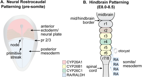
“This is followed by the later expression of Cyp26A1 in r2 and Cyp26C1 in r2 and r4 [18,21,139,144].”
BackgroundDuester, G. (2000) · European Journal of Biochemistry · doi:10.1046/j.1432-1327.2000.01497.x · Cited by 570
Vitamin A metabolism: An update
D’Ambrosio, D. N., Clugston, R. D., & Blaner, W. S. (2011) · Nutrients · doi:10.3390/nu3010063 · Cited by 562 · cited 1×
“Vitamin A and metabolites are lipophilic compounds that are generally found in association with serum and cellular binding proteins [24].”
BackgroundNiederreither, K., McCaffery, P., Dräger, U. C., Chambon, P., & Dollé, P. (1997) · Mechanisms of Development · doi:10.1016/s0925-4773(96)00653-3 · Cited by 523 · cited 3×
“Raldh2 is first expressed during gastrulation in the mesenchyme adjacent to the node and the primitive streak [148].”
BackgroundShenefelt, R. (1972) · Teratology · doi:10.1002/tera.1420050115 · Cited by 512 · cited 1×
“However, too much RA at critical stages can result in embryo lethality or malformation [117–120].”
BackgroundBaltus, A., Menke, D. B., Hu, Y.-C., Goodheart, M., Carpenter, A. E., de Rooij, D., & Page, D. (2006) · Nature Genetics · doi:10.1038/ng1919 · Cited by 501 · cited 1×
“female gonocytes [76].”
Developmental biology of the pancreas: A comprehensive review
Gittes, G. K. (2008) · Developmental Biology · doi:10.1016/j.ydbio.2008.10.024 · Cited by 465 · cited 1×
“Additional information regarding the role of RA in pancreas development can be found in several recent reviews [283,284].”
BackgroundVertebrate limb bud development: moving towards integrative analysis of organogenesis
Zeller, R., López-Rı́os, J., & Zúñiga, A. (2009) · Nature Reviews Genetics · doi:10.1038/nrg2681 · Cited by 463 · cited 1×
“RA has been proposed to play a role in patterning the developing limb by regulating the pattern of expression of genes such as Hand2 (activates SHH), Shh (patterns the anterior-to-posterior axis), and Meis1/2 (transcription factors that mark the proximal limb bud mesenchyme) [287,290–292].”
BackgroundT HE R OLE OF V ITAMIN A IN M AMMALIAN R EPRODUCTION AND E MBRYONIC D EVELOPMENT
Clagett-Dame, M., & DeLuca, H. F. (2002) · Annual Review of Nutrition · doi:10.1146/annurev.nutr.22.010402.102745e · Cited by 461 · cited 1×
“In the female, the effect of vitamin A deficiency on reproductive outcome is dependent upon the time when deficiency is imposed, as well as its severity [58].”
BackgroundSelf-citationMarshall, H., Nonchev, S., Sham, M. H., Muchamore, I., Lumsden, A., & Krumlauf, R. K. (1992) · Nature · doi:10.1038/360737a0 · Cited by 458 · cited 1×
“Hox gene expression appears to be major way in which retinoids affect positional information along the anteroposterior body axis [222–225].”
BackgroundSakai, Y., Meno, C., Fujii, H., Nishino, J., Shiratori, H., Saijoh, Y., Rossant, J., & Hamada, H. (2001) · Genes & Development · doi:10.1101/gad.851501 · Cited by 458 · cited 1×
“character [139,140], however, individual CYP26B1 and CYP26C1 mutants develop normally [143,144].”
BackgroundWhite, J. A., Beckett-Jones, B., Guo, Y.-D., Dilworth, F., Bonasoro, J., Jones, G., & Petkovich, M. (1997) · Journal of Biological Chemistry · doi:10.1074/jbc.272.30.18538 · Cited by 412 · cited 1×
“family (A1, B1 and C1) [17–23].”
Retinoic acid from the meninges regulates cortical neuron generation.
Siegenthaler, J. A., Ashique, A. M., Zarbalis, K., Patterson, K. P., Hecht, J. H., Kane, M. A., Folias, A. E., Choe, Y., May, S. R., Kume, T., et al. (2009) · Cell · doi:10.1016/j.cell.2009.10.004 · Cited by 402 · cited 1×
“division, resulting in the formation of neurons or intermediate progenitor cells [185].”
BackgroundButers, J., Sakai, S., Richter, T., Pineau, T., Alexander, D. L., Savas, U., Doehmer, J., Ward, J. M., Jefcoate, C., & Gonzalez, F. (1999) · Proceedings of the National Academy of Sciences of the United States of America · doi:10.1073/pnas.96.5.1977 · Cited by 400 · cited 1×
“Cyp1B1 can also generate RA and is expressed at known sites of RALDH-independent retinoid signaling [16], however, null mutant mice for this gene are viable [137].”
BackgroundA sonic hedgehog-independent, retinoid-activated pathway of neurogenesis in the ventral spinal cord.
Pierani, A., Brenner-Morton, S., Chiang, C., & Jessell, T. (1999) · Cell · doi:10.1016/s0092-8674(00)80802-8 · Cited by 398 · cited 1×
“motor neurons [190,191].”
THE BIOLOGICAL FUNCTION OF VITAMIN A ACID.
Dowling, J., & Wald, G. (1960) · Proceedings of the National Academy of Sciences of the United States of America · doi:10.1073/pnas.46.5.587 · Cited by 393 · cited 2×
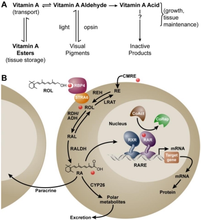
“(A) Metabolic scheme proposed by Dowling and Wald in 1960 [11]; (B) Mechanism ca.”
BackgroundMethodVitamin A and reproduction in rats
Thompson, J. N., Howell, J., & Pitt, G. A. (1964) · Proceedings of the Royal Society of London. Series B. Biological Sciences · doi:10.1098/rspb.1964.0017 · Cited by 381 · cited 1×
“If provided in adequate amounts, retinol will support reproduction and embryonic development in full [62].”
BackgroundFujii, H., Sato, T., Kaneko, S., Gotoh, O., Fujii-Kuriyama, Y., Osawa, K., Kato, S., & Hamada, H. (1997) · EMBO Journal · doi:10.1093/emboj/16.14.4163 · Cited by 374 · cited 2×
“This is followed by the later expression of Cyp26A1 in r2 and Cyp26C1 in r2 and r4 [18,21,139,144].”
BackgroundMatson, C. K., Murphy, M. W., Griswold, M. D., Yoshida, S., Bardwell, V. J., & Zarkower, D. A. (2010) · Developmental Cell · doi:10.1016/j.devcel.2010.09.010 · Cited by 368 · cited 1×
“Recent work supports the conclusion that the vitamin A metabolite, RA, is needed both for adult male spermatogonial differentiation (transition to A1) and the entrance into meiosis [45–47].”
ResultDissociation of the mouse testis and characterization of isolated spermatogenic cells.
Bellvé, A., Millette, C. F., Bhatnagar, Y., & O'Brien, D. A. (1977) · Journal of Histochemistry and Cytochemistry · doi:10.1177/25.7.893996 · Cited by 366 · cited 1×
“testis by postnatal day 6 [84] and spermatogenesis begins shortly thereafter [85].”
BackgroundFunction of retinoic acid receptors during embryonic development
Mark, M., Ghyselinck, N. B., & Chambon, P. (2009) · Nuclear Receptor Signaling · doi:10.1621/nrs.07002 · Cited by 363 · cited 1×
“importance of the RAR in mediating the actions of RA in developing embryos [99,101,102].”
BackgroundPasutto, F., Sticht, H., Hammersen, G., Gillessen-Kaesbach, G., FitzPatrick, D. R., Nürnberg, G., Brasch, F., Schirmer-Zimmermann, H., Tolmie, J. L., Chitayat, D., et al. (2007) · The American Journal of Human Genetics · doi:10.1086/512203 · Cited by 361 · cited 1×
“for a membrane RBP receptor [26,250], as well as to CDH-affected chromosome loci encoding for several other retinoid-related genes [251].”
BackgroundRetinoic acid, meiosis and germ cell fate in mammals
Bowles, J., & Koopman, P. (2007) · Development · doi:10.1242/dev.001107 · Cited by 356 · cited 1×
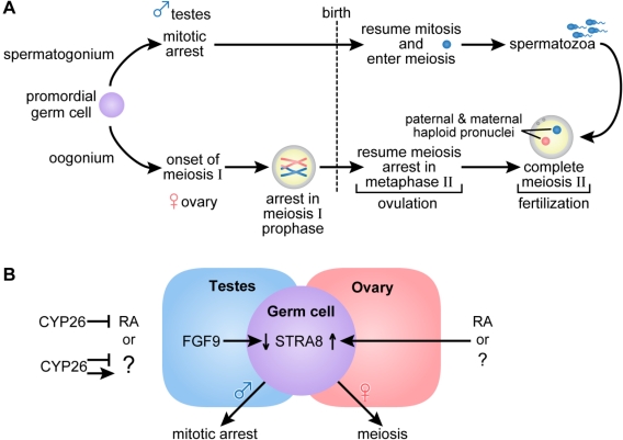
“male embryo, this pathway is blocked by the action of CYP26B1, but is enabled after birth [70–73].”
BackgroundVitamin A-deficient quail embryos have half a hindbrain and other neural defects.
Maden, M., Gale, E., Kostetskii, I., & Zile, M. (1996) · Current Biology · doi:10.1016/s0960-9822(02)00509-2 · Cited by 351 · cited 3×
“RA is also linked to the promotion of neurite outgrowth [157,162].”
BackgroundMethodNovitch, B. G., Wichterle, H., Jessell, T. M., & Sockanathan, S. (2003) · Neuron · doi:10.1016/j.neuron.2003.08.006 · Cited by 350 · cited 1×
“motor neurons [190,191].”
A population-based study of congenital diaphragmatic hernia.
Torfs, C., Curry, C. J. R., Bateson, T., & Honoré, L. H. (1992) · Teratology · doi:10.1002/tera.1420460605 · Cited by 346 · cited 1×
“Congenital diaphragmatic hernia (CDH) occurs in approximately one in 3000 births, and is associated with high neonatal mortality [248].”
BackgroundFrom carrot to clinic: an overview of the retinoic acid signaling pathway
Theodosiou, M., Laudet, V., & Schubert, M. (2010) · Cellular and Molecular Life Sciences · doi:10.1007/s00018-010-0268-z · Cited by 344 · cited 1×
“mice; some have been found to be dispensable, while roles for others have been revealed when animals are fed a vitamin A-restricted diet [12,27,28].”
BackgroundThe Necessity of Certain Lipins in the Diet during Growth.
Mccollum, E. V., & Davis, M. (1913) · Nutrition Reviews · doi:10.1111/j.1753-4887.1973.tb07065.x · Cited by 342 · cited 1×
“In 1913 McCollum and Davis reported that the addition of an ether extract from egg yolk or butter, but not lard or olive oil, could reinstate growth in rats maintained for several months on a purified ration of casein, carbohydrates and salt mixtures [1].”
BackgroundSandell, L. J., Sanderson, B. W., Moiseyev, G. P., Johnson, T., Mushegian, A. R., Young, K., Rey, J., Ma, J., Staehling‐Hampton, K., & Trainor, P. A. (2007) · Genes & Development · doi:10.1101/gad.1533407 · Cited by 341 · cited 2×
“Mice null for RALDH2 or RDH10, also have lung agenesis or hypoplastic phenotypes [122,258,259].”
BackgroundMercader, N., Leonardo, E., Piedra, M., Martı́nez-A, C., Ros, M. A., & Torres, M. (2000) · Development · doi:10.7892/boris.79628 · Cited by 339 · cited 1×
“RA has been proposed to play a role in patterning the developing limb by regulating the pattern of expression of genes such as Hand2 (activates SHH), Shh (patterns the anterior-to-posterior axis), and Meis1/2 (transcription factors that mark the proximal limb bud mesenchyme) [287,290–292].”
BackgroundNephron number, hypertension, renal disease, and renal failure.
Hoy, W., Hughson, M., Bertram, J., Douglas-Denton, R. N., & Amann, K. (2005) · Journal of the American Society of Nephrology · doi:10.1681/asn.2005020172 · Cited by 320 · cited 1×
“In kidney, the number of embryonic branching events determines the final number of nephrons an individual will have for life, and it has been proposed that suboptimal nephron number at birth increases susceptibility to acquired renal disease and essential hypertension later in life [243–246].”
BackgroundDupé, V., Matt, N., Garnier, J., Chambon, P., Mark, M. P., & Ghyselinck, N. B. (2003) · Proceedings of the National Academy of Sciences · doi:10.1073/pnas.2336223100 · Cited by 319 · cited 2×
“RA acting through the RAR is also necessary for the morphogenesis of other components of the respiratory tract including partitioning of the primitive foregut into oesophagus and trachea, and for the opening between the nasal and oropharyngeal cavities [67,131,209,256,258,261].”
BackgroundYashiro, K., Zhao, X., Uehara, M., Yamashita, K., Nishijima, M., Nishino, J., Saijoh, Y., Sakai, Y., & Hamada, H. (2004) · Developmental Cell · doi:10.1016/s1534-5807(04)00062-0 · Cited by 316 · cited 2×
“These studies show that controlled exclusion of RA from the limb bud is essential for proper limb morphology [142].”
BackgroundRetinoic acid signaling is required for a critical early step in zebrafish pancreatic development
Stafford, D., & Prince, V. E. (2002) · Current Biology · doi:10.1016/s0960-9822(02)00929-6 · Cited by 309 · cited 1×
“The effects of RA deficiency on pancreatic lineages appear to be due to the loss of pancreatic field specification within the endoderm [274].”
BackgroundPostnatal treatment with retinoic acid increases the number of pulmonary alveoli in rats.
Massaro, G., & Massaro, D. (1996) · American Journal of Physiology · doi:10.1152/ajplung.1996.270.2.l305 · Cited by 309 · cited 1×
“reports that exogenous RA can stimulate alveoli formation in immature rat and mouse lung [264–269].”
Backgrounddel Corral, R. D., & Storey, K. G. (2004) · BioEssays · doi:10.1002/bies.20080 · Cited by 295
Dorsal pancreas agenesis in retinoic acid-deficient Raldh2 mutant mice.
Martín, M., Gallego-Llamas, J., Ribes, V., Kedinger, M., Niederreither, K., Chambon, P., Dollé, P., & Gradwohl, G. (2005) · Developmental Biology · doi:10.1016/j.ydbio.2005.05.035 · Cited by 295 · cited 5×
“null mutant mice, a requirement for RA in pancreatic development has been proposed based on studies in RALDH null mutant mice [271,272] and in several other organisms in which retinoid signaling is deficient or inhibited, including Xenopus, zebrafish, chick, and quail [273–276].”
BackgroundInfluentialApoptotic extinction of germ cells in testes of Cyp26b1 knockout mice.
Maclean, G., Li, H., Metzger, D., Chambon, P., & Petkovich, M. (2007) · Endocrinology · doi:10.1210/en.2007-0492 · Cited by 291 · cited 2×
“5, and this is followed by apoptosis [73].”
BackgroundStem cells, signals and vertebrate body axis extension
Wilson, V., Olivera-Martinez, I., & Storey, K. G. (2009) · Development · doi:10.1242/dev.021246 · Cited by 290 · cited 1×
“comprises the caudal end of the node and the rostral end of the primitive streak [186].”
BackgroundCollins, M. D., & Mao, G. E. (1999) · The Annual Review of Pharmacology and Toxicology · doi:10.1146/annurev.pharmtox.39.1.399 · Cited by 290 · cited 1×
“However, too much RA at critical stages can result in embryo lethality or malformation [117–120].”
BackgroundVitamin A controls epithelial/mesenchymal interactions through Ret expression
Batourina, E., Gim, S., Bello, N. A., Shy, M., Clagett‐Dame, M., Srinivas, S., Costantini, F. D., & Mendelsohn, C. L. (2001) · Nature Genetics · doi:10.1038/83792 · Cited by 288 · cited 3×
“mutants rescues renal development, restoring ureteric bud growth and stromal cell patterning [237].”
BackgroundSelf-citationSignaling Gradients during Paraxial Mesoderm DevelopmentPreprint
Aulehla, A., & Pourquie, O. (2009) · Cold Spring Harbor Perspectives in Biology · doi:10.1101/cshperspect.a000869 · Cited by 277 · cited 2×
“RA plays a role in the initiation of differentiation of the anterior region of the presomitic mesoderm from which the new somites originate [217].”
BackgroundPLASMA RETINOL‐BINDING PROTEIN*
Goodman, D. S. (1980) · Annals of the New York Academy of Sciences · doi:10.1111/j.1749-6632.1980.tb21314.x · Cited by 276
Signaling Pathways Controlling Second Heart Field Development
Rochais, F., Mesbah, K., & Kelly, R. (2009) · Circulation Research · doi:10.1161/circresaha.109.194464 · Cited by 276 · cited 1×
“reviews [229,232,234].”
The key role of vitamin A in spermatogenesis.
Hogarth, C., & Griswold, M. (2010) · Journal of Clinical Investigation · doi:10.1172/jci41303 · Cited by 263 · cited 2×
“Recent work supports the conclusion that the vitamin A metabolite, RA, is needed both for adult male spermatogonial differentiation (transition to A1) and the entrance into meiosis [45–47].”
BackgroundResultDynamic and combinatorial control of gene expression by nuclear retinoic acid receptors (RARs)
Rochette‐Egly, C., & Germain, P. (2009) · Nuclear Receptor Signaling · doi:10.1621/nrs.07005 · Cited by 263 · cited 1×
“The nuclear RARs act as ligand-activated transcription factors to regulate gene transcription in a cell-type and tissue-specific manner [30].”
BackgroundAortic-arch and cardiac anomalies in the offspring of vitamin A deficient rats.
Wilson, J. G., & Warkany, J. (1949) · American Journal of Anatomy · doi:10.1002/aja.1000850106 · Cited by 261 · cited 1×
“Cardiac and aortic arch defects were observed as a part of the early vitamin A deficiency syndrome in rat embryos [93,94] and in RAR compound null mutant mouse embryos [98,102].”
BackgroundZhou, Q., Nie, R., Li, Y., Friel, P., Mitchell, D., Hess, R., Small, C., & Griswold, M. (2008) · Biology of Reproduction · doi:10.1095/biolreprod.107.066795 · Cited by 260 · cited 1×
“testis by postnatal day 6 [84] and spermatogenesis begins shortly thereafter [85].”
BackgroundRetinoic acid metabolism and signaling pathways in the adult and developing mouse testis.
Vernet, N., Dennefeld, C., Rochette-Egly, C., Oulad-Abdelghani, M., Chambon, P., Ghyselinck, N., & Mark, M. (2006) · Endocrinology · doi:10.1210/en.2005-0953 · Cited by 259 · cited 4×
“Within the normal tubule, the Sertoli cell is believed to generate RA by the action of Raldh1 [49,51,52], and possibly Raldh2 [50].”
BackgroundInfluentialRetinoic acid-dependent eye morphogenesis is orchestrated by neural crest cells
Matt, N., Dupé, V., Garnier, J., Dennefeld, C., Chambon, P., Mark, M. P., & Ghyselinck, N. B. (2005) · Development · doi:10.1242/dev.02031 · Cited by 256 · cited 3×
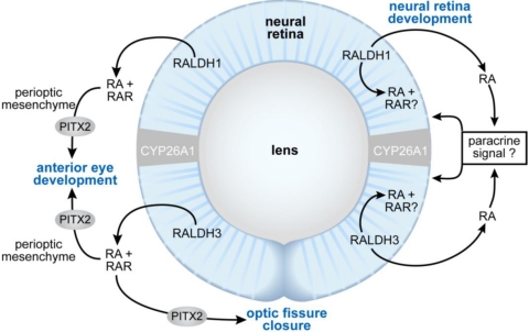
“A number of eye defects in RALDH3 and RALDH1/3 compound mutants [134,135,178] are also similar to those observed in late VAD rat embryos.”
BackgroundEndogenous distribution of retinoids during normal development and teratogenesis in the mouse embryo
Horton, C., & Maden, M. (1995) · Developmental Dynamics · doi:10.1002/aja.1002020310 · Cited by 256
Mild vitamin A deficiency leads to inborn nephron deficit in the rat.
Lelièvre-Pégorier, M., Vilar, J., Ferrier, M., Moreau, E., Freund, N., Gilbert, T., & Merlet-Bénichou, C. (1998) · Kidney International · doi:10.1046/j.1523-1755.1998.00151.x · Cited by 250
White, J. A., Ramshaw, H., Taimi, M., Stangle, W., Zhang, A., Everingham, S., Creighton, S., Tam, S.-P., Jones, G., & Petkovich, M. (2000) · Proceedings of the National Academy of Sciences · doi:10.1073/pnas.120161397 · Cited by 248 · cited 1×
“family (A1, B1 and C1) [17–23].”
FGF9 suppresses meiosis and promotes male germ cell fate in mice.
Bowles, J., Feng, C., Spiller, C. M., Davidson, T.-L., Jackson, A., & Koopman, P. (2010) · Developmental Cell · doi:10.1016/j.devcel.2010.08.010 · Cited by 247 · cited 1×
“(Adapted from [77,78]).”
Niederreither, K., Fraulob, V., Garnier, J.-M., Chambon, P., & Dollé, P. (2002) · Mechanisms of Development · doi:10.1016/s0925-4773(01)00561-5 · Cited by 246 · cited 1×
“In the embryonic kidney, Raldh2 is localized in stromal mesenchyme of the outer cortex and Raldh3 is expressed in the ureteric bud [130,237,238].”
BackgroundCyp26 enzymes generate the retinoic acid response pattern necessary for hindbrain development
Hernandez, R. E., Putzke, A. P., Myers, J. P., Margaretha, L., & Moens, C. B. (2007) · Development · doi:10.1242/dev.02706 · Cited by 245 · cited 9×
“Interestingly, in Cyp26 morpholino studies in zebrafish, restriction of the RA-responsive gene, Vhnff1, is determined by the posterior limit of Cyp26C1 at the r4/r5 boundary, a function that can be compensated for by CYP26B1 [173].”
BackgroundInfluentialMic, F. A., Molotkov, A., Benbrook, D. M., & Duester, G. (2003) · Proceedings of the National Academy of Sciences · doi:10.1073/pnas.1231422100 · Cited by 241 · cited 1×
“However, work from the Duester group indicates that ligand binding to the RAR is sufficient to transduce the signal, as a RAR- but not a RXR- selective ligand is able to rescue the developmental defects in RALDH2 null mutant embryos [108].”
BackgroundRole of the retinoic acid receptor beta (RARbeta) during mouse development.
Ghyselinck, N., Dupé, V., Dierich, A., Messaddeq, N., Garnier, J.-M., Rochette-Egly, C., Chambon, P., & Mark, M. (1997) · International Journal of Developmental Biology · doi:10.1387/ijdb.9240560 · Cited by 240 · cited 7×
“number of RAR mutants show both cervical anteriorizations and posteriorization at the cervical thoracic junction [97,209].”
BackgroundInfluentialMoore, T. S., II (1930) · Biochemical Journal · doi:10.1042/bj0240692 · Cited by 233 · cited 1×
“This problem was solved when Moore fed carotenoids to vitamin A-deficient (VAD) rats in amounts that enabled the animals to resume normal growth, and showed that only the ―colorless‖ form of vitamin A was found in the livers collected from these animals [5].”
BackgroundTeratogenic activity of retinoic acid.
Kochhar, D. (2009) · Acta pathologica et microbiologica Scandinavica · doi:10.1111/j.1699-0463.1967.tb01308.x · Cited by 231 · cited 1×
“However, too much RA at critical stages can result in embryo lethality or malformation [117–120].”
BackgroundKrężel, W., Dupé, V., Mark, M., Dierich, A., Kastner, P., & Chambon, P. (1996) · Proceedings of the National Academy of Sciences of the United States of America · doi:10.1073/pnas.93.17.9010 · Cited by 231 · cited 1×
“RAR/RXR heterodimers form the functional unit that transduces the RA signal, with one genomic copy of the Rxrα sufficient to support RXR function in embryogenesis [105].”
BackgroundFan, X., Molotkov, A., Manabe, S.-I., Donmoyer, C. M., Deltour, L., Foglio, M. H., Cuenca, A. E., Blaner, W. S., Lipton, S. A., & Duester, G. (2003) · Molecular and Cellular Biology · doi:10.1128/mcb.23.13.4637-4648.2003 · Cited by 230 · cited 1×
“Deletion of RALDH2 is lethal early in development, whereas RALDH3 null mutants die at birth [131], and RALDH1 mutants are viable [133].”
BackgroundRetinoic acid and pattern formation in vertebrates.
Conlon, R. A. (1995) · Trends in Genetics · doi:10.1016/s0168-9525(00)89089-7 · Cited by 225 · cited 1×
“However, too much RA at critical stages can result in embryo lethality or malformation [117–120].”
BackgroundMcGowan, S., Jackson, S. K., Jenkins-Moore, M., Dai, H.-H., Chambon, P., & Snyder, J. M. (2000) · American Journal of Respiratory Cell and Molecular Biology · doi:10.1165/ajrcmb.23.2.3904 · Cited by 216 · cited 1×
“reports that exogenous RA can stimulate alveoli formation in immature rat and mouse lung [264–269].”
BackgroundMolotkov, A., Molotkova, N., & Duester, G. (2005) · Developmental Dynamics · doi:10.1002/dvdy.20256 · Cited by 211 · cited 5×
“null mutant mice, a requirement for RA in pancreatic development has been proposed based on studies in RALDH null mutant mice [271,272] and in several other organisms in which retinoid signaling is deficient or inhibited, including Xenopus, zebrafish, chick, and quail [273–276].”
BackgroundInfluentialTaimi, M., Helvig, C., Wisniewski, J., Ramshaw, H., White, J., Amad, M., Korczak, B., & Petkovich, M. (2004) · Journal of Biological Chemistry · doi:10.1074/jbc.m308337200 · Cited by 211 · cited 1×
“family (A1, B1 and C1) [17–23].”
Molotkov, A., Molotkova, N., & Duester, G. (2006) · Development · doi:10.1242/dev.02328 · Cited by 210 · cited 2×
“A number of eye defects in RALDH3 and RALDH1/3 compound mutants [134,135,178] are also similar to those observed in late VAD rat embryos.”
BackgroundKieny, M., Mauger, A., & Sengel, P. (1972) · Developmental Biology · doi:10.1016/0012-1606(72)90133-9 · Cited by 209
Retinoids and Mammalian Development
Morriss‐Kay, G. M., & Wardt, S. J. (1999) · International review of cytology · doi:10.1016/s0074-7696(08)61566-1 · Cited by 208 · cited 2×
“Retinol-binding protein (RBP or RBP4) carries the majority of retinol in the circulation [25], and a membrane receptor, STRA6, binds to RBP to enable efficient retinol uptake by a number of cells [26].”
BackgroundMalformations in the genito-urinary tract induced by maternal vitamin A deficiency in the rat.
Wilson, J. G., & Warkany, J. (1948) · American Journal of Anatomy · doi:10.1097/00006254-194812000-00013 · Cited by 205 · cited 2×
“ureteric openings and other genitourinary tract defects have been reported in VAD rat fetuses [67,91].”
BackgroundHale, F. (1933) · Springer eBooks · doi:10.1007/978-94-011-6621-8_21 · Cited by 199 · cited 2×
“Recognition of the importance of vitamin A in eye development first came from the experiments of Hale in which piglets from deficient mothers were born blind [86,87].”
BackgroundMaternal Vitamin A Supplementation and Lung Function in Offspring
Checkley, W., West, K. P., Wise, R. A., Baldwin, M. R., Wu, L. S. F., LeClerq, S. C., Christian, P. S., Katz, J., Tielsch, J. M., Khatry, S. K., et al. (2010) · New England Journal of Medicine · doi:10.1056/nejmoa0907441 · Cited by 199
Retinol-induced stage synchronization in seminiferous tubules of the rat.
Morales, C., & Griswold, M. (1987) · Endocrinology · doi:10.1210/endo-121-1-432 · Cited by 194 · cited 2×
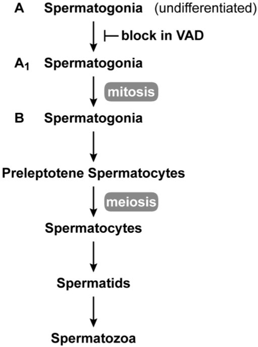
“In vitamin A deficiency, the transition from A to A1 spermatogonia is blocked [42–44].”
BackgroundRetinoid signalling and hindbrain patterning
Gavalas, A., & Krumlauf, R. K. (2000) · Current Opinion in Genetics & Development · doi:10.1016/s0959-437x(00)00100-3 · Cited by 193 · cited 1×
“the diverse range of nerves and craniofacial structures essential to hindbrain function [161].”
BackgroundMason, K. E. (1933) · doi:10.1002/aja.1000520202 · Cited by 190 · cited 1×
“Early work in the laboratories of Wolbach and Howe as well as Mason showed that in vitamin A deficiency, the epithelia of the epididymis, prostate, and seminal vesicle is replaced with stratified squamous keratinizing epithelium, and spermatogenesis ceases [2,38].”
ResultRetinoic acid treatment partially rescues failed septation in rats and in mice.
Massaro, G., & Massaro, D. (2000) · American Journal of Physiology - Lung cellular and Molecular Physiology · doi:10.1152/ajplung.2000.278.5.l955 · Cited by 186 · cited 1×
“reports that exogenous RA can stimulate alveoli formation in immature rat and mouse lung [264–269].”
BackgroundCongenital malformations induced in rats by maternal vitamin A deficiency; defects of the eye.
Warkany, J., & Schraffenberger, E. (1946) · A M A Archives of Ophthalmology · doi:10.1001/archopht.1946.00890200155008 · Cited by 184
Ang, H. L., Deltour, L., Hayamizu, T. F., Žgombić‐Knight, M., & Duester, G. (1996) · Journal of Biological Chemistry · doi:10.1074/jbc.271.16.9526 · Cited by 184 · cited 1×
“Adh 1, 3 and 4 enzyme family members are also expressed in developing embryos [126,127], however, mutation does not result in embryo lethality [128,129].”
BackgroundShifting boundaries of retinoic acid activity control hindbrain segmental gene expression
Sîrbu, I. O., Gresh, L., Barra, J., & Duester, G. (2005) · Development · doi:10.1242/dev.01845 · Cited by 184 · cited 1×
“developing hindbrain all include an important role for CYP26 family members in regulating the formation of waves or pulses of RA in this dynamic process [23,144,172,173].”
BackgroundDersch, H., & Zile, M. (1993) · Developmental Biology · doi:10.1006/dbio.1993.1318 · Cited by 183 · cited 2×
“When severe VAD is imposed in quail, the initiation of heart morphogenesis is disrupted [88,228,229].”
BackgroundWhite, J. C., Highland, M., Kaiser, M., & Clagett-Dame, M. (2000) · Developmental Biology · doi:10.1006/dbio.2000.9635 · Cited by 182 · cited 4×
“VAD rat embryos generated by severely restricting the amount of RA fed to deficient mothers at the beginning of pregnancy results in a loss of posterior rhombomere segmentation, with a significant shortening of the hindbrain [65].”
BackgroundInfluentialSelf-citationNon-cell-autonomous retinoid signaling is crucial for renal development
Rosselot, C., Spraggon, L., Chia, I. V., Batourina, E., Riccio, P., Lu, B., Niederreither, K., Dollé, P., Duester, G., Chambon, P., et al. (2010) · Development · doi:10.1242/dev.040287 · Cited by 181 · cited 1×
“Recent work shows that RA generated by RALDH2 in stromal mesenchyme acts in paracrine fashion to activate RA-receptor signaling and Ret expression in ureteric bud cells [242].”
BackgroundZhao, X.-L., Sirbu, I. O., Mic, F., Molotkova, N., Molotkov, A., Kumar, S., & Duester, G. (2009) · Current Biology · doi:10.1016/j.cub.2009.04.059 · Cited by 178 · cited 3×
“Raldh2 is largely responsible for RA synthesis in the mesonephros, with some contribution from Raldh3 in the region of the mesonephric duct [71,80].”
BackgroundMedium- and short-chain dehydrogenase/reductase gene and protein families
Parés, X., Farrés, J., Kedishvili, N. Y., & Duester, G. (2008) · Cellular and Molecular Life Sciences · doi:10.1007/s00018-008-8591-3 · Cited by 173
Chen, Y., Pan, F. C., Brandes, N., Afelik, S., Sölter, M., & Pieler, T. (2004) · Developmental Biology · doi:10.1016/j.ydbio.2004.03.030 · Cited by 172 · cited 2×
“Both defects in RA signaling and RA treatment have been shown to affect the expression of PDX-1 [271,272,275,278] an essential regulator of early pancreas development required for the”
BackgroundSirbu, I. O., & Duester, G. (2006) · Nature Cell Biology · doi:10.1038/ncb1374 · Cited by 171 · cited 2×
“In RALDH2 null mutants, the expression of Fgf8 mRNA in the primitive ectoderm (epiblast) is shifted anteriorly such that it enters the node ectoderm and neuroectoderm, and somite development is abnormal [170].”
BackgroundThe Relation of Vitamin a to Anophthalmos in Pigs
Hale, F. (1935) · American Journal of Ophthalmology · doi:10.1016/s0002-9394(35)90563-3 · Cited by 169 · cited 2×
“Recognition of the importance of vitamin A in eye development first came from the experiments of Hale in which piglets from deficient mothers were born blind [86,87].”
BackgroundIngram, J. L., Stodgell, C., Hyman, S. L., Figlewicz, D. A., WEITKAMP, L. R., & Rodier, P. M. (2000) · Teratology · doi:10.1002/1096-9926(200012)62:6 · Cited by 169
Self-citationRole of all‐trans retinoic acid in neurite outgrowth and axonal elongation
Clagett‐Dame, M., McNeill, E. M., & Muley, P. D. (2006) · Journal of Neurobiology · doi:10.1002/neu.20241 · Cited by 167 · cited 2×
“RA is also linked to the promotion of neurite outgrowth [157,162].”
BackgroundSelf-citationA retinoic acid synthesizing enzyme in ventral retina and telencephalon of the embryonic mouse.
Li, H., Wagner, E., McCaffery, P., Smith, D., Andreadis, A., & Dräger, U. C. (2000) · Mechanisms of Development · doi:10.1016/s0925-4773(00)00352-x · Cited by 167 · cited 1×
“5 [132,148,203], after which Raldh3 (also called V1 activity) is expressed in the ventral neural retina and pigmented neuroepithelium [134,204–206] and Raldh1 (also called ADH-2) is expressed in the dorsal retina from E9.”
BackgroundRegulation and perturbation of testicular functions by vitamin A.
Livera, G., Rouiller-Fabre, V., Pairault, C., Levacher, C., & Habert, R. (2002) · Reproduction · doi:10.1530/rep.0.1240173 · Cited by 166 · cited 3×
“Within the normal tubule, the Sertoli cell is believed to generate RA by the action of Raldh1 [49,51,52], and possibly Raldh2 [50].”
BackgroundRetinoic acid is required for the initiation of outgrowth in the chick limb bud.
Stratford, T., Horton, C., & Maden, M. (1996) · Current Biology · doi:10.1016/s0960-9822(02)70679-9 · Cited by 166 · cited 1×
“Inhibitors of RA synthesis and vitamin A deficiency inhibit limb outgrowth in the quail [285,286].”
BackgroundSpermatogenic response to vitamin A in vitamin A deficient rats.
Huang, H.-F.-S., & Hembree, W. (1979) · Biology of Reproduction · doi:10.1095/biolreprod21.4.891 · Cited by 165 · cited 1×
“Later work showed that in the VAD rat testes, undifferentiated spermatogonia, Sertoli cells and a small number of preleptotene spermatocytes remain [39–41], whereas in the mouse,”
BackgroundUehara, M., Yashiro, K., Mamiya, S., Nishino, J., Chambon, P., Dollé, P., & Sakai, Y. (2006) · Developmental Biology · doi:10.1016/j.ydbio.2006.09.045 · Cited by 164 · cited 7×
“This is followed by the later expression of Cyp26A1 in r2 and Cyp26C1 in r2 and r4 [18,21,139,144].”
BackgroundInfluentialBarrios, F., Filipponi, D., Pellegrini, M., Paronetto, M., Siena, S. D., Geremia, R., Rossi, P., Felici, M. D., Jannini, E., & Dolci, S. (2010) · Journal of Cell Science · doi:10.1242/jcs.057968 · Cited by 163 · cited 1×
“(Adapted from [77,78]).”
Benazet, J.-D., & Zeller, R. (2009) · Cold Spring Harbor Perspectives in Biology · doi:10.1101/cshperspect.a001339 · Cited by 163 · cited 1×
“RA has been proposed to play a role in patterning the developing limb by regulating the pattern of expression of genes such as Hand2 (activates SHH), Shh (patterns the anterior-to-posterior axis), and Meis1/2 (transcription factors that mark the proximal limb bud mesenchyme) [287,290–292].”
BackgroundMic, F. A., Molotkov, A., Fan, X., Cuenca, A. E., & Duester, G. (2000) · Mechanisms of Development · doi:10.1016/s0925-4773(00)00434-2 · Cited by 163 · cited 1×
“5 [132,148,203], after which Raldh3 (also called V1 activity) is expressed in the ventral neural retina and pigmented neuroepithelium [134,204–206] and Raldh1 (also called ADH-2) is expressed in the dorsal retina from E9.”
BackgroundMedium- and short-chain dehydrogenase/reductase gene and protein families
Eklund, H., & Ramaswamy, S. (2008) · Cellular and Molecular Life Sciences · doi:10.1007/s00018-008-8589-x · Cited by 158 · cited 1×
“The first and rate-limiting step in the production of RA from retinol results from the action of cytosolic alcohol dehydrogenases (ADH) and microsomal retinol dehydrogenases (RDH) yielding all-trans retinaldehyde [13].”
BackgroundDistal ureter morphogenesis depends on epithelial cell remodeling mediated by vitamin A and Ret
Batourina, E., Choi, C., Paragas, N., Bello, N., Hensle, T., Costantini, F., Schuchardt, A., Bacallao, R., & Mendelsohn, C. (2002) · Nature Genetics · doi:10.1038/ng952 · Cited by 157
Retinoic acid regulates morphogenesis and patterning of posterior foregut derivatives
Wang, Z., Dollé, P., Cardoso, W. V., & Niederreither, K. (2006) · Developmental Biology · doi:10.1016/j.ydbio.2006.05.019 · Cited by 156 · cited 2×
“RA acting through the RAR is also necessary for the morphogenesis of other components of the respiratory tract including partitioning of the primitive foregut into oesophagus and trachea, and for the opening between the nasal and oropharyngeal cavities [67,131,209,256,258,261].”
BackgroundRetinoic acid regulates endothelial cell proliferation during vasculogenesis
Lai, L., Bohnsack, B. L., Niederreither, K., & Hirschi, K. K. (2003) · Development · doi:10.1242/dev.00887 · Cited by 154 · cited 1×
“Raldh2 in the embryo, along with expression in the visceral endoderm (extraembryonic) is required for vascular development in both the embryo proper, as well as the yolk sac [150–152].”
BackgroundRALDH-independent generation of retinoic acid during vertebrate embryogenesis by CYP1B1
Chambers, D., Wilson, L. J., Maden, M., & Lumsden, A. (2007) · Development · doi:10.1242/dev.02815 · Cited by 154 · cited 2×
“Cyp1B1 can also generate RA and is expressed at known sites of RALDH-independent retinoid signaling [16], however, null mutant mice for this gene are viable [137].”
BackgroundDevelopmental roles of the retinoic acid receptors.
Lohnes, D., Mark, M., Mendelsohn, C., Dollé, P., Décimo, D., Lemeur, M., Dierich, A., Gorry, P., & Chambon, P. (1995) · Journal of Steroid Biochemistry and Molecular Biology · doi:10.1016/0960-0760(95)00094-g · Cited by 153 · cited 1×
“importance of the RAR in mediating the actions of RA in developing embryos [99,101,102].”
BackgroundTahayato, A., Dollé, P., & Petkovich, M. P. (2003) · Gene Expression Patterns · doi:10.1016/s1567-133x(03)00066-8 · Cited by 152 · cited 1×
“This is followed by the later expression of Cyp26A1 in r2 and Cyp26C1 in r2 and r4 [18,21,139,144].”
BackgroundDevelopmental expression of retinoic acid receptors (RARs)
Dollé, P. (2009) · Nuclear Receptor Signaling · doi:10.1621/nrs.07006 · Cited by 152 · cited 1×
“The Rars are widely expressed in development [100].”
BackgroundMolotkov, A., Fan, X., Deltour, L., Foglio, M. H., Martras, S., Farrés, J., Parés, X., & Duester, G. (2002) · Proceedings of the National Academy of Sciences · doi:10.1073/pnas.082093299 · Cited by 151 · cited 2×
“postnatal lethality if maintained for two generations on a VAD diet [129].”
BackgroundKumar, S., Chatzi, C., Brade, T., Cunningham, T. J., Zhao, X.-L., & Duester, G. (2011) · Nature Communications · doi:10.1038/ncomms1136 · Cited by 149 · cited 3×
“However, a recent report shows that female RALDH2 and RALDH2/3 knockout gonads express Stra8 and undergo meiosis in the absence of detectable RA activity as assessed using a β2-RARE-lacZ (RARE-lacZ) reporter [74].”
BackgroundMethodThe role of CYP26 enzymes in defining appropriate retinoic acid exposure during embryogenesis.
Pennimpede, T., Cameron, D., Maclean, G., Li, H., Abu-Abed, S., & Petkovich, M. (2010) · Birth defects research. Clinical and molecular teratology · doi:10.1002/bdra.20709 · Cited by 147 · cited 3×
“The rhombomere-specific expression pattern of the Cyp26 mRNAs led to the proposal that boundaries of RA activity are created by their expression [23,141,171] (Figure 4B).”
BackgroundRetinoic acid in the formation of the dorsoventral retina and its central projections.
Wagner, E., McCaffery, P., & Dräger, U. C. (2000) · Developmental Biology · doi:10.1006/dbio.2000.9719 · Cited by 146 · cited 1×
“5 [132,148,203], after which Raldh3 (also called V1 activity) is expressed in the ventral neural retina and pigmented neuroepithelium [134,204–206] and Raldh1 (also called ADH-2) is expressed in the dorsal retina from E9.”
BackgroundWhite, J. C., Shankar, V., Highland, M., Epstein, M., DeLuca, H., & Clagett-Dame, M. (1998) · Proceedings of the National Academy of Sciences of the United States of America · doi:10.1073/pnas.95.23.13459 · Cited by 146 · cited 2×
“Nervous system, cardiovascular, and axial patterning defects result from early deficiency [63–66,96], whereas a less well-developed nasal region,”
BackgroundSelf-citationRetinoic acid and hindbrain patterning
Glover, J. C., Renaud, J., & Rijli, F. M. (2006) · Journal of Neurobiology · doi:10.1002/neu.20272 · Cited by 145 · cited 1×
“One of the best-studied functions of RA is in the developing hindbrain where it contributes to the anteroposterior patterning of the neural plate [159].”
BackgroundA retinoic acid-dependent network in the foregut controls formation of the mouse lung primordium.
Chen, F., Cao, Y.-X., Qian, J., Shao, F., Niederreither, K., & Cardoso, W. (2010) · Journal of Clinical Investigation · doi:10.1172/jci40253 · Cited by 142 · cited 2×
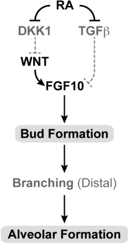
“of Tgfβ signaling [262].”
Sockanathan, S., Perlmann, T., & Jessell, T. (2003) · Neuron · doi:10.1016/s0896-6273(03)00532-4 · Cited by 142 · cited 1×
“RA generated from within the spinal cord is required for the differentiation of a subset of lateral motor column (LMC) neurons that extend axons into the limb [192,193].”
BackgroundReijntjes, S., Blentic, A., Gale, E. A., & Maden, M. (2005) · Developmental Biology · doi:10.1016/j.ydbio.2005.06.019 · Cited by 137 · cited 1×
“Interestingly, both the levels and domains of Cyp26 gene expression in the developing embryo are affected by retinoid status, whereas the Raldhs are unaffected [121].”
BackgroundMey, J., Babiuk, R. P., Clugston, R. D., Zhang, W., & Greer, J. (2003) · American Journal of Pathology · doi:10.1016/s0002-9440(10)63861-8 · Cited by 137 · cited 1×
“Similar defects in PPF development are seen in rat embryos treated with nitrofen and several other CDH-inducing teratogens that interfere with the synthesis of RA and RA signaling in vivo [253,254].”
BackgroundWarkany, J., & Roth, C. (1948) · Journal of Nutrition · doi:10.1093/jn/35.1.1 · Cited by 134
Synthesis of some Compounds Possessing Vitamin A Activity
Arens, J. F., & Dorp, D. A. V. (1946) · Nature · doi:10.1038/157190a0 · Cited by 133 · cited 1×
“In 1946, Arens and van Dorp synthesized vitamin A acid (retinoic acid), and reported it was as potent as vitamin A in supporting the growth of VAD rats but could not be converted back to vitamin A [8–10].”
BackgroundDhrs3a regulates retinoic acid biosynthesis through a feedback inhibition mechanism
Feng, L., Hernandez, R. E., Waxman, J. S., Yelon, D. L., & Moens, C. B. (2009) · Developmental Biology · doi:10.1016/j.ydbio.2009.10.029 · Cited by 132 · cited 1×
“member, DHRS3a, which catalyzes reduction of retinaldehyde to retinol serving as a RA-induced feedback inhibitor of RA biosynthesis [138].”
BackgroundClugston, R. D., Klattig, J., Englert, C., Clagett-Dame, M., Martinovic, J., Benachi, A., & Greer, J. (2006) · American Journal of Pathology · doi:10.2353/ajpath.2006.060445 · Cited by 131 · cited 1×
“5 in vitamin A-sufficient rat embryos, but it is abnormal in late VAD embryos [252].”
BackgroundSelf-citationQuadro, L., Hamberger, L., Gottesman, M. E., Wang, F., Colantuoni, V., Blaner, W. S., & Mendelsohn, C. L. (2005) · Endocrinology · doi:10.1210/en.2005-0158 · Cited by 130 · cited 6×
“Retinol-binding protein (RBP or RBP4) carries the majority of retinol in the circulation [25], and a membrane receptor, STRA6, binds to RBP to enable efficient retinol uptake by a number of cells [26].”
BackgroundInfluentialBåvik, C., Ward, S. J., & Chambon, P. (1996) · Proceedings of the National Academy of Sciences of the United States of America · doi:10.1073/pnas.93.7.3110 · Cited by 126 · cited 13×
“Retinol-binding protein (RBP or RBP4) carries the majority of retinol in the circulation [25], and a membrane receptor, STRA6, binds to RBP to enable efficient retinol uptake by a number of cells [26].”
BackgroundInfluentialActive repression of RAR signaling is required for head formationPreprint
Koide, T., Downes, M., Chandraratna, R. A. S., Blumberg, B., & Umesono, K. (2001) · Genes & Development · doi:10.1101/gad.908801 · Cited by 125 · cited 1×
“should remain off [175].”
Molotkova, N., Molotkov, A., & Duester, G. (2007) · Developmental Biology · doi:10.1016/j.ydbio.2006.11.035 · Cited by 124 · cited 2×
“Instead, Molotkova and colleagues propose that RA functions at later times in forebrain development, and provide evidence that RA generated by RALDH3 in the lateral ganglionic eminence is required for normal expression of the dopamine D2 receptor in the nucleus accumbens and medial striatum [180].”
BackgroundPrepubertal testis development relies on retinoic acid but not rexinoid receptors in Sertoli cells
Vernet, N., Dennefeld, C., Guillou, F., Chambon, P., Ghyselinck, N., & Mark, M. (2006) · EMBO Journal · doi:10.1038/sj.emboj.7601447 · Cited by 124 · cited 1×
“Expression of RARα is needed for differentiation of the spermatogonia during prepuberty [55].”
BackgroundRetinoic acid receptor-β: an endogenous inhibitor of the perinatal formation of pulmonary alveoli
Massaro, G., Massaro, D., Chan, W., Clerch, L., Ghyselinck, N., Chambon, P., & Chandraratna, R. (2000) · Physiological Genomics · doi:10.1152/physiolgenomics.2000.4.1.51 · Cited by 121 · cited 1×
“reports that exogenous RA can stimulate alveoli formation in immature rat and mouse lung [264–269].”
BackgroundUlven, S., Gundersen, T. E., Weedon, M. S., Landaas, V. Ø., Sakhi, A., Fromm, S., Geronimo, B., Moskaug, J. Ø., & Blomhoff, R. (2000) · Developmental Biology · doi:10.1006/dbio.2000.9634 · Cited by 121 · cited 1×
“Using direct identification by HPLC, all-trans retinaldehyde is detected in embryos as early as the egg cylinder or pre-primitive streak stage, whereas RA is detected between the mid-primitive streak stage and the late allantoic bud stage [146].”
MethodZur Kenntnis des Vitamins-A aus Fischtranen
Karrer, P., Morf, R., & Schopp, K. (1931) · Helvetica Chimica Acta · doi:10.1002/hlca.19310140511 · Cited by 121
Soprano, D. R., Soprano, K. J., & Goodman, D. S. (1986) · Proceedings of the National Academy of Sciences · doi:10.1073/pnas.83.19.7330 · Cited by 121 · cited 1×
“endoderm of the yolk sac visceral wall surrounding the embryo, and after definitive placentation, in the yolk sac membranes as well as in the uterus (decidua basalis) [111,112].”
BackgroundDupé, V., Ghyselinck, N., Thomázy, V. A., Nagy, L., Davies, P. J. A., Chambon, P., & Mark, M. (1999) · Developmental Biology · doi:10.1006/dbio.1998.9176 · Cited by 119 · cited 1×
“Webbed digits have been described in compound RAR mutant mice due to a loss of apoptosis [209,294].”
BackgroundHowell, J., Thompson, J. N., & Pitt, G. A. (1963) · Journal of Reproduction and Fertility · doi:10.1530/jrf.0.0050159 · Cited by 118 · cited 1×
“insufficient amounts of RA [68].”
Satre, M., & Kochhar, D. (1989) · Developmental Biology · doi:10.1016/0012-1606(89)90055-9 · Cited by 117 · cited 1×
“HPLC studies of embryonic tissues at later times confirm that all-trans retinol and RA are the primary retinoids detected, with retinol clearly the most abundant [153–155].”
BackgroundEndogenous retinoic acid regulates cardiac progenitor differentiation
Lin, S.-C., Dollé, P., Ryckebusch, L., Noseda, M., Zaffran, S., Schneider, M., & Niederreither, K. (2010) · Proceedings of the National Academy of Sciences of the United States of America · doi:10.1073/pnas.0910430107 · Cited by 115 · cited 1×
“heart tube patterning and looping, chamber and outflow tract septation, ventricular trabeculation, cardiomyocyte differentiation and coronary vessel development [231–233].”
BackgroundRibes, V., Roux, I. L., Rhinn, M., Schuhbaur, B., & Dollé, P. (2009) · Development · doi:10.1242/dev.016204 · Cited by 114 · cited 3×
“mesodermal markers are expanded at the expense of those representative of the prospective neuroepithelium [149].”
BackgroundA conserved role for retinoid signaling in vertebrate pancreas development
Stafford, D. A., Hornbruch, A., Mueller, P. R., & Prince, V. (2004) · Development, Genes and Evolution · doi:10.1007/s00427-004-0420-6 · Cited by 114 · cited 2×
“null mutant mice, a requirement for RA in pancreatic development has been proposed based on studies in RALDH null mutant mice [271,272] and in several other organisms in which retinoid signaling is deficient or inhibited, including Xenopus, zebrafish, chick, and quail [273–276].”
BackgroundBohnsack, B. L., Lai, L., Dollé, P., & Hirschi, K. K. (2004) · Genes & Development · doi:10.1101/gad.1184904 · Cited by 112 · cited 1×
“Raldh2 in the embryo, along with expression in the visceral endoderm (extraembryonic) is required for vascular development in both the embryo proper, as well as the yolk sac [150–152].”
BackgroundRequirement of mesodermal retinoic acid generated by Raldh2 for posterior neural transformation
Molotkova, N., Molotkov, A., Sîrbu, I. O., & Duester, G. (2004) · Mechanisms of Development · doi:10.1016/j.mod.2004.10.008 · Cited by 112 · cited 1×
“Although primary neurulation occurs in both RALDH2 mutant mice and VAD chick and rat embryos [65,123,187], the”
BackgroundRetinoids and spinal cord development.
Maden, M. (2006) · Journal of Neurobiology · doi:10.1002/neu.20248 · Cited by 111 · cited 1×
“Induction of more posterior domains occurs by the actions of RA, Wnts and FGFs, with RA specifying the posterior hindbrain and cervical spinal cord [160].”
BackgroundInduction and prepatterning of the zebrafish pectoral fin bud requires axial retinoic acid signaling
Gibert, Y., Gajewski, A., Meyer, A., & Begemann, G. (2006) · Development · doi:10.1242/dev.02438 · Cited by 110 · cited 2×
“, who found that axial retinoic acid signals played a permissive role in the induction of zebrafish pectoral fins [289].”
BackgroundRetinoic acid regulates programmed cell death through BMP signalling
Rodríguez-León, J., Merino, R., Macías, D., Gańan, Y., Santesteban, E., & Hurlé, J. (1999) · Nature Cell Biology · doi:10.1038/10098 · Cited by 110 · cited 1×
“The mechanism by which RA activates cell death is currently a subject of active investigation [294–297].”
BackgroundSnyder, E. M., Small, C., & Griswold, M. (2010) · Biology of Reproduction · doi:10.1095/biolreprod.110.085811 · Cited by 107 · cited 1×
“Recent work supports the conclusion that the vitamin A metabolite, RA, is needed both for adult male spermatogonial differentiation (transition to A1) and the entrance into meiosis [45–47].”
ResultKumar, S., & Duester, G. (2010) · Developmental Biology · doi:10.1016/j.ydbio.2010.01.027 · Cited by 106 · cited 2×
“Defects result from the loss of control of programmed cell death in this region, and by loss of RA-mediated Pitx2, Foxc1, Eya2 and Dkk2 expression, and subsequent loss of repression of Wnt/β-catenin signaling [134,135,212,214].”
BackgroundRetinoids control anterior and dorsal properties in the developing forebrain.
Halilagic, A., Ribes, V., Ghyselinck, N., Zile, M., Dollé, P., & Studer, M. (2007) · Developmental Biology · doi:10.1016/j.ydbio.2006.11.021 · Cited by 106 · cited 3×
“A number of eye defects in RALDH3 and RALDH1/3 compound mutants [134,135,178] are also similar to those observed in late VAD rat embryos.”
BackgroundMolotkov, A., Deltour, L., Foglio, M. H., Cuenca, A. E., & Duester, G. (2002) · Journal of Biological Chemistry · doi:10.1074/jbc.m112039200 · Cited by 105 · cited 1×
“Adh 1, 3 and 4 enzyme family members are also expressed in developing embryos [126,127], however, mutation does not result in embryo lethality [128,129].”
BackgroundHow degrading: Cyp26s in hindbrain development
White, R. J., & Schilling, T. F. (2008) · Developmental Dynamics · doi:10.1002/dvdy.21695 · Cited by 105 · cited 2×
“developing hindbrain all include an important role for CYP26 family members in regulating the formation of waves or pulses of RA in this dynamic process [23,144,172,173].”
BackgroundMatt, N., Ghyselinck, N., Pellerin, I., & Dupé, V. (2008) · Developmental Biology · doi:10.1016/j.ydbio.2008.04.039 · Cited by 103
MULTIPLE CONGENITAL MALFORMATIONS: A CONSIDERATION OF ETIOLOGIC FACTORS
Warkany, J., Roth, C., & Wilson, J. G. (1948) · Pediatrics · doi:10.1542/peds.1.4.462 · Cited by 103 · cited 1×
“Respiratory defects including left lung agenesis, bilateral lung hypoplasia, and agenesis of the esophagotracheal septum were described in early VAD syndrome embryos but were characterized as rare anomalies [90,95].”
BackgroundMic, F. A., Sîrbu, I. O., & Duester, G. (2004) · Journal of Biological Chemistry · doi:10.1074/jbc.m401920200 · Cited by 102 · cited 1×
“of RALDH2, murine forelimb buds do not develop and embryonic growth ceases prior to the stage when hindlimb buds are initiated [287,288].”
BackgroundIncreased Neonatal Mortality in Mice Lacking Cellular Retinol-binding Protein II
Xueping, E., Zhang, L., Lu, J., Tso, P., Blaner, W. S., Levin, M. S., & Li, E. (2002) · Journal of Biological Chemistry · doi:10.1074/jbc.m205519200 · Cited by 102 · cited 1×
“mice; some have been found to be dispensable, while roles for others have been revealed when animals are fed a vitamin A-restricted diet [12,27,28].”
BackgroundRetinol Status of Newborn Infants Is Associated With Congenital Diaphragmatic Hernia
Beurskens, L., Tibboel, D., Lindemans, J., Duvekot, J., Cohen-Overbeek, T., Veenma, D., de Klein, A., Greer, J., & Steegers-Theunissen, R. (2010) · Pediatrics · doi:10.1542/peds.2010-0521 · Cited by 102 · cited 1×
“A recent report shows that there is a relationship between low cord retinol and RBP levels and CDH in newborn infants [249].”
BackgroundDistinct roles for retinoic acid receptors alpha and beta in early lung morphogenesis
Desai, T., Chen, F. F., Lü, J., Qian, J., Niederreither, K., Dollé, P., Chambon, P., & Cardoso, W. V. (2006) · Developmental Biology · doi:10.1016/j.ydbio.2005.10.045 · Cited by 100
All-trans-retinol is a ligand for the retinoic acid receptors.
Repa, J., Hanson, K. K., & Clagett-Dame, M. (1993) · Proceedings of the National Academy of Sciences of the United States of America · doi:10.1073/pnas.90.15.7293 · Cited by 99 · cited 1×
“The all-trans isomer of RA is the highest affinity endogenous ligand for the RAR [31].”
BackgroundSelf-citationMic, F. A., Molotkov, A., Molotkova, N., & Duester, G. (2004) · Developmental Dynamics · doi:10.1002/dvdy.20128 · Cited by 96 · cited 1×
“5 [132,148,203], after which Raldh3 (also called V1 activity) is expressed in the ventral neural retina and pigmented neuroepithelium [134,204–206] and Raldh1 (also called ADH-2) is expressed in the dorsal retina from E9.”
BackgroundMitranond, V., Sobhon, P., Tosukhowong, P., & Chíndaduangrat, W. (1979) · Acta Anatomica · doi:10.1159/000145007 · Cited by 94
Delivery of retinoid-based therapies to target tissues
Moise, A. R., Noy, N., Palczewski, K., & Blaner, W. S. (2007) · Biochemistry · doi:10.1021/bi7003069 · Cited by 93 · cited 2×
“mice; some have been found to be dispensable, while roles for others have been revealed when animals are fed a vitamin A-restricted diet [12,27,28].”
BackgroundLi, H., Maclean, G., Cameron, D., Clagett-Dame, M., & Petkovich, M. (2009) · PLoS ONE · doi:10.1371/journal.pone.0007501 · Cited by 92 · cited 1×
“CYP26B1 is also required later in embryogenesis to maintain cells in an undifferentiated state [82].”
BackgroundSelf-citationStage-dependent responses of the developing lung to retinoic acid signaling.
Mollard, R., Ghyselinck, N., Wendling, O., Chambon, P., & Mark, M. (2000) · International Journal of Developmental Biology · doi:10.1387/ijdb.11032179 · Cited by 90 · cited 1×
“RA acting through the RAR is also necessary for the morphogenesis of other components of the respiratory tract including partitioning of the primitive foregut into oesophagus and trachea, and for the opening between the nasal and oropharyngeal cavities [67,131,209,256,258,261].”
BackgroundStrate, I., Grahn, T. H. M., Iliev, D., & Pera, E. M. (2009) · Development · doi:10.1242/dev.024901 · Cited by 88 · cited 1×
“Recent work in Xenopus shows that RA down regulates XRDH10 transcripts, and this may serve as an additional mechanism to regulate endogenous retinoid levels [125].”
BackgroundRetinoic acid synthesis for the developing telencephalon.
Smith, D., Wagner, E., Koul, O., McCaffery, P., & Dräger, U. C. (2001) · Cerebral Cortex · doi:10.1093/cercor/11.10.894 · Cited by 88 · cited 1×
“In the meninges covering the cortex, levels of Raldh2 increase in the mouse embryo from E13 to E14 onward, to a maximal level in the newborn brain [184].”
BackgroundVermot, J., Schuhbaur, B., Mouellic, H. L., McCaffery, P., Garnier, J., Hentsch, D., Brûlet, P., Niederreither, K., Chambon, P., Dollé, P., et al. (2005) · Development · doi:10.1242/dev.01718 · Cited by 86
Ang, H., Deltour, L., Žgombic-Knight, M., Wagner, M., & Duester, G. (1996) · Alcoholism: Clinical and Experimental Research · doi:10.1111/j.1530-0277.1996.tb01946.x · Cited by 86 · cited 1×
“Adh 1, 3 and 4 enzyme family members are also expressed in developing embryos [126,127], however, mutation does not result in embryo lethality [128,129].”
BackgroundMaternal Vitamin A Supplementation and Lung Function in Offspring
Shulman, L. (2010) · doi:10.1016/s1090-798x(10)79165-7 · Cited by 86 · cited 1×
“Additionally, they found that the period during which supplementation with vitamin A was most important was from gestation through a postnatal age of 6 months [270].”
BackgroundMaclean, G., Dollé, P., & Petkovich, M. (2009) · Developmental Dynamics · doi:10.1002/dvdy.21878 · Cited by 85 · cited 2×
“character [139,140], however, individual CYP26B1 and CYP26C1 mutants develop normally [143,144].”
BackgroundQuadro, L., Hamberger, L., Gottesman, M. E., Colantuoni, V., Ramakrishnan, R., & Blaner, W. S. (2004) · American Journal of Physiology-Endocrinology and Metabolism · doi:10.1152/ajpendo.00556.2003 · Cited by 84 · cited 2×
“RBP does not cross the placenta into the fetal circulation [113].”
BackgroundRetinoids and spermatogenesis: Lessons from mutant mice lacking the plasma retinol binding protein
Ghyselinck, N. B., Vernet, N., Dennefeld, C., Giese, N., Nau, H., Chambon, P., Viville, S., & Mark, M. P. (2006) · Developmental Dynamics · doi:10.1002/dvdy.20795 · Cited by 84 · cited 14×
“In the mouse, Raldh2 (also called V2 activity) is transiently expressed in the optic vesicle neuroepithelium at E8.”
BackgroundInfluentialSapin, V., Ward, S. J., Bronner, S., Chambon, P., & Dollé, P. (1997) · Developmental Dynamics · doi:10.1002/(sici)1097-0177(199702)208:2 · Cited by 84
The atRA‐responsive gene neuron navigator 2 functions in neurite outgrowth and axonal elongation
Muley, P. D., McNeill, E. M., Marzinke, M. A., Knobel, K. M., Barr, M. M., & Clagett‐Dame, M. (2008) · Developmental Neurobiology · doi:10.1002/dneu.20670 · Cited by 81 · cited 1×
“When NAV2 is knocked down, the neuroblastoma cell is no longer able to extend neurites in response to RA [201].”
BackgroundSelf-citationWagner, M., & Siddiqui, M. (2007) · Experimental biology and medicine · doi:10.3181/00379727-232-2320866 · Cited by 80 · cited 1×
“heart tube patterning and looping, chamber and outflow tract septation, ventricular trabeculation, cardiomyocyte differentiation and coronary vessel development [231–233].”
BackgroundBlood island formation in attached cultures of murine embryonic stem cells
Bautch, V. L., Stanford, W. L., Rapoport, R., Russell, S., Byrum, R. S., & Futch, T. A. (1996) · Developmental Dynamics · doi:10.1002/(sici)1097-0177(199601)205:1 · Cited by 79
Self-citationTulachan, S., Doi, R., Kawaguchi, Y., Tsuji, S., Nakajima, S., Masui, T., Koizumi, M., Toyoda, E., Mori, T., Ito, D., et al. (2003) · Diabetes · doi:10.2337/diabetes.52.1.76 · Cited by 78 · cited 2×
“Both defects in RA signaling and RA treatment have been shown to affect the expression of PDX-1 [271,272,275,278] an essential regulator of early pancreas development required for the”
BackgroundThe Meninges Is a Source of Retinoic Acid for the Late-Developing Hindbrain
Zhang, J., Smith, D. F., Yamamoto, M., Ma, L., & McCaffery, P. J. A. (2003) · Journal of Neuroscience · doi:10.1523/jneurosci.23-20-07610.2003 · Cited by 78 · cited 1×
“pontine nuclei [182].”
Wilson, J. G., & Warkany, J. (1950) · Pediatrics · doi:10.1542/peds.5.4.708 · Cited by 77 · cited 1×
“Cardiac and aortic arch defects were observed as a part of the early vitamin A deficiency syndrome in rat embryos [93,94] and in RAR compound null mutant mouse embryos [98,102].”
BackgroundLi, H., & Clagett‐Dame, M. (2009) · Biology of Reproduction · doi:10.1095/biolreprod.109.078808 · Cited by 77 · cited 1×
“There is clear in vivo evidence that vitamin A is required for the normal onset of meiotic prophase in ovarian germ cells [79].”
BackgroundSelf-citationRetinoic acid signalling specifies intermediate character in the developing telencephalon
Marklund, M., Sjödal, M., Beehler, B. C., Jessell, T. M., Edlund, H., & Gunhaga, L. (2004) · Development · doi:10.1242/dev.01308 · Cited by 76 · cited 1×
“RA was also reported in chick to contribute to regionalization of the telencephalon along the dorsoventral axis, and RALDH3 produced by the head ectoderm was proposed as the retinoid source [179].”
BackgroundClugston, R. D., Zhang, W., Álvarez, S., de Lera, Á. R., & Greer, J. J. (2009) · American Journal of Respiratory Cell and Molecular Biology · doi:10.1165/rcmb.2009-0076oc · Cited by 76 · cited 1×
“Similar defects in PPF development are seen in rat embryos treated with nitrofen and several other CDH-inducing teratogens that interfere with the synthesis of RA and RA signaling in vivo [253,254].”
BackgroundKarrer, P., Morf, R., & Schopp, K. (1931) · Helvetica Chimica Acta · doi:10.1002/hlca.19310140622 · Cited by 75
See, A. W., Kaiser, M. E., White, J. C., & Clagett‐Dame, M. (2007) · Developmental Biology · doi:10.1016/j.ydbio.2007.10.018 · Cited by 74 · cited 10×
“5 in the rat embryo, a time when the cardiac primordium has completed looping and the primitive vasculature is established, early cardiac and aortic arch defects are not observed [67].”
BackgroundInfluentialSelf-citationLi, H., Palczewski, K., Baehr, W., & Clagett-Dame, M. (2011) · Biology of Reproduction · doi:10.1095/biolreprod.110.086157 · Cited by 71 · cited 1×
“showed that vitamin A is required in the male neonate for germ cells to enter the first round of spermatogenesis [83].”
BackgroundSelf-citationMerrill, R. A., Plum, L., Kaiser, M., & Clagett-Dame, M. (2002) · Proceedings of the National Academy of Sciences of the United States of America · doi:10.1073/pnas.052017399 · Cited by 71 · cited 1×
“Recently, a number of genes including neuron navigator 2 (Nav2) and Nedd9 were discovered in a screen for RA-responsive genes in a neuroblastoma cell line that elaborates neurites in response to RA [198–200].”
BackgroundSelf-citationSee, A. W., & Clagett‐Dame, M. (2008) · Developmental Biology · doi:10.1016/j.ydbio.2008.09.030 · Cited by 71 · cited 4×
“the conjunctival sac, folding of the retina, absence of the vitreous body and the presence of a fibrous retrolenticular membrane [67,104].”
BackgroundInfluentialSelf-citation
Research works printed but not linked: 48
- Evans, 1928, The effects of inadequate vitamin A on the sexual physiology of the female, J. Biol. Chem., 77, 651, 10.1016/S0021-9258(20)74018-4 · DOI
- Carpenter, 1997, Experiments That Changed Nutritional Thinking, J. Nutr., 127, 1017S, 10.1093/jn/127.5.1017S · DOI
- Karrer, P. (1966). Nobel Lectures, Chemistry (1922-1941), Elsevier.
- Chithalen, 2002, HPLC-MS/MS analysis of the products generated from all-trans-retinoic acid using recombinant human CYP26A, J. Lipid Res., 43, 1133, 10.1194/jlr.M100343-JLR200 · DOI
- Sporn, M.B., Roberts, A.B., and Goodman, D.S. (1994). The Retinoids: Biology, Chemistry, and Medicine, Raven Press. [2nd].
- Sporn, M.B., Roberts, A.B., and Goodman, D.S. (1994). The Retinoids: Biology, Chemistry and Medicine, Raven Press. [2nd].
- Balmer, 2002, Gene expression regulation by retinoic acid, J. Lipid Res., 43, 1773, 10.1194/jlr.R100015-JLR200 · DOI · Paper page
- Unni, 1983, Histological & ultrastructural studies on the effect of vitamin A depletion & subsequent repletion with vitamin A on germ cells & Sertoli cells in rat testis, Indian J. Exp. Biol., 21, 180
- 1990, Synchronization of the seminiferous epithelium after vitamin A replacement in vitamin A-deficient mice, Biol. Reprod., 43, 363, 10.1095/biolreprod43.3.363 · DOI
- 1990, The origin of the synchronization of the seminiferous epithelium in vitamin A-deficient rats after vitamin A replacement, Biol. Reprod., 42, 677, 10.1095/biolreprod42.4.677 · DOI
- 1991, Retinoic acid is able to reinitiate spermatogenesis in vitamin A-deficient rats and high replicate doses support the full development of spermatogenic cells, Endocrinology, 128, 697, 10.1210/endo-128-2-697 · DOI
- Evans, 1922, On an invariable and characteristic disturbance of reproductive function in animals reared on a diet poor in fat soluble vitamine A, Anat. Rec., 23, 17
- Koubova, 2006, Retinoic acid regulates sex-specific timing of meiotic initiation in mice, Proc. Natl. Acad. Sci. USA, 103, 2474, 10.1073/pnas.0510813103 · DOI · Paper page
- Dickman, 1997, Temporally-regulated retinoic acid depletion produces specific neural crest, ocular and nervous system defects, Development, 124, 3111, 10.1242/dev.124.16.3111 · DOI
- Lohnes, 1994, Function of the retinoic acid receptors (RARs) during development (I). Craniofacial and skeletal abnormalities in RAR double mutants, Development, 120, 2723, 10.1242/dev.120.10.2723 · DOI
- Mendelsohn, 1994, Function of the retinoic acid receptors (RARs) during development (II). Multiple abnormalities at various stages of organogenesis in RAR double mutants, Development, 120, 2749, 10.1242/dev.120.10.2749 · DOI
- Mascrez, 1998, The RXRalpha ligand-dependent activation function 2 (AF-2) is important for mouse development, Development, 125, 4691, 10.1242/dev.125.23.4691 · DOI
- Farese, 1996, A novel function for apolipoprotein B: lipoprotein synthesis in the yolk sac is critical for maternal-fetal lipid transport in mice, J. Lipid Res., 37, 347, 10.1016/S0022-2275(20)37621-5 · DOI
- Mic, 2002, Novel retinoic acid generating activities in the neural tube and heart identified by conditional rescue of Raldh2 null mutant mice, Development, 129, 2271, 10.1242/dev.129.9.2271 · DOI
- Niederreither, 2000, Retinoic acid synthesis and hindbrain patterning in the mouse embryo, Development, 127, 75, 10.1242/dev.127.1.75 · DOI
- Begemann, 2001, The zebrafish neckless mutation reveals a requirement for raldh2 in mesodermal signals that pattern the hindbrain, Development, 128, 3081, 10.1242/dev.128.16.3081 · DOI
- Grandel, 2002, Retinoic acid signalling in the zebrafish embryo is necessary during pre-segmentation stages to pattern the anterior-posterior axis of the CNS and to induce a pectoral fin bud, Development, 129, 2851, 10.1242/dev.129.12.2851 · DOI
- Wendling, 2001, Roles of retinoic acid receptors in early embryonic morphogenesis and hindbrain patterning, Development, 128, 2031, 10.1242/dev.128.11.2031 · DOI
- Dupe, 1999, Key roles of retinoic acid receptors alpha and beta in the patterning of the caudal hindbrain, pharyngeal arches and otocyst in the mouse, Development, 126, 5051, 10.1242/dev.126.22.5051 · DOI
- Dupe, 2001, Hindbrain patterning involves graded responses to retinoic acid signalling, Development, 128, 2199, 10.1242/dev.128.12.2199 · DOI
- MacLean, 2001, Cloning of a novel retinoic-acid metabolizing cytochrome P450, Cyp26B1, and comparative expression analysis with Cyp26A1 during early murine developmen, Mech. Dev., 107, 195, 10.1016/S0925-4773(01)00463-4 · DOI
- Schneider, 2001, Local retinoid signaling coordinates forebrain and facial morphogenesis by maintaining FGF8 and SHH, Development, 128, 2755, 10.1242/dev.128.14.2755 · DOI
- Rohrer, 1991, Retinoic acid induces NGF-dependent survival response and high-affinity NGF receptors in immature chick sympathetic neurons, Development, 112, 813, 10.1242/dev.112.3.813 · DOI
- Plum, 1996, All-trans retinoic acid stimulates and maintains neurite outgrowth in nerve growth factor-supported developing chick embryonic sympathetic neurons, Dev. Biol., 205, 52
- McCaffery, 1991, Aldehyde dehydrogenase is a positional marker in the retina, Development, 112, 693, 10.1242/dev.112.3.693 · DOI
- Mori, 2001, Systematic immunolocalization of retinoid receptors in developing and adult mouse eyes, Invest. Ophthalmol. Vis. Sci., 42, 1312
- Kastner, 1994, Genetic analysis of RXR alpha developmental function: convergence of RXR and RAR signaling pathways in heart and eye morphogenesis, Cell, 78, 987, 10.1016/0092-8674(94)90274-7 · DOI
- Kastner, 1997, Genetic evidence that the retinoid signal is transduced by heterodimeric RXR/RAR functional units during mouse development, Development, 124, 313, 10.1242/dev.124.2.313 · DOI
- Gage, 1999, Dosage requirement of Pitx2 for development of multiple organs, Development, 126, 4643, 10.1242/dev.126.20.4643 · DOI
- Sawada, 2001, Fgf/MAPK signalling is a crucial positional cue in somite boundary formation, Development, 128, 4873, 10.1242/dev.128.23.4873 · DOI
- Kessel, 1992, Respecification of vertebral identities by retinoic acid, Development, 115, 487, 10.1242/dev.115.2.487 · DOI
- Niederreither, 2001, Embryonic retinoic acid synthesis is essential for heart morphogenesis in the mouse, Development, 128, 1019, 10.1242/dev.128.7.1019 · DOI
- Mendelsohn, 1999, Stromal cells mediate retinoid-dependent functions essential for renal development, Development, 126, 1139, 10.1242/dev.126.6.1139 · DOI
- Schuchardt, 1996, Renal agenesis and hypodysplasia in ret-k- mutant mice result from defects in ureteric bud development, Development, 122, 1919, 10.1242/dev.122.6.1919 · DOI
- Brenner, 1997, Nephron mass as a risk factor for progression of renal disease, Kidney Int. Suppl., 63, S124
- Keller, 2003, Nephron number in patients with primary hypertension, New Engl. J. Med., 348, 101, 10.1056/NEJMoa020549 · DOI
- Hind, 2002, Temporal/spatial expression of retinoid binding proteins and RAR isoforms in the postnatal lung, Am. J. Physiol. Lung Cell. Mol. Physiol., 282, L468, 10.1152/ajplung.00196.2001 · DOI
- Malpel, 2000, Regulation of retinoic acid signaling during lung morphogenesis, Development, 127, 3057, 10.1242/dev.127.14.3057 · DOI
- Mollard, 2000, Tissue-specific expression of retinoic acid receptor isoform transcripts in the mouse embryo, Mech. Dev., 94, 223, 10.1016/S0925-4773(00)00303-8 · DOI
- Massaro, 2003, Retinoic acid receptor-alpha regulates pulmonary alveolus formation in mice after, but not during, perinatal perio, Am. J. Physiol. Lung Cell. Mol. Physiol., 284, L431, 10.1152/ajplung.00245.2002 · DOI
- Ahlgren, 1996, The morphogenesis of the pancreatic mesenchyme is uncoupled from that of the pancreatic epithelium in IPF1/PDX1-deficient mice, Development, 122, 1409, 10.1242/dev.122.5.1409 · DOI · Paper page
- Offield, 1996, PDX-1 is required for pancreatic outgrowth and differentiation of the rostral duodenum, Development, 122, 983, 10.1242/dev.122.3.983 · DOI
- Niederreither, 2002, Embryonic retinoic acid synthesis is required for forelimb growth and anteroposterior patterning in the mouse, Development, 129, 3563, 10.1242/dev.129.15.3563 · DOI
Cited by
Sorted by citations
Micronutrient deficiencies in pregnancy worldwide: health effects and prevention
Gernand, A. D., Schulze, K. J., Stewart, C. P., West, K. P., & Christian, P. S. (2016) · Nature Reviews Endocrinology · doi:10.1038/nrendo.2016.37 · Cited by 760
Carazo, A., Macáková, K., Matoušová, K., Krčmová, L. K., Protti, M., & Mladěnka, P. (2021) · Nutrients · doi:10.3390/nu13051703 · Cited by 461
Vitamin A and retinoid signaling: genomic and nongenomic effects
Al Tanoury, Z., Piskunov, A., & Rochette-Egly, C. (2013) · Journal of Lipid Research · doi:10.1194/jlr.r030833 · Cited by 408
Scientific Opinion on Dietary Reference Values for vitamin A
EFSA Panel on Dietetic Products, N. (2015) · EFSA Journal · doi:10.2903/j.efsa.2015.4028 · Cited by 247
“The main symptoms observed in case of deficiency of vitamin A are intrauterine and post-natal growth retardation and a large array of congenital malformations collectively referred to as the fetal “vitamin A deficiency syndrome”, which is well documented in animals (Clagett-Dame and Knutson, 2011).”
BackgroundRetinoic acid signaling pathways in development and diseases
Das, B. C., Thapa, P., Karki, R., Das, S., Mahapatra, S., Liu, T.-C., Torregroza, I., Wallace, D. P., Kambhampati, S., Van Veldhuizen, P., et al. (2014) · Bioorganic & Medicinal Chemistry · doi:10.1016/j.bmc.2013.11.025 · Cited by 276
Metadata
- Authors
- Clagett-Dame, M., & Knutson, D.
- Affiliations
- Department of Biochemistry, University of Wisconsin-Madison, 433 Babcock Drive, Madison, WI 53706, USA
- University of Wisconsin–Madison (US)
- Venue
- Nutrients
- Publisher
- MDPI AG
- Published
- 2011-03-29
- Volume
- 3
- Issue
- 4
- Pages
- 385-428
- ISSN
- 2072-6643 (ISSN-L)
- PMC
- PMC3257687
- OpenAlex
- W2068902736
- Cited by
- 439
- References
- 309
- Citations by year
- 2026: 27 · 2025: 28 · 2024: 43 · 2023: 46 · 2022: 25 · 2021: 31 · 2020: 36 · 2019: 34 · 2018: 24 · 2017: 24 · 2016: 26 · 2015: 28 · 2014: 28 · 2013: 18 · 2012: 18
- FWCI
- 1.94
- Type
- journal article
- Language
- en
- Open access
- gold
- doi.org
- Licence
- Locations
- Nutrients (publishedVersion, cc-by)
- Europe PMC (PubMed Central) (publishedVersion, cc-by)
- PubMed Central (publishedVersion, cc-by)
- PubMed (publishedVersion)
- DOAJ (DOAJ: Directory of Open Access Journals) (submittedVersion, cc-by-sa)
- Funders
- National Institutes of Health (5p01dk014881-28)
- National Cancer Institute (CA49837, R01CA049837)
- National Institute of Diabetes and Digestive and Kidney Diseases (DK-14881, T32-DK-07665, T32DK007665)
- Topics
- Life Sciences › Biochemistry, Genetics and Molecular Biology › Molecular Biology › Retinoids in leukemia and cellular processes
- Health Sciences › Medicine › Ophthalmology › Retinal Development and Disorders
- Health Sciences › Medicine › Pediatrics, Perinatology and Child Health › Birth, Development, and Health
- Keywords
- vitamin A, all-trans retinoic acid, reproduction, embryonic development, vitamin A deficiency, retinoic acid receptor, spermatogenesis, male genital tract, meiosis initiation, organogenesis, carotene
- MeSH terms
- Retinoic Acid 4-Hydroxylase, Animals, Cytochrome P-450 Enzyme System, Diet, Eye, Female, Infertility, Male, Maternal-Fetal Exchange, Oocytes, Ovary, Pregnancy, Reproduction, Spermatogenesis, Vitamin A, Vitamin A Deficiency, Organogenesis, Embryonic Development, Rats
About these checks
Registry, retraction, Cites and statistics results are computed, not judged, with one exception: a printed reference that no index lists is read once by a model before it counts as a citation error. Claims are extracted by a model and kept only when their quote appears verbatim in the text; each cited source is then checked, with a model’s help, for whether it says what the claim relies on. Those verdicts can be wrong — read the claim’s quote and the cited source before relying on one.
Statistics and arithmetic are recomputed, not judged.
Confidence bands are shown only for fields calibrated on our annotated test set. None is calibrated yet, so no band is shown.
What counts as a claim ›How we measure confidence ›How we check this ›
Are you an author, or do you see a problem with this page? Request a review
