

The DOI matches its Crossref record; 2 of the 2 references it cites were found in a registry or an index.
Mast Cell Targeted Chimeric Toxin Can Be Developed as an Adjunctive Therapy in Colon Cancer Treatment
Abstract
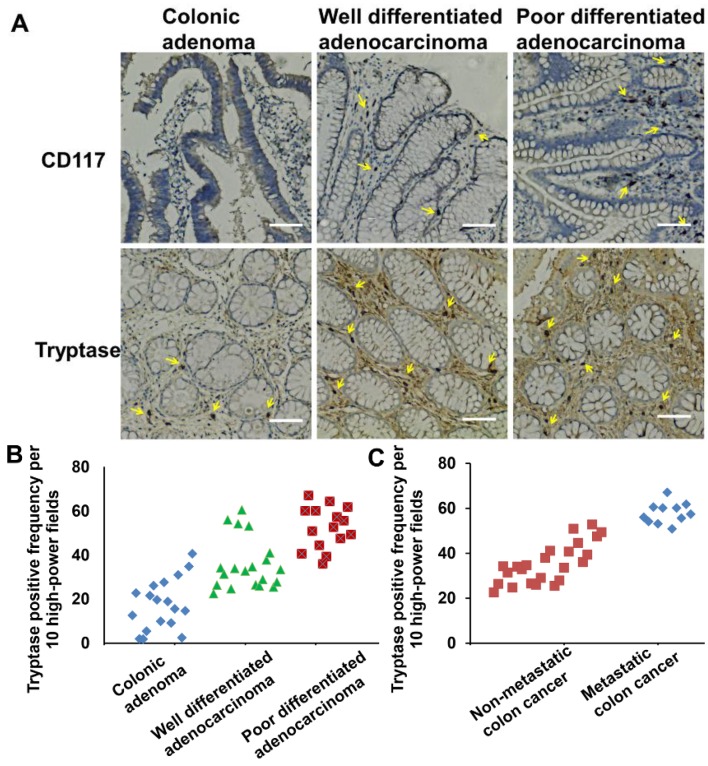
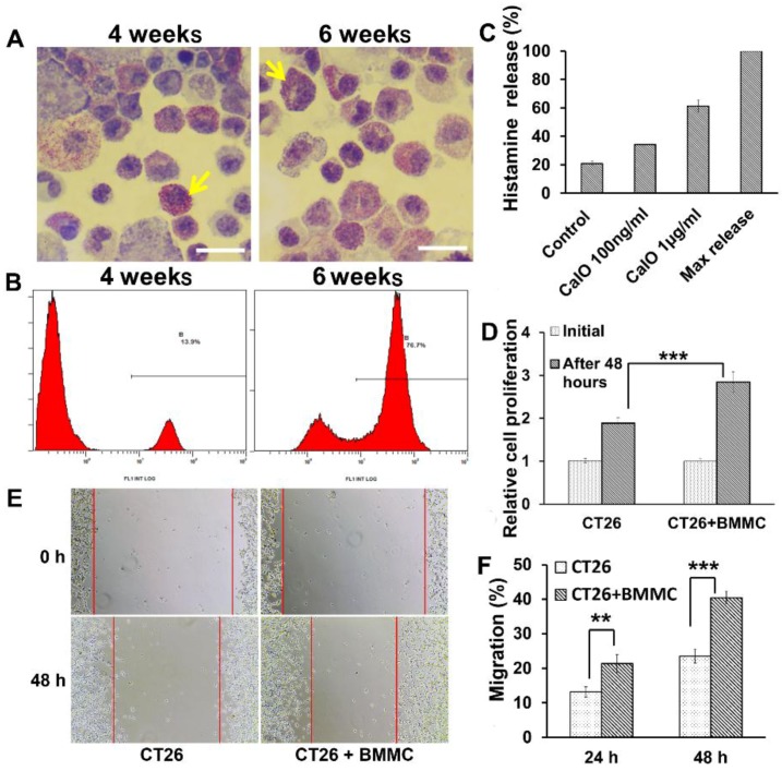
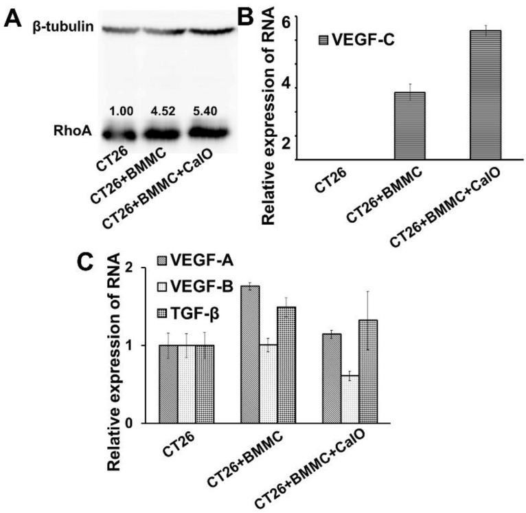
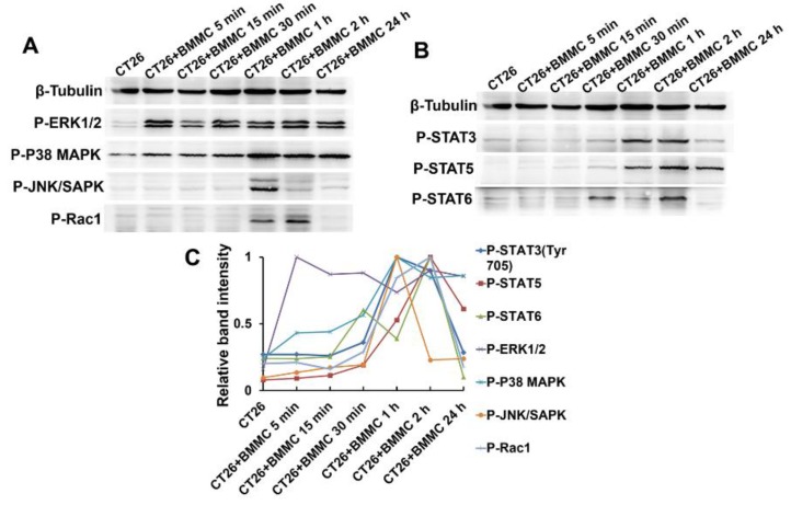
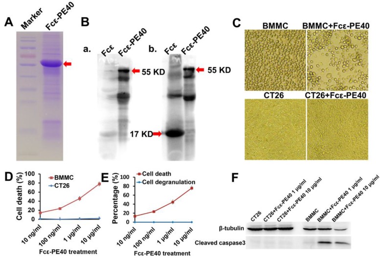
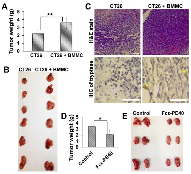
The association of colitis with colorectal cancer has become increasingly clear with mast cells being identified as important inflammatory cells in the process. In view of the relationship between mast cells and cancer, we studied the effect and mechanisms of mast cells in the development of colon cancer. Functional and mechanistic insights were gained from ex vivo and in vivo studies of cell interactions between mast cells and CT26 cells. Further evidence was reversely obtained in studies of mast cell targeted Fcε-PE40 chimeric toxin. Experiments revealed mast cells could induce colon tumor cell proliferation and invasion. Cancer progression was found to be related to the density of mast cells in colonic submucosa. The activation of MAPK, Rho-GTPase, and STAT pathways in colon cancer cells was triggered by mast cells during cell-to-cell interaction. Lastly, using an Fcε-PE40 chimeric toxin we constructed, we confirmed the promoting effect of mast cells in development of colon cancer. Mast cells are a promoting factor of colon cancer and thus also a potential therapeutic target. The Fcε-PE40 chimeric toxin targeting mast cells could effectively prevent colon cancer in vitro and in vivo. Consequently, these data may demonstrate a novel immunotherapeutic approach for the treatment of tumors.
Overview
The claims and statistics of this paper haven’t been checked yet.
Run the check — free, no account needed. It reads the open-access text and checks the sources it cites.
Integrity signals
Registry
Cites
Statistics
Figures
Verification
Claims the paper makes
The claims and statistics of this paper haven’t been checked yet.
Run the check — free, no account needed. It reads the open-access text and checks the sources it cites.
Claims other papers make about this one
Each is checked against this paper’s own text: does it say what the citing paper says it does?
In the citing paper
“In clinical and experimental studies alike, mast cells have been associated with both, beneficial and detrimental outcomes. This is true often even for the same tumor types, including pancreatic, 217 , 218 lung, 219 , 220 colorectal 221 , 222 and prostate cancer 223 , 224 , 225 as well as melanoma. 220 , 226 , 227 , 228 , 229 , 230 Other reports concluded they have no role at all. 231 , 232 , 233”
From Mast cell ontogeny: From fetal development to life‐long health and disease (2023) · cites this paper as [218]
Self-check
Percentages, sums and reported differences re-computed from the paper’s own numbers.
The self-check needs the paper’s full text.
Citations
Cites
Galli, S. J., & Tsai, M. (2010) · European Journal of Immunology · doi:10.1002/eji.201040559 · Cited by 403
Jumping the barrier: VE‐cadherin, VEGF and other angiogenic modifiers in cancer
Le Guelte, A., Dwyer, J., & Gavard, J. (2011) · Biology of the Cell · doi:10.1042/bc20110069 · Cited by 91
Research works printed but not linked: 65
- Eichten, 2006, Paradoxical roles of the immune system during cancer development, Nat. Rev. Cancer, 6, 24, 10.1038/nrc1782 · DOI
- Grivennikov, 2010, Immunity, inflammation, and cancer, Cell, 140, 883, 10.1016/j.cell.2010.01.025 · DOI · Paper page
- Quante, 2013, The gastrointestinal tumor microenvironment, Gastroenterology, 145, 63, 10.1053/j.gastro.2013.03.052 · DOI
- Wood, 2014, The role of the tumor-microenvironment in lung cancer-metastasis and its relationship to potential therapeutic targets, Cancer Treat. Rev., 40, 558, 10.1016/j.ctrv.2013.10.001 · DOI
- Riese, 2014, Epiregulin: Roles in normal physiology and cancer, Semin. Cell Dev. Biol., 28, 49, 10.1016/j.semcdb.2014.03.005 · DOI
- Loesch, 2008, The p38 mapk stress pathway as a tumor suppressor or more?, Front. Biosci., 13, 3581, 10.2741/2951 · DOI
- Kim, 2010, Pathological roles of mapk signaling pathways in human diseases, Biochim. Biophys. Acta, 1802, 396, 10.1016/j.bbadis.2009.12.009 · DOI · Paper page
- Zhang, 2013, Interleukin-6 is required for pancreatic cancer progression by promoting mapk signaling activation and oxidative stress resistance, Cancer Res., 73, 6359, 10.1158/0008-5472.CAN-13-1558-T · DOI
- Taylor, C.A., Zheng, Q., Liu, Z., and Thompson, J.E. (2013). Role of p38 and jnk mapk signaling pathways and tumor suppressor p53 on induction of apoptosis in response to ad-eif5a1 in a549 lung cancer cells. Mol. Cancer. · DOI
- Sanders, 2010, The type ii transmembrane serine protease, matriptase-2: Possible links to cancer?, Anti-Cancer Agents Med. Chem., 10, 64, 10.2174/1871520611009010064 · DOI
- Chia, 2014, Breast cancer cell invasion mediated by galpha12 signaling involves expression of interleukins-6 and -8, and matrix metalloproteinase-2, J. Mol. Signal., 9, 1, 10.1186/1750-2187-9-6 · DOI
- Lin, 2007, Il-6 induces ags gastric cancer cell invasion via activation of the c-src/rhoa/rock signaling pathway, Int. J. Cancer., 120, 2600, 10.1002/ijc.22599 · DOI
- Geng, Y., Chandrasekaran, S., Hsu, J.W., Gidwani, M., Hughes, A.D., and King, M.R. (2013). Phenotypic switch in blood: Effects of pro-inflammatory cytokines on breast cancer cell aggregation and adhesion. PLoS ONE, 8. · DOI
- Gacche, 2013, Targeting tumor micro-environment for design and development of novel anti-angiogenic agents arresting tumor growth, Prog. Biophys. Mol. Biol., 113, 333, 10.1016/j.pbiomolbio.2013.10.001 · DOI
- Allavena, 2012, Immunology in the clinic review series; focus on cancer: Tumour-associated macrophages: Undisputed stars of the inflammatory tumour microenvironment, Clin. Exp. Immunol., 167, 195, 10.1111/j.1365-2249.2011.04515.x · DOI
- Sakurai, 2012, Targeting of tak1 in inflammatory disorders and cancer, Trends Pharmacol. Sci., 33, 522, 10.1016/j.tips.2012.06.007 · DOI
- Vaiopoulos, 2010, Colon carcinogenesis: Learning from nf-kappab and ap-1, Int. J. Biochem. Cell Biol., 42, 1061, 10.1016/j.biocel.2010.03.018 · DOI
- Verstovsek, 2013, Molecular pathways: Jak/stat pathway: Mutations, inhibitors, and resistance, Clin. Cancer Res., 19, 1933, 10.1158/1078-0432.CCR-12-0284 · DOI
- Laveti, 2013, Anti-inflammatory treatments for chronic diseases: A review, Inflamm. Allergy Drug Targets, 12, 349, 10.2174/18715281113129990053 · DOI
- Yu, 2009, Stats in cancer inflammation and immunity: A leading role for stat3, Nat. Rev. Cancer, 9, 798, 10.1038/nrc2734 · DOI · Paper page
- Crivellato, 2003, Paul ehrlich’s doctoral thesis: A milestone in the study of mast cells, Br. J. Haematol., 123, 19, 10.1046/j.1365-2141.2003.04573.x · DOI
- Wedemeyer, 2000, Roles of mast cells and basophils in innate and acquired immunity, Curr. Opin. Immunol., 12, 624, 10.1016/S0952-7915(00)00154-0 · DOI
- Ali, 2010, Regulation of human mast cell and basophil function by anaphylatoxins c3a and c5a, Immunol. Lett., 128, 36, 10.1016/j.imlet.2009.10.007 · DOI
- Huang, 2008, Scf-mediated mast cell infiltration and activation exacerbate the inflammation and immunosuppression in tumor microenvironment, Blood, 112, 1269, 10.1182/blood-2008-03-147033 · DOI
- Kanbe, 1999, Human mast cells produce matrix metalloproteinase 9, Eur. J. Immunol., 29, 2645, 10.1002/(SICI)1521-4141(199908)29:08<2645::AID-IMMU2645>3.0.CO;2-1 · DOI
- Crivellato, 2005, Piecemeal degranulation in human tumour pheochromocytes, J. Anat., 206, 47, 10.1111/j.0021-8782.2005.00365.x · DOI
- Caruso, 2005, Degranulation patterns of eosinophils in advanced gastric carcinoma: An electron microscopic study, Ultrastruct. Pathol., 29, 29, 10.1080/019131290882303 · DOI
- Theoharides, 2004, Mast cells: The jekyll and hyde of tumor growth, Trends Immunol., 25, 235, 10.1016/j.it.2004.02.013 · DOI
- McNeil, 2007, Mast cell-restricted tryptases: Structure and function in inflammation and pathogen defense, J. Biol. Chem., 282, 20785, 10.1074/jbc.R700017200 · DOI
- Coussens, 2000, Mmp-9 supplied by bone marrow-derived cells contributes to skin carcinogenesis, Cell, 103, 481, 10.1016/S0092-8674(00)00139-2 · DOI · Paper page
- Maltby, 2009, Mast cells in tumor growth: Angiogenesis, tissue remodelling and immune-modulation, Biochim. Biophys. Acta, 1796, 19
- Crivellato, 2008, Mast cells and tumour angiogenesis: New insight from experimental carcinogenesis, Cancer Lett., 269, 1, 10.1016/j.canlet.2008.03.031 · DOI
- Ribatti, 2013, Mast cells and macrophages exert beneficial and detrimental effects on tumor progression and angiogenesis, Immunol. Lett., 152, 83, 10.1016/j.imlet.2013.05.003 · DOI
- Adams, 1994, Short-course cimetidine and survival with colorectal cancer, Lancet, 344, 1768, 10.1016/S0140-6736(94)92907-6 · DOI
- Matsumoto, 1995, Cimetidine and survival with colorectal cancer, Lancet, 346, 115, 10.1016/S0140-6736(95)92136-2 · DOI
- Matsumoto, 2002, Cimetidine increases survival of colorectal cancer patients with high levels of sialyl lewis-x and sialyl lewis-a epitope expression on tumour cells, Br. J. Cancer, 86, 161, 10.1038/sj.bjc.6600048 · DOI
- Khazaie, 2011, The significant role of mast cells in cancer, Cancer Metastasis Rev., 30, 45, 10.1007/s10555-011-9286-z · DOI
- Galinsky, 2008, Mast cells and cancer—No longer just basic science, Crit. Rev. Oncol. Hematol., 68, 115, 10.1016/j.critrevonc.2008.06.001 · DOI
- Nonomura, 2007, Decreased number of mast cells infiltrating into needle biopsy specimens leads to a better prognosis of prostate cancer, Br. J. Cancer, 97, 952, 10.1038/sj.bjc.6603962 · DOI
- Strouch, 2010, Crosstalk between mast cells and pancreatic cancer cells contributes to pancreatic tumor progression, Clin. Cancer Res., 16, 2257, 10.1158/1078-0432.CCR-09-1230 · DOI
- Goffredo, 2013, Tryptase serum levels in patients suffering from hepatocellular carcinoma undergoing intra-arterial chemoembolization: Possible predictive role of response to treatment, Mol. Clin. Oncol., 1, 385, 10.3892/mco.2013.59 · DOI
- Tan, 2005, P53 and c-kit (cd117) protein expression as prognostic indicators in breast phyllodes tumors: A tissue microarray study, Mod. Pathol., 18, 1527, 10.1038/modpathol.3800488 · DOI
- Gulubova, 2009, Prognostic significance of mast cell number and microvascular density for the survival of patients with primary colorectal cancer, J. Gastroenterol. Hepatol., 24, 1265, 10.1111/j.1440-1746.2007.05009.x · DOI
- Yodavudh, 2008, Prognostic significance of microvessel density and mast cell density for the survival of thai patients with primary colorectal cancer, J. Med. Assoc. Thail., 91, 723
- Acikalin, 2005, Tumour angiogenesis and mast cell density in the prognostic assessment of colorectal carcinomas, Dig. Liver Dis., 37, 162, 10.1016/j.dld.2004.09.028 · DOI
- Fishman, 1997, Targeted elimination of cells expressing the high-affinity receptor for ige (fc epsilon ri) by a pseudomonas exotoxin-based chimeric protein, Eur. J. Immunol., 27, 486, 10.1002/eji.1830270220 · DOI
- Fishman, 2000, Targeted fc2′-3-pe40 chimeric protein abolishes passive cutaneous anaphylaxis in mice, Clin. Exp. Immunol., 119, 398, 10.1046/j.1365-2249.2000.01151.x · DOI
- Kruger, 1974, Structural features of histamine release in rat peritoneal mast cells. A study with toluidine blue, Int. Arch. Allergy Appl. Immunol., 46, 740, 10.1159/000231174 · DOI
- Nitta, 2009, Image analysis of mast cell degranulation in a concentration gradient of stimuli formed in the channel between a glass plate and a silicon substrate, Eur. J. Cell Biol., 88, 541, 10.1016/j.ejcb.2009.03.004 · DOI
- Kovarikova, 2014, Methods for studying tumor cell migration and invasiveness, Klin. Onkol., 27, S22, 10.14735/amko20141S22 · DOI
- Hill, 1996, Stem cell factor enhances immunoglobulin e-dependent mediator release from cultured rat bone marrow-derived mast cells: Activation of previously unresponsive cells demonstrated by a novel elispot assay, Immunology, 87, 326, 10.1046/j.1365-2567.1996.455545.x · DOI
- Oliveira, 2002, Stem cell factor induces eosinophil activation and degranulation: Mediator release and gene array analysis, Blood, 100, 4291, 10.1182/blood.V100.13.4291 · DOI
- Mantovani, 2008, Cancer-related inflammation, Nature, 454, 436, 10.1038/nature07205 · DOI · Paper page
- Grimbaldeston, 2004, Association between melanoma and dermal mast cell prevalence in sun-unexposed skin, Br. J. Dermatol., 150, 895, 10.1111/j.1365-2133.2004.05966.x · DOI
- Anuradha, A., Kiran Kumar Naik, B., Vijay Srinivas, G., Devi, R.S., and Puneet, H.K. (2014). Incidence of mast cells in oral squamous cell carcinoma: A short study. J. Oncol. · DOI
- Rabenhorst, 2012, Mast cells play a protumorigenic role in primary cutaneous lymphoma, Blood, 120, 2042, 10.1182/blood-2012-03-415638 · DOI
- Ribatti, 2009, The controversial role of mast cells in tumor growth, Int. Rev. Cell Mol. Biol., 275, 89, 10.1016/S1937-6448(09)75004-X · DOI
- Marichal, 2013, Mast cells: Potential positive and negative roles in tumor biology, Cancer Immunol. Res., 1, 269, 10.1158/2326-6066.CIR-13-0119 · DOI
- Shantikumar, 2013, Immune system: A double-edged sword in cancer, Inflamm. Res., 62, 823, 10.1007/s00011-013-0645-9 · DOI · Paper page
- Noguchi, 2013, Antibody-based therapy in colorectal cancer, Immunotherapy, 5, 533, 10.2217/imt.13.35 · DOI
- Brinkmann, 1998, Role of caspases in immunotoxin-induced apoptosis of cancer cells, Biochemistry, 37, 16934, 10.1021/bi980995m · DOI
- Perentesis, 1997, Induction of apoptosis in multidrug-resistant and radiation-resistant acute myeloid leukemia cells by a recombinant fusion toxin directed against the human granulocyte macrophage colony-stimulating factor receptor, Clin. Cancer Res., 3, 347
- Turcotte, 2011, Immunotherapy for metastatic solid cancers, Adv. Surg., 45, 341, 10.1016/j.yasu.2011.04.003 · DOI · Paper page
- Tartour, 2011, Cancer immunotherapy: Recent breakthroughs and perspectives, Med. Sci., 27, 833
- Dalton, 2012, The roles of mast cells in anticancer immunity. Cancer immunology, immunotherapy, Cancer Immunol. Immunother., 61, 1511, 10.1007/s00262-012-1246-0 · DOI
Cited by
Sorted by citations
Mast cell ontogeny: From fetal development to life‐long health and disease
Chia, S. L., Kapoor, S., Carvalho, C., Bajénoff, M., & Gentek, R. (2023) · Immunological Reviews · doi:10.1111/imr.13191 · Cited by 60
Metadata
- Authors
- Wang, S., Li, L., Shi, R., Liu, X., Zhang, J., Zou, Z., Hao, Z., & Tao, A.
- Affiliations
- The Second Affiliated Hospital of Guangzhou Medical University, Guangdong Provincial Key Laboratory of Allergy & Clinical Immunology, The State Key Clinical Specialty in Allergy, The State Key Laboratory of Respiratory Disease; Guangzhou 510260, China
- The Second Affiliated Hospital of Guangzhou Medical University, Guangzhou 510260, China
- Venue
- Toxins
- Publisher
- MDPI AG
- Published
- 2016-03-11
- Volume
- 8
- Issue
- 3
- Pages
- 71
- ISSN
- 2072-6651
- PMC
- PMC4810216
- Cited by
- 17
- References
- 2
- Language
- en
- Funders
- the National Natural Science Foundation of China (81373128, 81402025)
- Guangdong provincal public welfare research and capacity building special fundation (2014A020212363)
About these checks
Registry, retraction, Cites and statistics results are computed, not judged, with one exception: a printed reference that no index lists is read once by a model before it counts as a citation error. Claims are extracted by a model and kept only when their quote appears verbatim in the text; each cited source is then checked, with a model’s help, for whether it says what the claim relies on. Those verdicts can be wrong — read the claim’s quote and the cited source before relying on one.
Statistics and arithmetic are recomputed, not judged.
Confidence bands are shown only for fields calibrated on our annotated test set. None is calibrated yet, so no band is shown.
What counts as a claim ›How we measure confidence ›How we check this ›
Are you an author, or do you see a problem with this page? Request a review
How to cite
Cite CoChat for the checks on this page, and the paper for its own findings.
- This page
- CoChat. (2026). Citation check: Mast Cell Targeted Chimeric Toxin Can Be Developed as an Adjunctive Therapy in Colon Cancer Treatment. https://app.cochat.ai/papers/10.3390/toxins8030071
- The paper
- Wang, S., Li, L., Shi, R., Liu, X., Zhang, J., Zou, Z., Hao, Z., & Tao, A. (2016). Mast Cell Targeted Chimeric Toxin Can Be Developed as an Adjunctive Therapy in Colon Cancer Treatment. Toxins. https://doi.org/10.3390/toxins8030071
