

The DOI matches its Crossref record; 28 of the 28 references it cites were found in a registry or an index.
Comparison of Four SARS-CoV-2 Neutralization Assays
Open accessAbstract
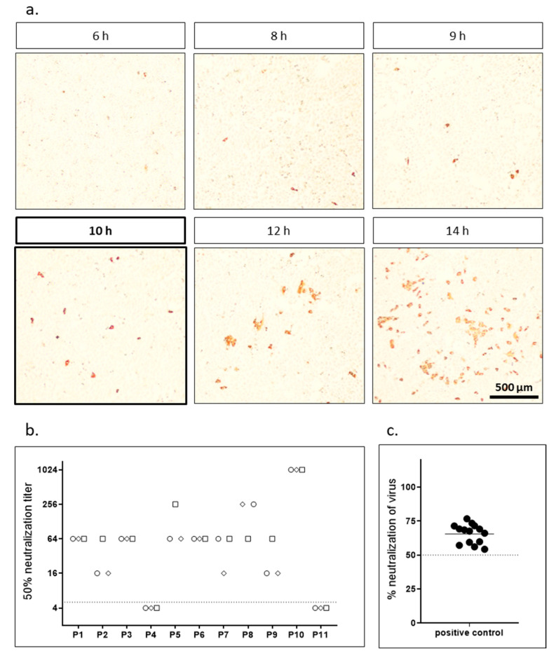
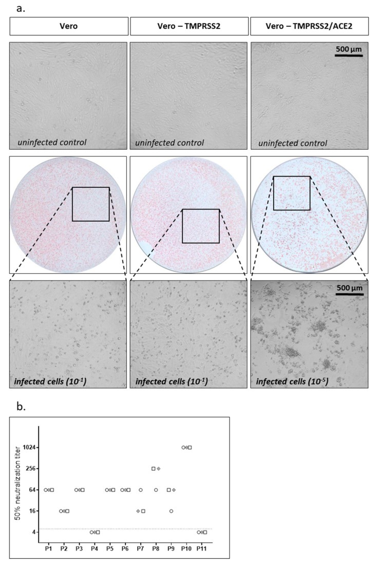
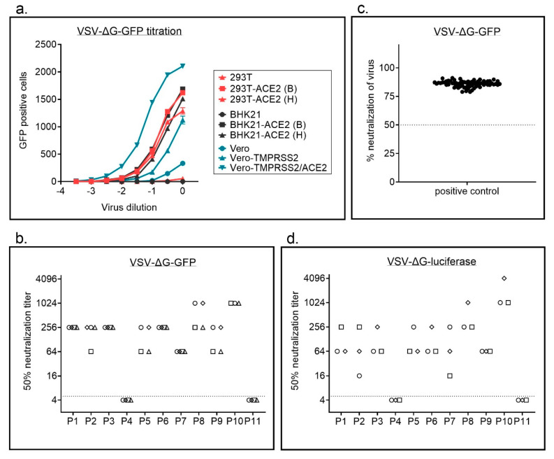
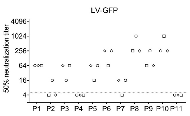
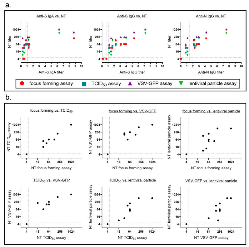
Neutralizing antibodies are a major correlate of protection for many viruses including the novel coronavirus SARS-CoV-2. Thus, vaccine candidates should potently induce neutralizing antibodies to render effective protection from infection. A variety of in vitro assays for the detection of SARS-CoV-2 neutralizing antibodies has been described. However, validation of the different assays against each other is important to allow comparison of different studies. Here, we compared four different SARS-CoV-2 neutralization assays using the same set of patient samples. Two assays used replication competent SARS-CoV-2, a focus forming assay and a TCID50-based assay, while the other two assays used replication defective lentiviral or vesicular stomatitis virus (VSV)-based particles pseudotyped with SARS-CoV-2 spike. All assays were robust and produced highly reproducible neutralization titers. Titers of neutralizing antibodies correlated well between the different assays and with the titers of SARS-CoV-2 S-protein binding antibodies detected in an ELISA. Our study showed that commonly used SARS-CoV-2 neutralization assays are robust and that results obtained with different assays are comparable.
Overview
The claims and statistics of this paper haven’t been checked yet.
Run the check — free, no account needed. It reads the open-access text and checks the sources it cites.
Integrity signals
Registry
Cites
Statistics
Figures
Verification
Claims the paper makes
The claims and statistics of this paper haven’t been checked yet.
Run the check — free, no account needed. It reads the open-access text and checks the sources it cites.
Claims other papers make about this one
Each is checked against this paper’s own text: does it say what the citing paper says it does?
In the citing paper
“This system faithfully mimics key aspects of SARS-CoV-2 entry into cells, including ACE2 engagement, priming of the S protein by TMPRSS2, and antibody-mediated neutralization (Hoffmann et al., 2020b; Riepler et al., 2020; Schmidt et al., 2020).”
From SARS-CoV-2 variants B.1.351 and P.1 escape from neutralizing antibodies (2021) · cites this paper as Riepler et al., 2020
Self-check
Percentages, sums and reported differences re-computed from the paper’s own numbers.
The self-check needs the paper’s full text.
Citations
Cites
Hoffmann, M., Kleine-Weber, H., Schroeder, S., Krüger, N., Herrler, T., Erichsen, S., Schiergens, T. S., Herrler, G., Wu, N.-H., Nitsche, A., et al. (2020) · Cell · doi:10.1016/j.cell.2020.02.052 · Cited by 20,286
WHO Coronavirus Disease (COVID-19) Dashboard
e, C. R. D. I. (2020) · Bangladesh Physiotherapy Journal · doi:10.46945/bpj.10.1.03.01 · Cited by 5,465
Antibody responses to SARS-CoV-2 in patients with COVID-19
Long, Q.-X., Liu, B.-Z., Deng, H.-J., Wu, G.-C., Deng, K., Chen, Y.-K., Liao, P., Qiu, J.-F., Lin, Y., Cai, X.-F., et al. (2020) · Nature Medicine · doi:10.1038/s41591-020-0897-1 · Cited by 2,876
Antibody Responses to SARS-CoV-2 in Patients With Novel Coronavirus Disease 2019
Zhao, J., Yuan, Q., Wang, H., Liu, W., Liao, X., Su, Y., Wang, X., Yuan, J., Li, T., Li, J., et al. (2020) · Clinical Infectious Diseases · doi:10.1093/cid/ciaa344 · Cited by 2,607
Neuropilin-1 facilitates SARS-CoV-2 cell entry and infectivity
Cantuti-Castelvetri, L., Ojha, R., Pedro, L. D., Djannatian, M., Franz, J., Kuivanen, S., van der Meer, F., Kallio, K., Kaya, T., Anastasina, M., et al. (2020) · Science · doi:10.1126/science.abd2985 · Cited by 1,925
Hoffmann, M., Kleine-Weber, H., & Pöhlmann, S. (2020) · Molecular Cell · doi:10.1016/j.molcel.2020.04.022 · Cited by 1,852
Escape from neutralizing antibodies by SARS-CoV-2 spike protein variants
Weisblum, Y., Schmidt, F., Zhang, F., DaSilva, J., Poston, D., Lorenzi, J. C., Muecksch, F., Rutkowska, M., Hoffmann, H.-H., Michailidis, E., et al. (2020) · eLife · doi:10.7554/elife.61312 · Cited by 1,308
Baum, A., Fulton, B. O., Wloga, E., Copin, R., Pascal, K. E., Russo, V., Giordano, S., Lanza, K., Negron, N., Ni, M., et al. (2020) · Science · doi:10.1126/science.abd0831 · Cited by 1,253
To, K. K.-W., Hung, I. F.-N., Ip, J. D., Chu, A. W.-H., Chan, W.-M., Tam, A. R., Fong, C. H.-Y., Yuan, S., Tsoi, H.-W., Ng, A. C.-K., et al. (2021) · Clinical Infectious Diseases · doi:10.1093/cid/ciaa1275 · Cited by 746
Crawford, K. H. D., Eguia, R. T., Dingens, A. S., Loes, A. N., Malone, K. D., Wolf, C. R., Chu, H. Y., Tortorici, M. A., Veesler, D., Murphy, M., et al. (2020) · Viruses · doi:10.3390/v12050513 · Cited by 744
Cited by
Sorted by citations
Multiple SARS-CoV-2 variants escape neutralization by vaccine-induced humoral immunity
Garcia-Beltran, W. F., Lam, E. C., St. Denis, K., Nitido, A. D., Garcia, Z. H., Hauser, B. M., Feldman, J., Pavlovic, M. N., Gregory, D. J., Poznansky, M. C., et al. (2021) · Cell · doi:10.1016/j.cell.2021.03.013 · Cited by 1,353
mRNA-based COVID-19 vaccine boosters induce neutralizing immunity against SARS-CoV-2 Omicron variant
Garcia-Beltran, W. F., St. Denis, K. J., Hoelzemer, A., Lam, E. C., Nitido, A. D., Sheehan, M. L., Berrios, C., Ofoman, O., Chang, C. C., Hauser, B. M., et al. (2022) · Cell · doi:10.1016/j.cell.2021.12.033 · Cited by 1,045
The biological and clinical significance of emerging SARS-CoV-2 variants
Tao, K., Tzou, P. L., Nouhin, J., Gupta, R. K., de Oliveira, T., Kosakovsky Pond, S. L., Fera, D., & Shafer, R. W. (2021) · Nature Reviews Genetics · doi:10.1038/s41576-021-00408-x · Cited by 1,009
SARS-CoV-2 variants B.1.351 and P.1 escape from neutralizing antibodies
Hoffmann, M., Arora, P., Groß, R., Seidel, A., Hörnich, B. F., Hahn, A. S., Krüger, N., Graichen, L., Hofmann-Winkler, H., Kempf, A., et al. (2021) · Cell · doi:10.1016/j.cell.2021.03.036 · Cited by 897
Hoffmann, M., Krüger, N., Schulz, S., Cossmann, A., Rocha, C., Kempf, A., Nehlmeier, I., Graichen, L., Moldenhauer, A.-S., Winkler, M. S., et al. (2022) · Cell · doi:10.1016/j.cell.2021.12.032 · Cited by 889
Metadata
- Authors
- Riepler, L., Rössler, A., Falch, A., Volland, A., Borena, W., von Laer, D., & Kimpel, J.
- Affiliations
- Innsbruck Medical University (AT)
- Department of Hygiene, Microbiology and Public Health, Institute of Virology, Medical University of Innsbruck, 6020 Innsbruck, Austria
- Corresponding author
- Kimpel, Janine
- Venue
- Vaccines
- Publisher
- MDPI AG
- Published
- 2020-12-28
- Volume
- 9
- Issue
- 1
- Pages
- 13
- ISSN
- 2076-393X (ISSN-L)
- PMC
- PMC7824240
- OpenAlex
- W3114250596
- Cited by
- 117
- References
- 26
- Citations by year
- 2026: 2 · 2025: 10 · 2024: 17 · 2023: 19 · 2022: 37 · 2021: 32
- FWCI
- 2.89
- Type
- journal article
- Language
- en
- Open access
- gold
- doi.org
- Licence
- Locations
- Vaccines (publishedVersion, cc-by)
- Europe PMC (PubMed Central) (publishedVersion, other-oa)
- PubMed Central (publishedVersion, cc-by)
- PubMed (publishedVersion)
- DOAJ (DOAJ: Directory of Open Access Journals) (submittedVersion, cc-by-sa)
- Topics
- Health Sciences › Medicine › Infectious Diseases › SARS-CoV-2 and COVID-19 Research
- Health Sciences › Medicine › Infectious Diseases › SARS-CoV-2 detection and testing
- Life Sciences › Agricultural and Biological Sciences › Animal Science and Zoology › Animal Virus Infections Studies
- Keywords
- SARS-CoV-2, neutralizing antibodies, assay validation, TCID50, vaccine-induced immunity, assay reproducibility
About these checks
Registry, retraction, Cites and statistics results are computed, not judged, with one exception: a printed reference that no index lists is read once by a model before it counts as a citation error. Claims are extracted by a model and kept only when their quote appears verbatim in the text; each cited source is then checked, with a model’s help, for whether it says what the claim relies on. Those verdicts can be wrong — read the claim’s quote and the cited source before relying on one.
Statistics and arithmetic are recomputed, not judged.
Confidence bands are shown only for fields calibrated on our annotated test set. None is calibrated yet, so no band is shown.
What counts as a claim ›How we measure confidence ›How we check this ›
Are you an author, or do you see a problem with this page? Request a review
