

The DOI matches its Crossref record; 106 of the 107 references it cites were found in a registry or an index.
Potential toxicity of superparamagnetic iron oxide nanoparticles (SPION)
Open accessAbstract
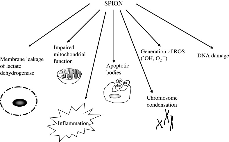
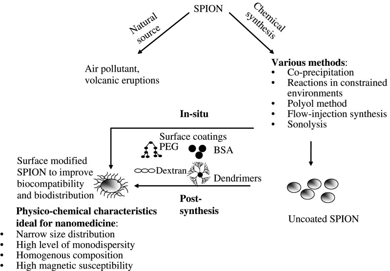
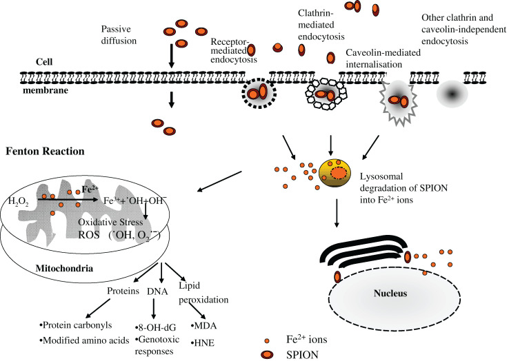
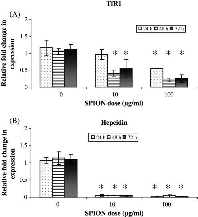
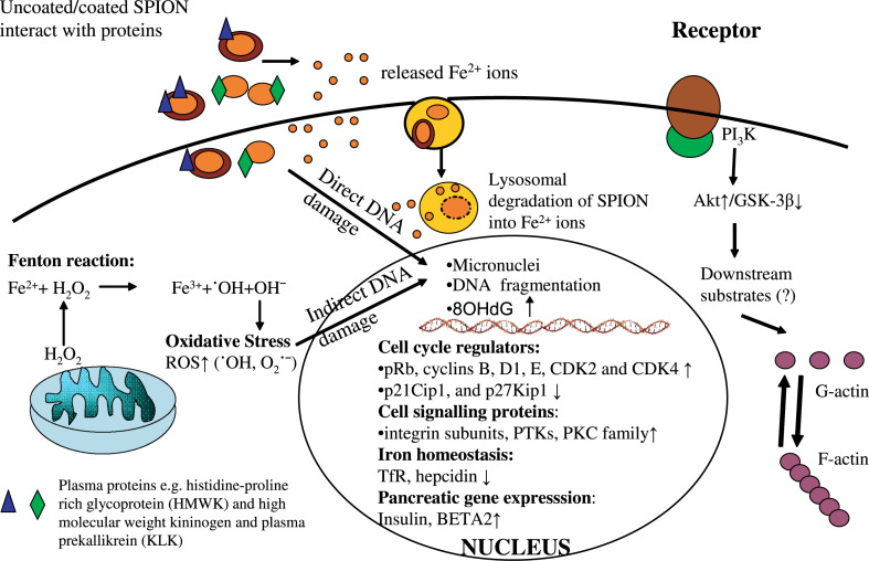
Superparamagnetic iron oxide nanoparticles (SPION) are being widely used for various biomedical applications, for example, magnetic resonance imaging, targeted delivery of drugs or genes, and in hyperthermia. Although, the potential benefits of SPION are considerable, there is a distinct need to identify any potential cellular damage associated with these nanoparticles. Besides focussing on cytotoxicity, the most commonly used determinant of toxicity as a result of exposure to SPION, this review also mentions the importance of studying the subtle cellular alterations in the form of DNA damage and oxidative stress. We review current studies and discuss how SPION, with or without different surface coating, may cause cellular perturbations including modulation of actin cytoskeleton, alteration in gene expression profiles, disturbance in iron homeostasis and altered cellular responses such as activation of signalling pathways and impairment of cell cycle regulation. The importance of protein-SPION interaction and various safety considerations relating to SPION exposure are also addressed.
Overview
The claims and statistics of this paper haven’t been checked yet.
Run the check — free, no account needed. It reads the open-access text and checks the sources it cites.
Integrity signals
Registry
Cites
Statistics
Figures
Verification
Claims the paper makes
The claims and statistics of this paper haven’t been checked yet.
Run the check — free, no account needed. It reads the open-access text and checks the sources it cites.
Claims other papers make about this one
Each is checked against this paper’s own text: does it say what the citing paper says it does?
The source backs part of the claim, not all of it — or backs it for a different population, condition or comparison.
In the citing paper
“Metallic NPs are valuable for their theragnostic capabilities but face significant barriers to translation due to potential long-term toxicity and bioaccumulation, which limit clinical progress [102, 103].”
From Targeted Nanoparticles for Drug Delivery Across the Blood–Brain Barrier in Early and Late Stages of Alzheimer's Disease: A Review (2026) · cites this paper as [102]
Experimental — accuracy not yet measuredIn the citing paper
“In vitro, upon prolonged incubation, surface-coated SPION are typically nontoxic at iron concentration up to and 10 μg/ml, while slight cytotoxicity is seen at 100 μg/ml.”
From Iron oxide nanoparticles: Diagnostic, therapeutic and theranostic applications (2019) · cites this paper as [120]
In the citing paper
“In general, SPION toxicity depends on a variety of factors, including - besides dosing and exposure time - also the nature of the coating material, the hydrodynamic diameter of the particle and SPION-protein interactions [119–122].”
From Iron oxide nanoparticles: Diagnostic, therapeutic and theranostic applications (2019) · cites this paper as [120]
Self-check
Percentages, sums and reported differences re-computed from the paper’s own numbers.
The self-check needs the paper’s full text.
Citations
Cites
Free radicals in biology and medicine
Gutteridge, J. M. C. (1991) · General Pharmacology The Vascular System · doi:10.1016/0306-3623(91)90478-o · Cited by 17,178
Free radicals and antioxidants in normal physiological functions and human disease
Valko, M., Leibfritz, D., Moncol, J., Cronin, M. T. D., Mazur, M., & Telser, J. (2007) · The International Journal of Biochemistry & Cell Biology · doi:10.1016/j.biocel.2006.07.001 · Cited by 14,269
Toxic potential of materials at the nanolevel
Nel, A., Xia, T., Mädler, L., & Li, N. (2006) · Science · doi:10.1126/science.1114397 · Cited by 9,324
Free Radicals in Biology and Medicine
Halliwell, B., & Gutteridge, J. M. C. (2015) · doi:10.1093/acprof:oso/9780198717478.001.0001 · Cited by 8,990
Synthesis and surface engineering of iron oxide nanoparticles for biomedical applications
Gupta, A. K., & Gupta, M. S. (2004) · Biomaterials · doi:10.1016/j.biomaterials.2004.10.012 · Cited by 6,906
Laurent, S., Forge, D., Port, M., Roch, A., Robic, C., Vander Elst, L., & Muller, R. N. (2008) · Chemical Reviews · doi:10.1021/cr068445e · Cited by 6,852
Hepcidin Regulates Cellular Iron Efflux by Binding to Ferroportin and Inducing Its Internalization
Nemeth, E., Tuttle, M. S., Powelson, J., Vaughn, M. B., Donovan, A., Ward, D. M., Ganz, T., & Kaplan, J. (2004) · Science · doi:10.1126/science.1104742 · Cited by 4,803
Biodegradable nanoparticles for drug and gene delivery to cells and tissue
Panyam, J., & Labhasetwar, V. D. (2003) · Advanced Drug Delivery Reviews · doi:10.1016/s0169-409x(02)00228-4 · Cited by 3,417
Fischer, D., Li, Y., Ahlemeyer, B., Krieglstein, J., & Kissel, T. (2003) · Biomaterials · doi:10.1016/s0142-9612(02)00445-3 · Cited by 2,329
In vitro toxicity of nanoparticles in BRL 3A rat liver cells
Hussain, S. M., Hess, K. L., Gearhart, J. M., Geiss, K. T., & Schlager, J. J. (2005) · Toxicology in Vitro · doi:10.1016/j.tiv.2005.06.034 · Cited by 2,025
Research works printed but not linked: 3
- Gould P, 2006, Nano Today, 1, 34
- 10.1016/j.biomaterials.2004.10.012 · DOI
- Sodum RS, 1988, Cancer Res, 48, 320
Cited by
Sorted by citations
Items 301–400 of 1,000
Wu, Q., Xu, Z., Duan, Y., Zhu, Y., Ou, M., & Xu, X. (2017) · RSC Advances · doi:10.1039/c7ra03174b · Cited by 48
The role of nanocomposites against biofilm infections in humans
Varma, A., Warghane, A. J., Dhiman, N. K., Paserkar, N., Upadhye, V. J., Modi, A., & Saini, R. (2023) · Frontiers in Cellular and Infection Microbiology · doi:10.3389/fcimb.2023.1104615 · Cited by 48
Calcium phosphate-based nanosystems for advanced targeted nanomedicine
Esposti, L. D., Carella, F., Adamiano, A., Tampieri, A., & Iafisco, M. (2018) · Drug Development and Industrial Pharmacy · doi:10.1080/03639045.2018.1451879 · Cited by 47
Saengruengrit, C., Ritprajak, P., Wanichwecharungruang, S. P., Sharma, A., Salvan, G., Zahn, D. R. T., & Insin, N. (2018) · Journal of Colloid and Interface Science · doi:10.1016/j.jcis.2018.03.008 · Cited by 47
Assessment of oxidative damage induced by iron oxide nanoparticles on different nervous system cells
Fernández‐Bertólez, N., Costa, C., Bessa, M. J., Park, M. V. D. Z., Carrière, M., Dussert, F., Teixeira, J. P., Pásaro, E., Laffón, B., & Valdiglesias, V. (2018) · Mutation Research/Genetic Toxicology and Environmental Mutagenesis · doi:10.1016/j.mrgentox.2018.11.013 · Cited by 47
Zhang, T., Xu, Q., Huang, T., Ling, D., & Gao, J. (2020) · Small · doi:10.1002/smll.202001588 · Cited by 47
Thermoseeds for interstitial magnetic hyperthermia: from bioceramics to nanoparticles
Baeza, A., Arcos, D., & Vallet‐Regí, M. (2013) · Journal of Physics Condensed Matter · doi:10.1088/0953-8984/25/48/484003 · Cited by 47
Male reproductive toxicity of inorganic nanoparticles in rodent models: A systematic review
Dantas, G. D. P. F., Ferraz, F. S., de Andrade, L. M., & Costa, G. M. J. (2022) · Chemico-Biological Interactions · doi:10.1016/j.cbi.2022.110023 · Cited by 46
Interaction of Size-Tailored PEGylated Iron Oxide Nanoparticles with Lipid Membranes and Cells
Gal, N., Lassenberger, A., Nogareda, L. H., Scheberl, A., Charwat, V., Kasper, C., & Reimhult, E. (2016) · ACS Biomaterials Science & Engineering · doi:10.1021/acsbiomaterials.6b00311 · Cited by 46
Perspective: Magnetoresistive sensors for biomedicine
Giouroudi, I., & Hristoforou, E. V. (2018) · Journal of Applied Physics · doi:10.1063/1.5027035 · Cited by 46
Sachdeva, V., Monga, A., Vashisht, R., Singh, D., Singh, A., & Bedi, N. (2022) · Journal of Drug Delivery Science and Technology · doi:10.1016/j.jddst.2022.103585 · Cited by 45
Atabaev, T. S., Lee, J.-H., Lee, J.-J., Han, D., Hwang, Y., Kim, H.-K., & Hong, N. H. (2013) · Nanotechnology · doi:10.1088/0957-4484/24/34/345603 · Cited by 45
Effective delivery of immunosuppressive drug molecules by silica coated iron oxide nanoparticles
Hwang, J., Lee, E., Kim, J.-E., Seo, Y., Lee, K. H., Hong, J. W., Gilad, A. A., Park, H., & Choi, J. H. (2016) · Colloids and Surfaces B Biointerfaces · doi:10.1016/j.colsurfb.2016.01.040 · Cited by 45
The prospective opportunities offered by magnetic scaffolds for bone tissue engineering: a review
Ortolani, A., Bianchi, M., Mosca, M., Caravelli, S., Fuiano, M., Marcacci, M., & Russo, A. (2016) · Joints · doi:10.11138/jts/2016.4.4.228 · Cited by 45
Nanoparticle Biokinetics in Mice and Nonhuman Primates
Chiarelli, P. A., Revia, R. A., Stephen, Z. R., Wang, K., Jeon, M., Nelson, V., Kievit, F. M., Sham, J. G., Ellenbogen, R. G., Kiem, H., et al. (2017) · ACS Nano · doi:10.1021/acsnano.7b05377 · Cited by 45
Gupta, A., Ahmad, A., Singh, H., Kaur, S., Neethu, K. M., Ansari, M. M., Jayamurugan, G., & Khan, R. (2018) · Biomacromolecules · doi:10.1021/acs.biomac.7b01607 · Cited by 45
Fathi, M., Bahmanpour, S., Barshidi, A., Rasouli, H., Kiani, F. K., Khesht, A. M. S., Izadi, S., Rashidi, B., Kermanpour, S., Mokhtarian, R., et al. (2021) · International Immunopharmacology · doi:10.1016/j.intimp.2021.108288 · Cited by 45
Kasten, A., Grüttner, C., Kühn, J., Bader, R., Pasold, J., & Frerich, B. (2014) · PLoS ONE · doi:10.1371/journal.pone.0108055 · Cited by 44
Zhu, Y., Kekalo, K. A., Ndong, C., Huang, Y., Shubitidze, F., Griswold, K. E., Baker, I. A., & Zhang, J. X. J. (2016) · Advanced Functional Materials · doi:10.1002/adfm.201504176 · Cited by 44
Zhou, Q., & Wei, Y. (2016) · Chemical Research in Toxicology · doi:10.1021/acs.chemrestox.6b00298 · Cited by 44
New developments in breast cancer therapy: role of iron oxide nanoparticles
Thoidingjam, S., & Tiku, A. B. (2017) · Advances in Natural Sciences Nanoscience and Nanotechnology · doi:10.1088/2043-6254/aa5e33 · Cited by 44
Kamalzare, S., Noormohammadi, Z., Rahimi, P., Atyabi, F., Irani, S., Tekie, F. S. M., & Mottaghitalab, F. (2019) · Journal of Cellular Physiology · doi:10.1002/jcp.28655 · Cited by 44
Biomedical Applications of Nanoparticles
Bharathala, S., & Sharma, P. (2019) · Elsevier eBooks · doi:10.1016/b978-0-12-818823-1.00008-9 · Cited by 44
Mehmood, Z., Sadiq, M. B., & Khan, M. R. (2020) · Journal of Food Safety · doi:10.1111/jfs.12814 · Cited by 44
Malekshah, R. E., Fahimirad, B., Aallaei, M., & Khaleghian, A. (2020) · Drug Delivery · doi:10.1080/10717544.2020.1801890 · Cited by 44
3D Bioprinted Bacteriostatic Hyperelastic Bone Scaffold for Damage-Specific Bone Regeneration
Shokouhimehr, M., Theus, A. S., Kamalakar, A., Ning, L., Cao, C., Tomov, M. L., Kaiser, J. M., Goudy, S. L., Willett, N. J., Jang, H. W., et al. (2021) · Polymers · doi:10.3390/polym13071099 · Cited by 44
Huang, J.-Y., Chen, M., Kuo, W., Sun, Y., & Lin, F. (2014) · Ceramics International · doi:10.1016/j.ceramint.2014.10.054 · Cited by 44
Pre‐Programmed Rod‐Shaped Microgels to Create Multi‐Directional Anisogels for 3D Tissue Engineering
Braunmiller, D. L., Babu, S., Gehlen, D. B., Seuß, M., Haraszti, T., Falkenstein, A., Eigen, J., Laporte, L. D., & Crassous, J. J. (2022) · Advanced Functional Materials · doi:10.1002/adfm.202202430 · Cited by 43
Ecophysiological perspectives on engineered nanomaterial toxicity in fish and crustaceans
Callaghan, N. I., & MacCormack, T. J. (2016) · Comparative Biochemistry and Physiology Part C Toxicology & Pharmacology · doi:10.1016/j.cbpc.2016.12.007 · Cited by 43
The toxicity mechanism of different sized iron nanoparticles on human breast cancer (MCF7) cells
Zhang, Y., Hai, Y., Miao, Y., Qi, X., Xue, W., Luo, Y., Fan, H., & Yue, T. (2020) · Food Chemistry · doi:10.1016/j.foodchem.2020.128263 · Cited by 43
Yuan, M., Feng, X., Yan, T., Chen, J., Ma, X., Cunha, P. A., Lan, S., Li, Y., Zhou, H.-C. “., & Wang, Y. (2022) · Advanced Composites and Hybrid Materials · doi:10.1007/s42114-022-00444-z · Cited by 42
Nanocarriers for combating biofilms: Advantages and challenges
Zhang, Y., Lin, S., Fu, J., Zhang, W., Shu, G., Lin, J., Li, H., Xu, F., Tang, H., Peng, G., et al. (2022) · Journal of Applied Microbiology · doi:10.1111/jam.15640 · Cited by 42
Bi‐Functional Mechanism in Degradation of Toxic Water Pollutants by Catalytic Amorphous Metals
Das, S., Garrison, S., & Mukherjee, S. (2015) · Advanced Engineering Materials · doi:10.1002/adem.201500239 · Cited by 42
Chen, C.-C. V., Ku, M., Jayaseema, D. M., Lai, J.-S., Hueng, D., & Chen, C. (2013) · PLoS ONE · doi:10.1371/journal.pone.0056125 · Cited by 42
Faraj, A. A., Shaik, A. P., & Shaik, A. S. (2014) · Nanotoxicology · doi:10.3109/17435390.2014.980450 · Cited by 42
Biotinylated chitosan-based SPIONs with potential in blood-contacting applications
Bălan, V., Petrache, I. A., Popa, M., Butnaru, M., Barbu, E., Tsibouklis, J., & Vereștiuc, L. (2012) · Journal of Nanoparticle Research · doi:10.1007/s11051-012-0730-y · Cited by 42
Aday, S., Zoldan, J., Besnier, M., Carreto, L. C., Saif, J., Fernandes, R., Santos, T., Bernardino, L. I., Langer, R., Emanueli, C., et al. (2017) · Nature Communications · doi:10.1038/s41467-017-00746-7 · Cited by 42
Toxicological assessment of silica-coated iron oxide nanoparticles in human astrocytes
Fernández‐Bertólez, N., Costa, C., Brandão, F., Kılıç, G., Duarte, J. A., Teixeira, J. P., Pásaro, E., Valdiglesias, V., & Laffón, B. (2018) · Food and Chemical Toxicology · doi:10.1016/j.fct.2018.04.058 · Cited by 42
Dheyab, M. A., Aziz, A. A., Jameel, M. S., Noqta, O. A., Khaniabadi, P. M., & Mehrdel, B. (2020) · Materials Today Communications · doi:10.1016/j.mtcomm.2020.101368 · Cited by 42
Dumitrache, F. V., Morjan, I., Fleaca, C. T., Badoi, A., Manda, G., Pop, S., Marta, D. S., Huminic, G., Huminic, A., Vékás, L., et al. (2014) · Applied Surface Science · doi:10.1016/j.apsusc.2014.12.098 · Cited by 42
Oxidative removal of phenol by HRP-immobilized beads and its environmental toxicology assessment
Wang, S., Fang, H., Yi, X., Xu, Z., Xie, X., Tang, Q., Ou, M., & Xu, X. (2016) · Ecotoxicology and Environmental Safety · doi:10.1016/j.ecoenv.2016.04.022 · Cited by 42
Deng, M., Huang, Z., Zou, Y., Yin, G., Liu, J., & Gu, J. (2014) · Colloids and Surfaces B Biointerfaces · doi:10.1016/j.colsurfb.2014.01.021 · Cited by 41
Piché, D., Tavernaro, I., Fleddermann, J., Lozano, J. G., Varambhia, A., Maguire, M. L., Koch, M., Ukai, T., Hernández-Rodríguez, A. J., Jones, L., et al. (2019) · ACS Applied Materials & Interfaces · doi:10.1021/acsami.8b17162 · Cited by 41
Świętek, M., Brož, A., Tarasiuk, J., Wroński, S., Tokarz, W., Kozieł, A., Błażewicz, M., & Bačáková, L. (2019) · Materials Science and Engineering C · doi:10.1016/j.msec.2019.109913 · Cited by 41
Electron paramagnetic resonance: a powerful tool to support magnetic resonance imaging research
Danhier, P., & Gallez, B. (2014) · Contrast Media & Molecular Imaging · doi:10.1002/cmmi.1630 · Cited by 41
Skopalík, J., Poláková, K., Havrdová, M., Justan, I., Magro, M., Milde, D., Knopfová, L., Šmarda, J., Polakova, H., Gabrielová, E., et al. (2014) · International Journal of Nanomedicine · doi:10.2147/ijn.s66986 · Cited by 41
Scharf, A. M., Holmes, S. P., Thoresen, M., Mumaw, J. L., Stumpf, A., & Peroni, J. F. (2015) · Contrast Media & Molecular Imaging · doi:10.1002/cmmi.1642 · Cited by 40
Polylactide-based Magnetic Spheres as Efficient Carriers for Anticancer Drug Delivery
Mhlanga, N., Ray, S. S., Lemmer, Y., & Wesley‐Smith, J. (2015) · ACS Applied Materials & Interfaces · doi:10.1021/acsami.5b07567 · Cited by 40
Facilitating Translational Nanomedicine via Predictive Safety Assessment
Mirshafiee, V., Jiang, W., Sun, B., Wang, X., & Xia, T. (2017) · Molecular Therapy · doi:10.1016/j.ymthe.2017.03.011 · Cited by 40
Gokduman, K., Bestepe, F., Li, L., Yarmush, M. L., & Usta, O. B. (2018) · Nanomedicine · doi:10.2217/nnm-2017-0387 · Cited by 40
Neuronanotechnology for brain regeneration
Liaw, K., Zhang, Z., & Kannan, S. (2019) · Advanced Drug Delivery Reviews · doi:10.1016/j.addr.2019.04.004 · Cited by 40
Zhang, H., Li, S., Liu, Y., Yu, Y., Lin, S., Wang, Q., Miao, L., Wei, H., & Sun, W. (2020) · Biomaterials Science · doi:10.1039/d0bm00906g · Cited by 40
Janani, B. J., Al-Mohaimeed, A. M., Raju, L. L., Farraj, D. A. A., Thomas, A. M., & Khan, S. S. (2021) · Journal of Environmental Health Science and Engineering · doi:10.1007/s40201-021-00612-1 · Cited by 40
Saravanan, M., Suganya, R., Ramesh, M., Poopal, R.-K., Gopalan, N. K., & Ponpandian, N. (2015) · Journal of Nanoparticle Research · doi:10.1007/s11051-015-3082-6 · Cited by 40
Tian, M., Keshavarz, M., Demirçalı, A. A., Han, B., & Yang, G. (2024) · Small · doi:10.1002/smll.202408813 · Cited by 39
Translational nanorobotics breaking through biological membranes
Ressnerova, A., Heger, Z., & Pumera, M. (2025) · Chemical Society Reviews · doi:10.1039/d4cs00483c · Cited by 39
Brain Tumor Diagnostics and Therapeutics with Superparamagnetic Ferrite Nanoparticles
Hyder, F., & Hoque, S. M. (2017) · Contrast Media & Molecular Imaging · doi:10.1155/2017/6387217 · Cited by 39
Palacios-Hernández, T., Diaz-Diestra, D., Nguyen, A. K., Skoog, S. A., Chikkaveeraiah, B. V., Tang, X., Wu, Y. C., Petrochenko, P. E., Sussman, E. M., & Goering, P. L. (2020) · Journal of Applied Toxicology · doi:10.1002/jat.3953 · Cited by 39
Liu, Y., Ding, M., Guo, K., Wang, Z., Zhang, C., & Shubhra, Q. T. H. (2022) · Smart Materials in Medicine · doi:10.1016/j.smaim.2022.05.003 · Cited by 39
Shirangi, A., Mottaghitalab, F., Dinarvand, S., & Atyabi, F. (2022) · International Journal of Biological Macromolecules · doi:10.1016/j.ijbiomac.2022.09.020 · Cited by 39
RetractedWang, D., Ya, G., Lin, Z., Yao, Z., & Zhang, W. (2014) · Aquatic Toxicology · doi:10.1016/j.aquatox.2014.05.023 · Cited by 39
Pershina, A. G., Demin, A. M., Ivanov, V. V., Nevskaya, K. V., Shevelev, O. B., Минин, А. С., Бызов, И. В., Сазонов, А. Э., Krasnov, V. P., & Ogorodova, L. M. (2016) · International Journal of Nanomedicine · doi:10.2147/ijn.s111880 · Cited by 38
Huang, C., Liao, Z., Lu, H.-M., Pan, W., Wan, W., Chen, C.-C., & Sung, H. (2016) · Chemistry of Materials · doi:10.1021/acs.chemmater.6b03905 · Cited by 38
Rahdar, A., Taboada, P., Aliahmad, M., Hajinezhad, M. R., & Sadeghfar, F. (2018) · Journal of Molecular Structure · doi:10.1016/j.molstruc.2018.06.098 · Cited by 38
Gomes, A. C., & Sárria, M. P. (2018) · InTech eBooks · doi:10.5772/65837 · Cited by 38
Mechanisms for cellular uptake of nanosized clinical MRI contrast agents
Guggenheim, E. J., Rappoport, J. Z., & Lynch, I. (2020) · Nanotoxicology · doi:10.1080/17435390.2019.1698779 · Cited by 38
Škrátek, M., Dvurečenskij, A., Kluknavský, M., Barta, A., Bališ, P., Mičurová, A., Cigáň, A., Andicsová, A. E., Maňka, J., & Bernátová, I. (2020) · Nanomaterials · doi:10.3390/nano10101993 · Cited by 38
Golovin, Y. I., Golovin, D. Y., Vlasova, K. Y., Veselov, M. M., Usvaliev, A. D., Kabanov, A. V., & Klyachko, N. L. (2021) · Nanomaterials · doi:10.3390/nano11092255 · Cited by 38
Hybrid Molecularly Imprinted Polymers: The Future of Nanomedicine?
Garnier, M., Sabbah, M., Ménager, C., & Griffete, N. (2021) · Nanomaterials · doi:10.3390/nano11113091 · Cited by 38
Semeano, A. T. S., de Araújo, F. T., Corrêa-Velloso, J. C., Santos, A. P. D. J., Oliveira‐Giacomelli, Á., Cardoso, R. R., Pessoa, M. A., da Rocha, E. L., Ribeiro, G., Ferrari, M., et al. (2022) · Stem Cell Reviews and Reports · doi:10.1007/s12015-022-10332-0 · Cited by 38
Nanocluster-Based Drug Delivery and Theranostic Systems: Towards Cancer Therapy
Zumaya, A. L. V., Mincheva, R., Raquez, J., & Hassouna, F. (2022) · Polymers · doi:10.3390/polym14061188 · Cited by 38
Diagnostic and therapeutic roles of iron oxide nanoparticles in biomedicine
Lu, C.-H., & Hsiao, J. (2022) · Tzu Chi Medical Journal · doi:10.4103/tcmj.tcmj_65_22 · Cited by 39
Application of Iron Nanoparticle-Based Materials in the Food Industry
Góral, D., Marczuk, A., Góral, M., Koval, I., & Andrejko, D. (2023) · Materials · doi:10.3390/ma16020780 · Cited by 38
Magnetite Nanoparticles Induce Genotoxicity in the Lungs of Mice via Inflammatory Response
Totsuka, Y., Ishino, K., Kato, T., Goto, S., Tada, Y., Nakae, D., Watanabe, M., & Wakabayashi, K. (2014) · Nanomaterials · doi:10.3390/nano4010175 · Cited by 39
Cano, M., Núñez‐Lozano, R., Lumbreras, R., González-Rodríguez, V., Delgado-García, A., Jiménez-Hoyuela, J. M., & Mendez, G. A. (2016) · Nanoscale · doi:10.1039/c6nr07462f · Cited by 37
Nasr, S. H., Tonson, A., El‐Dakdouki, M. H., Zhu, D. C., Agnew, D., Wiseman, R., Qian, C., & Huang, X. (2018) · ACS Applied Materials & Interfaces · doi:10.1021/acsami.7b19708 · Cited by 37
Pancorbo, P. M., Thummavichai, K., Clark, L. M., Tabish, T. A., Mansfield, J., Gardner, B., Chang, H., Stone, N., & Zhu, Y. (2019) · Advanced Functional Materials · doi:10.1002/adfm.201903549 · Cited by 37
Application of nanoparticles in cancer therapy with an emphasis on cell cycle
Almanghadim, H. G., Nourollahzadeh, Z., Khademi, N. S., Tezerjani, M. D., Sehrig, F. Z., Estelami, N., Shirvaliloo, M., Sheervalilou, R., & Sargazi, S. (2021) · Cell Biology International · doi:10.1002/cbin.11658 · Cited by 37
Bassiony, H., Sabet, S., El-Din, T. A. S., Mohamed, M. M., & El-Ghor, A. A. (2014) · PLoS ONE · doi:10.1371/journal.pone.0111960 · Cited by 37
Iron Oxide Nanoparticles: An Insight into their Biomedical Applications
Couto, D., Freitas, M., Carvalho, F. D., & Fernandes, E. G. R. (2015) · Current Medicinal Chemistry · doi:10.2174/0929867322666150311151403 · Cited by 37
Shrestha, S., Jiang, P., Sousa, M. H., Morais, P. C., Mao, Z., & Gao, C. (2015) · Journal of Materials Chemistry B · doi:10.1039/c5tb02007g · Cited by 37
Faraj, A. A., Shaik, A. S., Pureza, M. A., Alnafea, M., & Halwani, R. (2014) · PLoS ONE · doi:10.1371/journal.pone.0090829 · Cited by 36
Portnoy, E., Polyak, B., Inbar, D., Kenan, G., Rai, A., Wehrli, S. L., Roberts, T. P. L., Bishara, A., Mann, A., Shmuel, M., et al. (2016) · Nanomedicine Nanotechnology Biology and Medicine · doi:10.1016/j.nano.2016.01.018 · Cited by 36
Sabareeswaran, A., Beeran, A. E., Varma, P. R. V. H., Valappil, M. P., & Kumary, T. V. (2016) · Nanomedicine Nanotechnology Biology and Medicine · doi:10.1016/j.nano.2016.02.018 · Cited by 36
Experimental Research into Metallic and Metal Oxide Nanoparticle Toxicity In Vivo
Katsnelson, B. A., Privalova, L. I., Sutunkova, M. P., Minigalieva, I. A., Gurvich, V. B., Shur, V. Y., Shishkina, E. V., Makeyev, O. H., Valamina, I. E., Вараксин, А. Н., et al. (2017) · Nanomedicine and nanotoxicology · doi:10.1007/978-981-10-5864-6_11 · Cited by 36
CEST‐MRI for glioma pH quantification in mouse model: Validation by immunohistochemistry
Ferrauto, G., Gregorio, E. D., Auboiroux, V., Petit, M., Berger, F. R., Aime, S., & Lahrech, H. (2018) · NMR in Biomedicine · doi:10.1002/nbm.4005 · Cited by 36
Nwoko, K. C., Raab, A., Cheyne, L., Dawson, D. K., Krupp, E. M., & Feldmann, J. (2019) · Journal of Chromatography B · doi:10.1016/j.jchromb.2019.06.029 · Cited by 36
Tabei, Y., Fukui, H., Nishioka, A., Hagiwara, Y., Sato, K., YONEDA, T., Koyama, T., & Horie, M. (2019) · Scientific Reports · doi:10.1038/s41598-019-38598-4 · Cited by 36
Ecotoxicity of nanosized magnetite to crustacean Daphnia magna and duckweed Lemna minor
Blinova, I., Kanarbik, L., Irha, N., & Kahru, A. (2015) · Hydrobiologia · doi:10.1007/s10750-015-2540-6 · Cited by 35
Bashir, S., Hanumandla, P., Huang, H. S., & Liu, J. L. (2018) · Springer eBooks · doi:10.1007/978-3-662-56364-9_18 · Cited by 35
Yu, B., Wang, Z., Almutairi, L. A., Huang, S. D., & Kim, M. (2020) · Nanomedicine Nanotechnology Biology and Medicine · doi:10.1016/j.nano.2020.102158 · Cited by 35
Sanati, M., Aminyavari, S., Khodagholi, F., Hajipour, M. J., Sadeghi, P., Noruzi, M., Moshtagh, A., Behmadi, H., & Sharifzadeh, M. S. (2021) · NeuroToxicology · doi:10.1016/j.neuro.2021.05.013 · Cited by 37
Yan, S. R., Hu, K., Zhang, M., Sheng, J., Xu, X., Tang, S., Li, Y., Yang, S. L., Si, G., Mao, Y., et al. (2022) · Bioactive Materials · doi:10.1016/j.bioactmat.2022.04.024 · Cited by 35
Xia, X., Yin, Z., Yang, Y., Li, S., Wang, L., Cai, X., Xu, Y., Ma, C., Qiu, Y., Chen, Z., et al. (2022) · ACS Nano · doi:10.1021/acsnano.2c03911 · Cited by 35
Kulkarni, V. M., Bodas, D., & Paknikar, K. M. (2015) · RSC Advances · doi:10.1039/c5ra02731d · Cited by 35
Diana, V., Bossolasco, P., Moscatelli, D., Silani, V., & Cova, L. (2013) · PLoS ONE · doi:10.1371/journal.pone.0078435 · Cited by 35
Tong, H.-I., Kang, W., Shi, Y., Zhou, G., & Lu, Y. (2016) · International Journal of Pharmaceutics · doi:10.1016/j.ijpharm.2016.03.028 · Cited by 35
Lima, S. A. C., Gaspar, A. S., Reis, S., & Durães, L. M. R. (2017) · Materials Science and Engineering C · doi:10.1016/j.msec.2017.03.049 · Cited by 35
Estévez, M., Cicuéndez, M., Crespo, J., Serrano‐López, J., Colilla, M., Fernández-Acevedo, C., Oroz-Mateo, T., Rada-Leza, A., González, B., Izquierdo‐Barba, I., et al. (2023) · Journal of Colloid and Interface Science · doi:10.1016/j.jcis.2023.07.009 · Cited by 34
Effect of Fe3O4Nanoparticles on Skin Tumor Cells and Dermal Fibroblasts
Alili, L., Chapiro, S., Marten, G. U., Schmidt, A. M., Zanger, K., & Brenneisen, P. (2015) · BioMed Research International · doi:10.1155/2015/530957 · Cited by 34
Metadata
- Authors
- Singh, N., Jenkins, G. J. S., Asadi, R., & Doak, S. H.
- Affiliations
- Swansea University (GB)
- Cardiff University (GB)
- Corresponding author
- Singh, Neenu
- Venue
- Nano Reviews
- Publisher
- Informa UK Limited
- Published
- 2010-01-01
- Volume
- 1
- Issue
- 1
- Pages
- 5358
- ISSN
- 2000-5121 (ISSN-L)
- PMC
- PMC3215220
- OpenAlex
- W2045080998
- Cited by
- 1,112
- References
- 107
- Citations by year
- 2026: 30 · 2025: 63 · 2024: 69 · 2023: 70 · 2022: 87 · 2021: 109 · 2020: 91 · 2019: 78 · 2018: 92 · 2017: 83 · 2016: 83 · 2015: 92 · 2014: 57 · 2013: 60 · 2012: 33
- FWCI
- 16.82
- Type
- journal article
- Language
- en
- Open access
- hybrid
- doi.org
- Licence
- cc-by-nc
- Locations
- Nano Reviews (publishedVersion, cc-by-nc)
- Europe PMC (PubMed Central) (publishedVersion, cc-by-nc)
- PubMed Central (publishedVersion, cc-by-nc)
- PubMed (publishedVersion)
- DOAJ (DOAJ: Directory of Open Access Journals) (submittedVersion)
- Topics
- Physical Sciences › Materials Science › Materials Chemistry › Nanoparticles: synthesis and applications
- Physical Sciences › Engineering › Biomedical Engineering › Characterization and Applications of Magnetic Nanoparticles
- Physical Sciences › Materials Science › Biomaterials › Nanoparticle-Based Drug Delivery
- Keywords
- superparamagnetic iron oxide nanoparticles, nanoparticle toxicity, cytotoxicity, DNA damage, oxidative stress, actin cytoskeleton, gene expression profiling, iron homeostasis, signaling pathways, cell cycle regulation, nanoparticle-protein interactions, surface coating, MRI contrast agents, drug delivery, hyperthermia
About these checks
Registry, retraction, Cites and statistics results are computed, not judged, with one exception: a printed reference that no index lists is read once by a model before it counts as a citation error. Claims are extracted by a model and kept only when their quote appears verbatim in the text; each cited source is then checked, with a model’s help, for whether it says what the claim relies on. Those verdicts can be wrong — read the claim’s quote and the cited source before relying on one.
Statistics and arithmetic are recomputed, not judged.
Confidence bands are shown only for fields calibrated on our annotated test set. None is calibrated yet, so no band is shown.
What counts as a claim ›How we measure confidence ›How we check this ›
Are you an author, or do you see a problem with this page? Request a review
