

The DOI matches its Crossref record; 106 of the 107 references it cites were found in a registry or an index.
Potential toxicity of superparamagnetic iron oxide nanoparticles (SPION)
Open accessAbstract
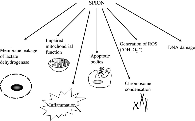
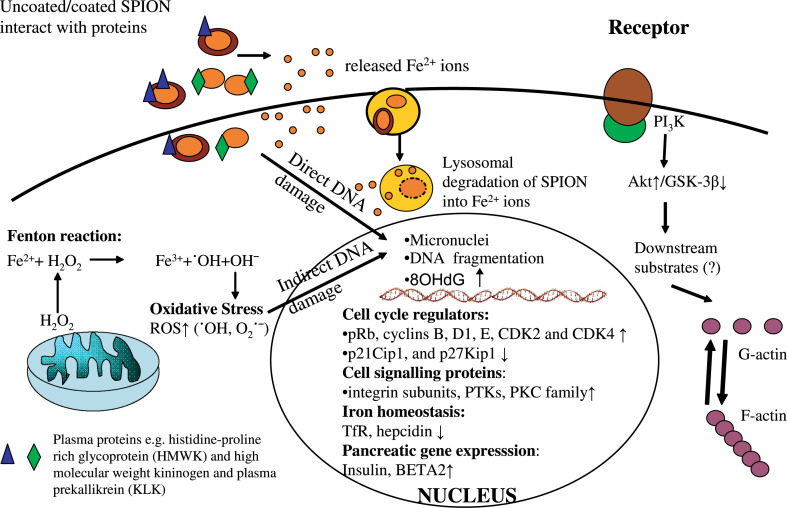
Superparamagnetic iron oxide nanoparticles (SPION) are being widely used for various biomedical applications, for example, magnetic resonance imaging, targeted delivery of drugs or genes, and in hyperthermia. Although, the potential benefits of SPION are considerable, there is a distinct need to identify any potential cellular damage associated with these nanoparticles. Besides focussing on cytotoxicity, the most commonly used determinant of toxicity as a result of exposure to SPION, this review also mentions the importance of studying the subtle cellular alterations in the form of DNA damage and oxidative stress. We review current studies and discuss how SPION, with or without different surface coating, may cause cellular perturbations including modulation of actin cytoskeleton, alteration in gene expression profiles, disturbance in iron homeostasis and altered cellular responses such as activation of signalling pathways and impairment of cell cycle regulation. The importance of protein-SPION interaction and various safety considerations relating to SPION exposure are also addressed.
Overview
The claims and statistics of this paper haven’t been checked yet.
Run the check — free, no account needed. It reads the open-access text and checks the sources it cites.
Integrity signals
Registry
Cites
Statistics
Figures
Verification
Claims the paper makes
The claims and statistics of this paper haven’t been checked yet.
Run the check — free, no account needed. It reads the open-access text and checks the sources it cites.
Claims other papers make about this one
Each is checked against this paper’s own text: does it say what the citing paper says it does?
The source backs part of the claim, not all of it — or backs it for a different population, condition or comparison.
In the citing paper
“Metallic NPs are valuable for their theragnostic capabilities but face significant barriers to translation due to potential long-term toxicity and bioaccumulation, which limit clinical progress [102, 103].”
From Targeted Nanoparticles for Drug Delivery Across the Blood–Brain Barrier in Early and Late Stages of Alzheimer's Disease: A Review (2026) · cites this paper as [102]
Experimental — accuracy not yet measuredIn the citing paper
“In vitro, upon prolonged incubation, surface-coated SPION are typically nontoxic at iron concentration up to and 10 μg/ml, while slight cytotoxicity is seen at 100 μg/ml.”
From Iron oxide nanoparticles: Diagnostic, therapeutic and theranostic applications (2019) · cites this paper as [120]
In the citing paper
“In general, SPION toxicity depends on a variety of factors, including - besides dosing and exposure time - also the nature of the coating material, the hydrodynamic diameter of the particle and SPION-protein interactions [119–122].”
From Iron oxide nanoparticles: Diagnostic, therapeutic and theranostic applications (2019) · cites this paper as [120]
Self-check
Percentages, sums and reported differences re-computed from the paper’s own numbers.
The self-check needs the paper’s full text.
Citations
Cites
Free radicals in biology and medicine
Gutteridge, J. M. C. (1991) · General Pharmacology The Vascular System · doi:10.1016/0306-3623(91)90478-o · Cited by 17,178
Free radicals and antioxidants in normal physiological functions and human disease
Valko, M., Leibfritz, D., Moncol, J., Cronin, M. T. D., Mazur, M., & Telser, J. (2007) · The International Journal of Biochemistry & Cell Biology · doi:10.1016/j.biocel.2006.07.001 · Cited by 14,269
Toxic potential of materials at the nanolevel
Nel, A., Xia, T., Mädler, L., & Li, N. (2006) · Science · doi:10.1126/science.1114397 · Cited by 9,324
Free Radicals in Biology and Medicine
Halliwell, B., & Gutteridge, J. M. C. (2015) · doi:10.1093/acprof:oso/9780198717478.001.0001 · Cited by 8,990
Synthesis and surface engineering of iron oxide nanoparticles for biomedical applications
Gupta, A. K., & Gupta, M. S. (2004) · Biomaterials · doi:10.1016/j.biomaterials.2004.10.012 · Cited by 6,906
Laurent, S., Forge, D., Port, M., Roch, A., Robic, C., Vander Elst, L., & Muller, R. N. (2008) · Chemical Reviews · doi:10.1021/cr068445e · Cited by 6,852
Hepcidin Regulates Cellular Iron Efflux by Binding to Ferroportin and Inducing Its Internalization
Nemeth, E., Tuttle, M. S., Powelson, J., Vaughn, M. B., Donovan, A., Ward, D. M., Ganz, T., & Kaplan, J. (2004) · Science · doi:10.1126/science.1104742 · Cited by 4,803
Biodegradable nanoparticles for drug and gene delivery to cells and tissue
Panyam, J., & Labhasetwar, V. D. (2003) · Advanced Drug Delivery Reviews · doi:10.1016/s0169-409x(02)00228-4 · Cited by 3,417
Fischer, D., Li, Y., Ahlemeyer, B., Krieglstein, J., & Kissel, T. (2003) · Biomaterials · doi:10.1016/s0142-9612(02)00445-3 · Cited by 2,329
In vitro toxicity of nanoparticles in BRL 3A rat liver cells
Hussain, S. M., Hess, K. L., Gearhart, J. M., Geiss, K. T., & Schlager, J. J. (2005) · Toxicology in Vitro · doi:10.1016/j.tiv.2005.06.034 · Cited by 2,025
Research works printed but not linked: 3
- Gould P, 2006, Nano Today, 1, 34
- 10.1016/j.biomaterials.2004.10.012 · DOI
- Sodum RS, 1988, Cancer Res, 48, 320
Cited by
Sorted by citations
Items 501–600 of 1,000
Endogenous doesn’t always mean innocuous: a scoping review of iron toxicity by inhalation
Morgan, J., Bell, R. G., & Jones, A. L. (2020) · Journal of Toxicology and Environmental Health Part B · doi:10.1080/10937404.2020.1731896 · Cited by 24
Iron oxide nanoparticle-induced hematopoietic and immunological response in rats
Gaharwar, U. S., Kumar, S. R., & Rajamani, P. (2020) · RSC Advances · doi:10.1039/d0ra05901c · Cited by 24
Cicha, I., Lyer, S., Alexiou, C., & Garlichs, C. D. (2013) · Nanotechnology Reviews · doi:10.1515/ntrev-2013-0009 · Cited by 24
Genetically Encoded Circuit for Remote Regulation of Cell Migration by Magnetic Fields
Mosabbir, A. A., & Truong, K. (2018) · ACS Synthetic Biology · doi:10.1021/acssynbio.7b00415 · Cited by 24
Attallah, A. H., Abdulwahid, F. S., Abdulrahman, H. J., Haider, A. J., & Ali, Y. A. (2024) · Plasmonics · doi:10.1007/s11468-024-02383-5 · Cited by 23
Rehman, F. U., Jiang, H., Selke, M., & Wang, X. (2018) · Journal of Materials Chemistry B · doi:10.1039/c8tb01955j · Cited by 23
Zhang, T.-G., Zhang, Y., Zhou, Q., Wang, X., & Zhan, L. (2020) · Journal of Applied Toxicology · doi:10.1002/jat.3933 · Cited by 23
Hernandes, E. P., Bini, R. D., Endo, K. M., de Oliveira, V. A., de Almeida, I. V., Dias, G. S., dos Santos, I. A., Oliveira, P. F., Vicentini, V. E. P., & Cótica, L. F. (2022) · Magnetochemistry · doi:10.3390/magnetochemistry8100114 · Cited by 23
Magnetic Nanoparticles Mediated Thrombolysis–A Review
Zhang, B., & Jiang, X. (2023) · IEEE Open Journal of Nanotechnology · doi:10.1109/ojnano.2023.3273921 · Cited by 23
Sart, S., Bejarano, F. C., Baird, M. A., Yan, Y., Rosenberg, J. T., Ma, T., Grant, S. C., & Li, Y. (2014) · Cytotherapy · doi:10.1016/j.jcyt.2014.09.008 · Cited by 23
Zhang, L., Wang, X., Zou, J., Liu, Y., & Wang, J. (2015) · Journal of Nanobiotechnology · doi:10.1186/s12951-014-0063-3 · Cited by 23
Prosen, L., Hudoklin, S., Čemažar, M., Stimac, M., Tratar, U. L., Ota, M., Ščančar, J., Romih, R., & Serša, G. (2016) · Nanomedicine · doi:10.2217/nnm.16.4 · Cited by 23
Geometrical and magnetic structure of iron oxide clusters (FeO) for n> 10
Gutsev, G. L., Belay, K., Gutsev, L. G., & Ramachandran, B. R. (2017) · Computational Materials Science · doi:10.1016/j.commatsci.2017.05.028 · Cited by 23
Wu, Q., Miao, T., Feng, T., Yang, C. H., Guo, Y., & Li, H. (2018) · Molecular Medicine Reports · doi:10.3892/mmr.2018.8972 · Cited by 23
Urandur, S., Marwaha, D., Gautam, S., Banala, V. T., Sharma, M., & Mishra, P. R. (2018) · Therapeutic Delivery · doi:10.4155/tde-2018-0038 · Cited by 23
Molecular Imaging of a New Multimodal Microbubble for Adhesion Molecule Targeting
Ahmed, M., Gustafsson, B. I., Aldi, S., Dusart, P., Egri, G., Butler, L. M., BONE, D., Dähne, L., Hedin, U., & Caidahl, K. (2018) · Cellular and Molecular Bioengineering · doi:10.1007/s12195-018-00562-z · Cited by 23
Miao, C., Hu, F., Rui, Y., Duan, Y., & Gu, H. (2019) · Journal of Materials Chemistry B · doi:10.1039/c9tb00002j · Cited by 23
Ferraz, F. S., López, J. L., Lacerda, S. M. D. S. N., Procópio, M. S., Figueiredo, A. F. A., Martins, E. M. N., Guimarães, P. P. G., Ladeira, L. O., Kitten, G. T., Dias, F. F., et al. (2020) · Journal of Inorganic Biochemistry · doi:10.1016/j.jinorgbio.2020.111017 · Cited by 23
Jahanbani, J., Ghotbi, M., Shahsavari, F., Seydi, E., Rahimi, S., & Pourahmad, J. (2020) · Journal of Biochemical and Molecular Toxicology · doi:10.1002/jbt.22557 · Cited by 23
Janik-Olchawa, N., Dróźdż, A., Ryszawy, D., Pudełek, M., Planeta, K., Setkowicz, Z., Śniegocki, M., Żądło, A. C., Ostachowicz, B., & Chwiej, J. G. (2020) · Scientific Reports · doi:10.1038/s41598-020-72414-8 · Cited by 23
Transient cell stiffening triggered by magnetic nanoparticle exposure
Perez, J. E., Fage, F., Pereira, D., Abou‐Hassan, A., Asnacios, S., Asnacios, A., & Wilhelm, C. (2021) · Journal of Nanobiotechnology · doi:10.1186/s12951-021-00790-y · Cited by 23
Magnetic Nanoparticles for Targeted Cancer Diagnosis and Therapy
Williams, M. J., & Cussen, S. A. (2013) · Frontiers of nanoscience · doi:10.1016/b978-0-08-098338-7.00002-9 · Cited by 23
Srikanth, K., Trindade, T., Duarte, A. C., & Pereira, E. (2016) · Environmental Science and Pollution Research · doi:10.1007/s11356-016-7870-z · Cited by 23
Ultrasmall superparamagnetic iron oxide nanoparticles for enhanced tumor penetration
Xue, F., Xue, Y., Gonca, S., Ji, K., Zhang, M., García–García, F. R., Li, Q., Huang, Y., Kamenev, K. V., & Chen, X. (2023) · Journal of Materials Chemistry B · doi:10.1039/d2tb02630a · Cited by 22
Merino, J. J., & Cabaña-Muñoz, M. E. (2023) · Micromachines · doi:10.3390/mi14112068 · Cited by 23
Mohn, F., Scheffler, K., Ackers, J., Weimer, A., Wegner, F., Thieben, F., Ahlborg, M., Vogel, P., Graeser, M., & Knopp, T. (2024) · Physics in Medicine and Biology · doi:10.1088/1361-6560/ad5828 · Cited by 22
García, K. P., Zarschler, K., Barreto, J. A., Hesse, J., Spiccia, L., Graham, B., & Stephan, H. (2013) · RSC Advances · doi:10.1039/c3ra43726d · Cited by 22
Hematopoietic and mesenchymal stem cells: polymeric nanoparticle uptake and lineage differentiation
Brüstle, I., Simmet, T., Nienhaus, G. U., Landfester, K., & Mailänder, V. (2015) · Beilstein Journal of Nanotechnology · doi:10.3762/bjnano.6.38 · Cited by 22
Fe3O4/Salicylic acid nanoparticles behavior on chick CAM vasculature
Mihăiescu, D. E., Buteică, S. A., Neamţu, J., Istrati, D., & Mîndrilă, I. (2013) · Journal of Nanoparticle Research · doi:10.1007/s11051-013-1857-1 · Cited by 22
Zhang, L., Wang, X., Zou, J., Liu, Y., & Wang, J. (2015) · Chemical Research in Toxicology · doi:10.1021/acs.chemrestox.5b00161 · Cited by 22
Silva, A. H., Lima, E., Mansilla, M. V., Zysler, R. D., Mojica‐Pisciotti, M. L., Locatelli, C., Rajoli, R. K. R., Owen, A., Creczynski‐Pasa, T. B., & Siccardi, M. (2017) · European Journal of Nanomedicine · doi:10.1515/ejnm-2017-0001 · Cited by 22
Molecular imaging in stem cell-based therapies of cardiac diseases
Li, X., & Hacker, M. (2017) · Advanced Drug Delivery Reviews · doi:10.1016/j.addr.2017.07.012 · Cited by 22
Minaeva, O. V., Brodovskaya, E. P., Анатольевич, П. М., Gerasimov, M. V., Жарков, М. Н., Yurlov, I. A., Kulikov, O. A., Kotlyarov, A. A., Балыкова, Л. А., Kokorev, A. V., et al. (2017) · Journal of Physics Conference Series · doi:10.1088/1742-6596/784/1/012038 · Cited by 21
Duan, H., Wang, L., Wang, S., & He, Y. (2023) · Nanomedicine · doi:10.2217/nnm-2023-0164 · Cited by 21
A review of design criteria for cancer-targeted, nanoparticle-based MRI contrast agents
Rahmati, S., & David, A. E. (2024) · Applied Materials Today · doi:10.1016/j.apmt.2024.102087 · Cited by 21
Chelluri, L. K., Verma, V. K., Kamaraju, S. R., Kancherla, R., Kona, L. K., Beevi, S. S., Debnath, T., Shalini, P. U., Arbab, S. A., & Vadapalli, R. M. (2015) · International Journal of Nanomedicine · doi:10.2147/ijn.s75445 · Cited by 21
Lanas, S. G., Valiente, M., Tolazzi, M., & Melchior, A. (2018) · Journal of Thermal Analysis and Calorimetry · doi:10.1007/s10973-018-7763-0 · Cited by 21
Gokduman, K. (2019) · Nanomedicine · doi:10.2217/nnm-2019-0126 · Cited by 21
Nasrin, S., Chowdhury, F.-U.-Z., Hossen, M. M., Islam, A., Kumar, A., & Hoque, S. M. (2022) · Journal of Magnetism and Magnetic Materials · doi:10.1016/j.jmmm.2022.170065 · Cited by 21
Toxicity of bare and surfaced functionalized iron oxide nanoparticles towards microalgae
Toh, P. Y., Tai, W. Y., Ahmad, A. L., Lim, J., & Chan, D. J. C. (2015) · International Journal of Phytoremediation · doi:10.1080/15226514.2015.1086300 · Cited by 21
Therapeutic advances of magnetic nanomaterials in chronic wound healing
Li, J., Yang, Y., Zhang, G., Sun, J., Li, Y., & Song, B. (2024) · Nano Today · doi:10.1016/j.nantod.2024.102554 · Cited by 20
Wiart, M., Tavakoli, C., Hubert, V., Hristovska, I., Dumot, C., Parola, S., Lerouge, F., Chauveau, F., Canet‐Soulas, E., Pascual, O., et al. (2022) · Journal of Neuroscience Methods · doi:10.1016/j.jneumeth.2022.109729 · Cited by 20
Umar, H., Aliyu, M. R., & Ozsahin, D. U. (2024) · Biomedical Physics & Engineering Express · doi:10.1088/2057-1976/ad3646 · Cited by 20
Jafari, N., Hamishehkar, H., & Mohammadpourfard, M. (2024) · Algal Research · doi:10.1016/j.algal.2024.103560 · Cited by 20
Novel sol–gel-derived hardystonite-based biomagnetic nanoparticles for hyperthermia applications
Farzin, A., Emadi, R., & Fathi, M. (2016) · Journal of Sol-Gel Science and Technology · doi:10.1007/s10971-016-4100-6 · Cited by 20
Molecular Imaging of Angiogenesis in Cardiac Regeneration
Mandic, L., Traxler, D., Gugerell, A., Zlabinger, K., Lukovic, D., Pávó, N., Goliasch, G., Spannbauer, A., Winkler, J., & Gyöngyösi, M. (2016) · Current Cardiovascular Imaging Reports · doi:10.1007/s12410-016-9389-6 · Cited by 20
He, J. H., Hu, H., Zeng, X., Lan, F., Wu, F., & Wu, Y. (2017) · Regenerative Biomaterials · doi:10.1093/rb/rbw039 · Cited by 20
Mekonnen, T. W., Darge, H. F., Tsai, H., Birhan, Y. S., Hanurry, E. Y., Gebrie, H. T., Chou, H., Lai, J., Lin, S., Harn, H., et al. (2022) · Colloids and Surfaces B Biointerfaces · doi:10.1016/j.colsurfb.2022.112391 · Cited by 20
Iron oxide nanoparticles for biomedical applications: an updated patent review (2015–2021)
Mao, Z., Li, X., Wang, P., & Yan, H. (2022) · Expert Opinion on Therapeutic Patents · doi:10.1080/13543776.2022.2109413 · Cited by 20
A value-added exopolysaccharide as a coating agent for MRI nanoprobes
Palma, S., Rodrigues, C. A. V., Carvalho, A., Morales, M. P., Freitas, F., Fernandes, A. R., Cabral, J. M. S., & Roque, A. C. A. (2015) · Nanoscale · doi:10.1039/c5nr01979f · Cited by 20
Treatment of Anemia Progression via Magnetite and Folate NanoparticlesIn Vivo
Elsayed, H. H., Al-Sherbini, A.-S., Abd-Elhady, E., & Ahmed, K. A. (2014) · ISRN Nanotechnology · doi:10.1155/2014/287575 · Cited by 20
Superparamagnetic iron oxide nanocolloids in MRI studies of neuroinflammation
Ugga, L., Romeo, V., Tedeschi, E., Brunetti, A., & Quarantelli, M. (2018) · Journal of Neuroscience Methods · doi:10.1016/j.jneumeth.2018.06.008 · Cited by 20
Nanotoxicity: Sources and Effects on Environment
Sarkar, A., Sarkar, D., & Poddar, K. (2019) · Nanotechnology in the life sciences · doi:10.1007/978-3-030-16534-5_9 · Cited by 20
Kojima, K., Takahashi, S., Saito, S., Endo, Y., Nittami, T., Nozaki, T., Sobti, R. C., & Watanabe, M. (2018) · Applied Sciences · doi:10.3390/app8010134 · Cited by 19
Vijay, P., Batchelor, W. J., & Saito, K. (2022) · Carbohydrate Polymers · doi:10.1016/j.carbpol.2021.119060 · Cited by 19
Dechsupa, N., Kosintarajit, P., Kamkan, K., Khanjina, T., Sirikul, C., Innuan, P., Suwan, A., Anukul, N., & Kantapan, J. (2022) · Nanomaterials · doi:10.3390/nano12162776 · Cited by 19
Carvalho, T. S. S., Ribeiro, N., Torres, P. M. C., Almeida, J. C., Belo, J. H., Araújo, J. P., Ramos, A. M., Oliveira, M. S. A., & Olhero, S. M. (2022) · Materials Chemistry and Physics · doi:10.1016/j.matchemphys.2022.127175 · Cited by 19
Estévez, M., Cicuéndez, M., Colilla, M., Vallet‐Regí, M., González, B., & Izquierdo‐Barba, I. (2024) · Journal of Colloid and Interface Science · doi:10.1016/j.jcis.2024.03.043 · Cited by 19
Biocompatible gelatin-coated ferrite nanoparticles: A magnetic approach to advanced drug delivery
Shakeel, V., Gul, I. H., John, P. C. L., & Bhatti, A. (2024) · Saudi Pharmaceutical Journal · doi:10.1016/j.jsps.2024.102066 · Cited by 19
Javaid, A., Munir, N., Abideen, Z., Duarte, B., Siddiqui, Z. S., Haq, R., & Naz, S. (2025) · Plant Nano Biology · doi:10.1016/j.plana.2025.100145 · Cited by 19
Novotná, B., Turnovcová, K., Veverka, P., Rössner, P., Bagryantseva, Y., Herynek, V., Žvátora, P., Vosmanská, M., Klementová, M., Syková, E., et al. (2015) · Nanotoxicology · doi:10.3109/17435390.2015.1107144 · Cited by 19
Ma, J., Li, D., Zhong, L., Du, F., Tan, J., Yang, J., & Peng, X. (2018) · Carbohydrate Polymers · doi:10.1016/j.carbpol.2018.07.003 · Cited by 19
Roy, D. G., Bell, J. C., & Bourgeois‐Daigneault, M. (2020) · Biochemical and Biophysical Research Communications · doi:10.1016/j.bbrc.2020.03.135 · Cited by 19
Challenges of implementing nano-specific safety and safe-by-design principles in academia
Soeteman‐Hernández, L. G., Blab, G. A., Carattino, A., Dekker, F., Dekkers, S., van der Linden, M., van Silfhout, A. M., & Noorlander, C. W. (2020) · NanoImpact · doi:10.1016/j.impact.2020.100243 · Cited by 19
Magnetic Nanodiscs—A New Promising Tool for Microsurgery of Malignant Neoplasms
Zamay, T. N., Prokopenko, V. S., Сергеевич, З. С., Lukyanenko, K. A., Kolovskaya, O. S., Orlov, V. A., Сергеевна, З. Г., Galeev, R. G., Аркадьевич, Н. А., & Kichkailo, A. S. (2021) · Nanomaterials · doi:10.3390/nano11061459 · Cited by 19
Kumar, M., Gupta, G., Nuzaiba, P. M., Karthik, R., Varghese, T., Srivastava, P. P., Bhushan, S., Shukla, S. P., Krishna, G., & Gupta, S. (2022) · Aquaculture International · doi:10.1007/s10499-022-00870-2 · Cited by 19
Nanotherapy based on magneto‐mechanochemical modulation of tumor redox state
Orel, V. B., Papazoglou, Α. S., Tsagkaris, C., Moysidis, D. V., Papadakos, S. P., Galkin, A. Y., Vé, O., & Syvak, L. A. (2022) · Wiley Interdisciplinary Reviews Nanomedicine and Nanobiotechnology · doi:10.1002/wnan.1868 · Cited by 19
Drug Delivery Systems in Bone Regeneration and Implant Dentistry
Anil, S., Al-Sulaimani, A. F., Beeran, A. E., Chalisserry, E. P., R, H. V. P., & Amri, M. D. A. (2015) · InTech eBooks · doi:10.5772/60003 · Cited by 19
Structure and properties of iron oxide clusters: From Fe 6 to Fe 6 O 20 and from Fe 7 to Fe 7 O 24
Gutsev, G. L., Belay, K., Gutsev, L. G., & Ramachandran, B. R. (2016) · Journal of Computational Chemistry · doi:10.1002/jcc.24478 · Cited by 19
Catalano, E., Miola, M., Ferraris, S., Novak, S., Oltolina, F., Cochis, A., Prat, M., Verné, E., Rimondini, L., & Follenzi, A. (2017) · Biomedical Physics & Engineering Express · doi:10.1088/2057-1976/aa62cc · Cited by 19
Updates on the applications of iron-based nanoplatforms in tumor theranostics
Wang, W., Huang, Z., Huang, Y., Pan, X., & Wu, C. (2020) · International Journal of Pharmaceutics · doi:10.1016/j.ijpharm.2020.119815 · Cited by 19
Iron oxide-silica nanocomposites yielded by chemical route and sol–gel method
Puscasu, E., Sacarescu, L., Lupu, N., Grigoras, M., Oanca, G., Balasoiu, M., & Creanga, D. (2016) · Journal of Sol-Gel Science and Technology · doi:10.1007/s10971-016-3996-1 · Cited by 18
Haro-Martínez, E., Muscolino, E., Moral, N., Durán, J., & Fornaguera, C. (2025) · Drug Delivery and Translational Research · doi:10.1007/s13346-025-01887-9 · Cited by 18
Raj, A. F. P. A. M., Bauman, M., Dimitrušev, N., Ali, L. M. A., Onofre, M., Gary‐Bobo, M., Durand, J., Lobnik, A., & Košak, A. (2023) · International Journal of Molecular Sciences · doi:10.3390/ijms241210072 · Cited by 18
Baskar, G., Palaniyandi, T., Ravi, M., Viswanathan, S., Wahab, M. R. A., Surendran, H., Govindaraj, M., Sugumaran, A., Almutairi, M. H., & Almutairi, B. O. (2024) · Process Biochemistry · doi:10.1016/j.procbio.2024.03.010 · Cited by 18
Magnetoresponsive Anisotropic Fiber‐Integrating Hydrogels for Neural Tissue Regeneration
Sousa, J. P. M., Monteiro, C. F., Deus, I. A., Completo, A., Stratakis, E. I., Mano, J. F., & Marques, P. A. A. P. (2024) · Small Structures · doi:10.1002/sstr.202400213 · Cited by 18
Faraj, A. A. (2013) · Journal of Nanoparticle Research · doi:10.1007/s11051-013-1797-9 · Cited by 18
Peacock, A. K., Cauët, S. I., Taylor, A., Murray, P. A., Williams, S. R., Weaver, J. V. M., Adams, D. J., & Rosseinsky, M. J. (2012) · Chemical Communications · doi:10.1039/c2cc34420c · Cited by 18
Maurizi, L., Papa, A., Boudon, J., Sudhakaran, S., Pruvot, B., Vandroux, D., Chluba, J., Lizard, G., & Millot, N. (2018) · InTech eBooks · doi:10.5772/intechopen.71833 · Cited by 18
Smart Polymersomes as Intelligent Nanomedicines in Cancer Treatment
Alibolandi, M., Shahriari, M., & Ramezani, M. (2019) · Elsevier eBooks · doi:10.1016/b978-0-12-816963-6.00016-9 · Cited by 18
Sanz-Ortega, L., Rojas, J. M., & Barber, D. F. (2020) · Pharmaceutics · doi:10.3390/pharmaceutics12090812 · Cited by 18
Sánchez, J., Rodríguez-Reyes, M., Cortés-Hernández, D. A., Ávila‐Orta, C. A., & Reyes-Rodríguez, P. Y. (2020) · Colloids and Surfaces A Physicochemical and Engineering Aspects · doi:10.1016/j.colsurfa.2020.125986 · Cited by 18
Preparation and Characterization of Dextran Coated Iron Oxide Nanoparticles Thin Layers
Predoi, G., Ciobanu, C. S., Iconaru, S. L., Predoi, D., Dreghici, D. B., Groza, A., Bărbuceanu, F., Cimpeanu, C. L., Badea, M. L., Bărbuceanu, Ș., et al. (2021) · Polymers · doi:10.3390/polym13142351 · Cited by 18
Chen, Y., Zhang, W., Dong, C., Hutchinson, S. M., & Feng, H. D. (2021) · Journal of Hazardous Materials · doi:10.1016/j.jhazmat.2021.127212 · Cited by 18
Metal-based nanoparticles for cardiovascular disease diagnosis and therapy
Li, L., Zeng, Y., & Liu, G. (2022) · Particuology · doi:10.1016/j.partic.2022.03.002 · Cited by 18
Lee, J. H., & Hur, W. (2013) · Biotechnology and Bioengineering · doi:10.1002/bit.25156 · Cited by 18
Rajan, B., Shanmugam, S., Balakumar, S., & Devaki, T. (2015) · Environmental Toxicology and Pharmacology · doi:10.1016/j.etap.2015.01.018 · Cited by 18
Paolini, A., Porredón, C., Ramos‐López, D., de Lapuente, J., Lascialfari, A., Guari≠, Y., Larionova≠, J., Long, J., & Nano, R. (2015) · Journal of Applied Toxicology · doi:10.1002/jat.3273 · Cited by 18
Dutta, S., Kumar, P., Yadav, S., Sharma, R. D., Shivaprasad, P., Vimaleswaran, K. S., Srivastava, A., & Sharma, R. K. (2023) · Catalysis Communications · doi:10.1016/j.catcom.2023.106615 · Cited by 17
Magnetic force-based cell manipulation for in vitro tissue engineering
Hu, H., Krishaa, L., & Fong, E. L. S. (2023) · APL Bioengineering · doi:10.1063/5.0138732 · Cited by 17
Li, J., Liu, Y., Cha, R., Ran, B., Mou, K., Wang, H., Xie, Q., Sun, J., & Jiang, X. (2016) · RSC Advances · doi:10.1039/c5ra25729h · Cited by 17
Deng, D., Fu, S., Cai, Z., Fu, X., Jin, R., & Ai, H. (2022) · Regenerative Biomaterials · doi:10.1093/rb/rbac018 · Cited by 17
Asgari, S., Nikkam, N., & Saniee, P. (2022) · Talanta Open · doi:10.1016/j.talo.2022.100129 · Cited by 17
Zarei, M., Esmaeili, A., Zarrabi, A., & Zarepour, A. (2022) · Pharmaceutics · doi:10.3390/pharmaceutics14122692 · Cited by 17
Ranking the in vivo toxicity of nanomaterials in Drosophila melanogaster
Vecchio, G., Galeone, A., Malvindi, M. A., Cingolani, R., & Pompa, P. P. (2013) · Journal of Nanoparticle Research · doi:10.1007/s11051-013-1936-3 · Cited by 17
Liu, X., Zhang, H., Chang, L., Yu, B., Liu, Q., Wu, J., Miao, Y., Ma, P., Fan, D., & Fan, H. (2015) · Nanoscale Research Letters · doi:10.1186/s11671-015-0752-3 · Cited by 17
Creanga, D. E., Oprica, L. A., Nădejde, C., Andries, M., Puscasu, E., & Balasoiu, M. (2015) · Environmental Engineering and Management Journal · doi:10.30638/eemj.2015.063 · Cited by 17
Chen, Y., Zhou, Q., Li, X., Wang, F., Heist, K., Kuick, R. D., Owens, S. R., & Wang, T. D. (2017) · Bioconjugate Chemistry · doi:10.1021/acs.bioconjchem.7b00501 · Cited by 17
Inorganic nanoparticles for theranostic use
Esposti, L. D., Carella, F., & Iafisco, M. (2018) · Elsevier eBooks · doi:10.1016/b978-0-08-101745-6.00017-7 · Cited by 17
Çiğerci, İ. H., Ali, M. M., Kaygısız, Ş. Y., Kaya, B., & Liman, R. (2018) · Bulletin of Environmental Contamination and Toxicology · doi:10.1007/s00128-018-2364-y · Cited by 17
Metadata
- Authors
- Singh, N., Jenkins, G. J. S., Asadi, R., & Doak, S. H.
- Affiliations
- Swansea University (GB)
- Cardiff University (GB)
- Corresponding author
- Singh, Neenu
- Venue
- Nano Reviews
- Publisher
- Informa UK Limited
- Published
- 2010-01-01
- Volume
- 1
- Issue
- 1
- Pages
- 5358
- ISSN
- 2000-5121 (ISSN-L)
- PMC
- PMC3215220
- OpenAlex
- W2045080998
- Cited by
- 1,112
- References
- 107
- Citations by year
- 2026: 30 · 2025: 63 · 2024: 69 · 2023: 70 · 2022: 87 · 2021: 109 · 2020: 91 · 2019: 78 · 2018: 92 · 2017: 83 · 2016: 83 · 2015: 92 · 2014: 57 · 2013: 60 · 2012: 33
- FWCI
- 16.82
- Type
- journal article
- Language
- en
- Open access
- hybrid
- doi.org
- Licence
- cc-by-nc
- Locations
- Nano Reviews (publishedVersion, cc-by-nc)
- Europe PMC (PubMed Central) (publishedVersion, cc-by-nc)
- PubMed Central (publishedVersion, cc-by-nc)
- PubMed (publishedVersion)
- DOAJ (DOAJ: Directory of Open Access Journals) (submittedVersion)
- Topics
- Physical Sciences › Materials Science › Materials Chemistry › Nanoparticles: synthesis and applications
- Physical Sciences › Engineering › Biomedical Engineering › Characterization and Applications of Magnetic Nanoparticles
- Physical Sciences › Materials Science › Biomaterials › Nanoparticle-Based Drug Delivery
- Keywords
- superparamagnetic iron oxide nanoparticles, nanoparticle toxicity, cytotoxicity, DNA damage, oxidative stress, actin cytoskeleton, gene expression profiling, iron homeostasis, signaling pathways, cell cycle regulation, nanoparticle-protein interactions, surface coating, MRI contrast agents, drug delivery, hyperthermia
About these checks
Registry, retraction, Cites and statistics results are computed, not judged, with one exception: a printed reference that no index lists is read once by a model before it counts as a citation error. Claims are extracted by a model and kept only when their quote appears verbatim in the text; each cited source is then checked, with a model’s help, for whether it says what the claim relies on. Those verdicts can be wrong — read the claim’s quote and the cited source before relying on one.
Statistics and arithmetic are recomputed, not judged.
Confidence bands are shown only for fields calibrated on our annotated test set. None is calibrated yet, so no band is shown.
What counts as a claim ›How we measure confidence ›How we check this ›
Are you an author, or do you see a problem with this page? Request a review
