

The DOI matches its Crossref record; 22 of the 24 references it cites were found in a registry or an index.
The limbic system
Open accessAbstract
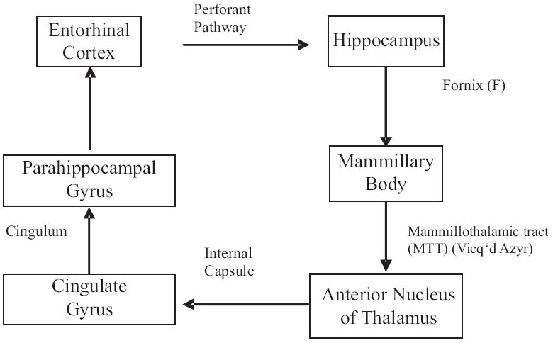
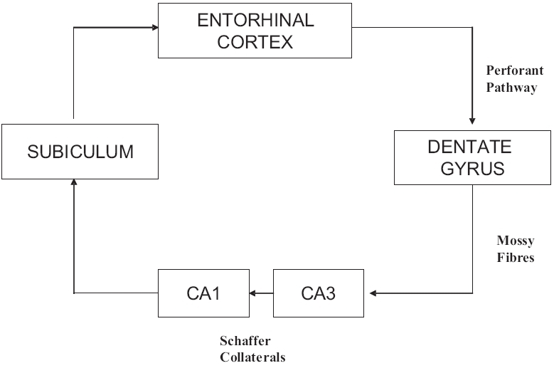
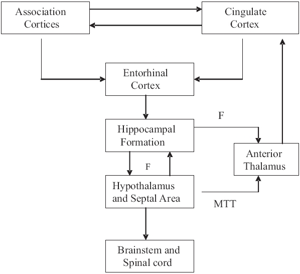
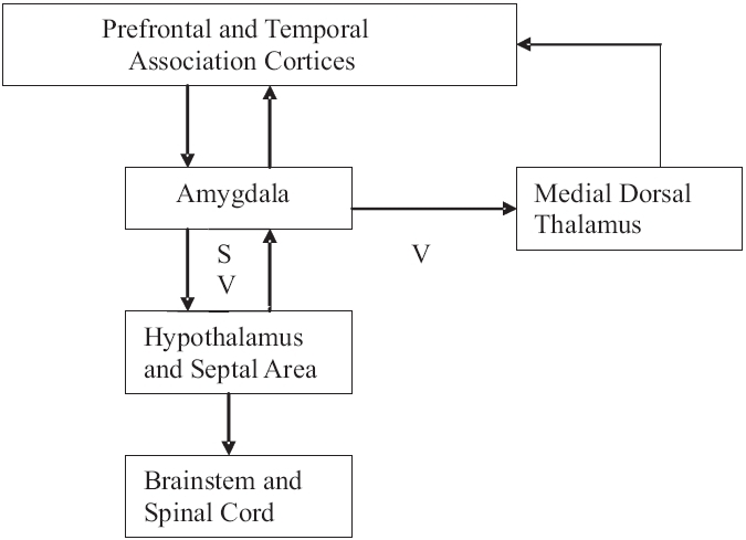
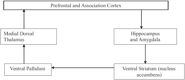
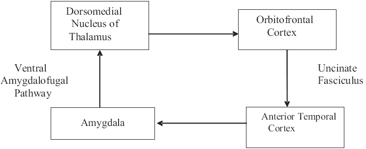
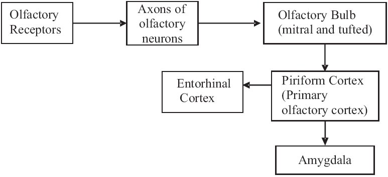
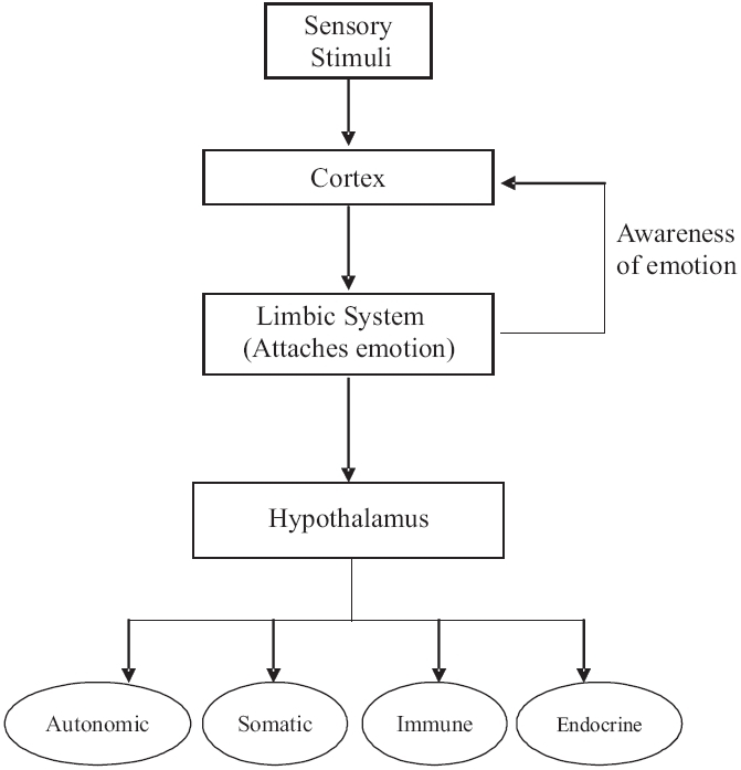
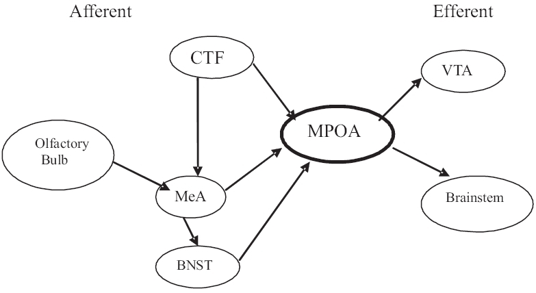
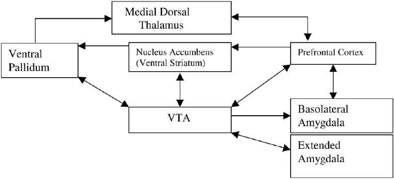
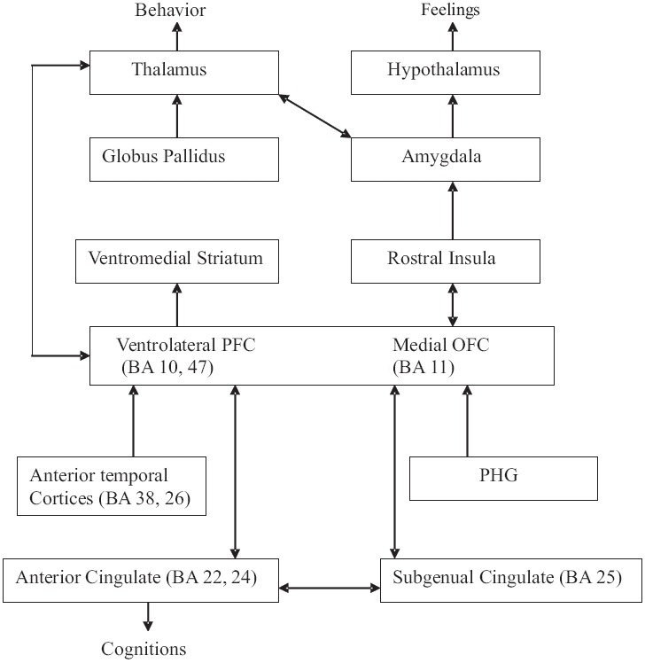
“The hypothalamus, the anterior thalamic nucleus, the cingulate gyrus, the hippocampus and their interconnections, constitute a harmonious mechanism which may elaborate the functions of central emotion as well as participate in the emotional expression.” James Papez, 1937 The limbic system consists of the phylogenetically old limbic lobe and other subcortical structures and their connections. Although not empirically proven, the limbic system is a functional concept which may be employed to explain various brain functions.[1] HISTORY Paul Pierre Broca in 1878 spoke of ‘le grand lobe limbique’ or the great limbic lobe and applied the term “limbic” (from the Latin limbus for border) to the curved rim of the cortex which incudes the cingulate and the parahippocampal gyri. However, its putative role in emotion was elaborated by the American physician, James Papez in 1937 in the seminal paper titled ‘A proposed mechanism of emotion’. This anatomical model is referred to as the Papez circuit.[2] Yakovlev in 1948 proposed Yakovlev's circuit in the control of emotion involving the orbitofrontal, insular and anterior temporal lobe cortex, the amygdala and the dorsomedial nucleus of thalamus.[3] In 1952, Paul D. MacLean coined the term “limbic system” to describe Broca's limbic lobe and related subcortical nuclei as the collective neural substrate for emotion.[1] MacLean was also instrumental in proposing and defining the Triune concept of the brain. MacLean's evolutionary “Triune brain theory” proposed that the human brain was in reality three brains in one: the R-complex (reptilian complex), the limbic system and the neocortex.[4] The concept of the limbic system has since been further expanded and developed by Nauta, Heimer and others. COMPONENTS OF THE LIMBIC SYSTEM There is no universal agreement on the total list of structures, which comprise the limbic system. The brain regions that constitute the limbic system are: Limbic cortex Cingulate gyrus Parahippocampal gyrus Hippocampal formation The dentate gyrus Hippocampus Subicular Complex Amygdala Septal area Hypothalamus These structures form a complex network for controlling emotion.[5] Limbic lobe The limbic lobe situated at the inferomedial aspect of the cerebral hemispheres, consists of two concentric gyri surrounding the corpus callosum. Broca proposed that the larger outer gyrus be named “limbic gyrus” and the smaller inner one “the intralimbic gyrus”. The limbic gyrus (limbic lobe) consists of the isthmus of the cingulate gyrus, the parahippocampal gyrus (both of which are continuous via a bundle of white matter called “cingulum”) and the subcallosal area.[6] The cingulate gyrus (Latin = Belt ridge) dorsal to the corpus callosum is heavily interconnected with the association areas of the cerebral cortex. The parahippocampal gyrus in the medial temporal lobe contains several distinct regions, the most important being the entorhinal cortex (ERC). The ERC funnels highly processed cortical information to the hippocampal formation and serves as its major output pathway.[5] The hippocampal formation Hippocampal formation in the temporal lobe has three distinct zones: The dentate gyrus The hippocampus proper The subiculum Embryologically, the hippocampal formation is an extension of the medial edge of the temporal lobe. The entire hippocampal formation has a length of about 5 cm from its anterior end at the amygdala to its tapering posterior end near the splenium of the corpus callosum.[5] Dentate gyrus The dentate gyrus is composed of three layers: an outer acellular molecular layer, a granular middle layer and an inner polymorphic layer. Hippocampus The word hippocampus means “Sea Horse” in Greek. Hippocampus is a trilaminate structure with an outer molecular layer, a middle pyramidal layer and an inner polymorphic layer. On the basis of differences in cytoarchitecture and connectivity, the hippocampus has four fields (named by Lorente de No in 1934): CA1, CA2, CA3 and CA4 (CA: Cornu Ammonis). To embryologic enthusiasts, CA4 is not part of the Cornu Ammonis but is a separate structure—the dentate hilus. The thin layer of fibers adjacent to the polymorphic layer of the hippocampus is known as the alveus. These fibres coalesce to form the fimbria and subsequently, the crura of the fornix (main efferent pathway of the hippocampal formation). The crura of the fornix converge to form the body of the fornix, which later forms the columns of the fornix and pass through the hypothalamus into the mammillary bodies.[57] Subicular complex The subicular complex has three components: the presubiculum, the parasubiculum and subiculum. Subiculum is the transitional zone between the six-layered, entorhinal cortex and the three-layered hippocampus.[5] Amygdala Identified by Burdach in the early 19th century, amygdala, an almond-shaped structure deep within the temporal lobe, is a collection of nuclei lying beneath the uncus. Lying at the anterior end of the hippocampal formati
Overview
The claims and statistics of this paper haven’t been checked yet.
Run the check — free, no account needed. It reads the open-access text and checks the sources it cites.
Integrity signals
Registry
Cites
Statistics
Figures
Verification
Claims the paper makes
The claims and statistics of this paper haven’t been checked yet.
Run the check — free, no account needed. It reads the open-access text and checks the sources it cites.
Claims other papers make about this one
Each is checked against this paper’s own text: does it say what the citing paper says it does?
The source backs part of the claim, not all of it — or backs it for a different population, condition or comparison.
In the citing paper
“so when the connection between the two is impaired, emotional lability can occur.”
From Approach to the patient with acquired hypothalamic syndrome (2026) · cites this paper as [86]
Experimental — accuracy not yet measuredThe cited source says what the claim attributes to it.
In the citing paper
“The limbic system affects emotional regulation and is integrally connected to the hypothalamus (86), so when the connection between the two is impaired, emotional lability can occur.”
From Approach to the patient with acquired hypothalamic syndrome (2026) · cites this paper as [86]
Self-check
Percentages, sums and reported differences re-computed from the paper’s own numbers.
The self-check needs the paper’s full text.
Citations
Cites
A PROPOSED MECHANISM OF EMOTION
PAPEZ, J. W. (1937) · Archives of Neurology And Psychiatry · doi:10.1001/archneurpsyc.1937.02260220069003 · Cited by 3,563
The Neural Basis of Addiction: A Pathology of Motivation and Choice
Kalivas, P. W., & Volkow, N. D. (2005) · American Journal of Psychiatry · doi:10.1176/appi.ajp.162.8.1403 · Cited by 2,757
The role of the hypothalamic-pituitary-adrenal axis in neuroendocrine responses to stress
Smith, S. M., & Vale, W. W. (2006) · Dialogues in Clinical Neuroscience · doi:10.31887/dcns.2006.8.4/ssmith · Cited by 1,948
Cognitive neuroscience of emotional memory
LaBar, K. S., & Cabeza, R. (2006) · Nature Reviews Neuroscience · doi:10.1038/nrn1825 · Cited by 1,834
The Amygdaloid Complex: Anatomy and Physiology
SAH, P., FABER, E. S. L., LOPEZ DE ARMENTIA, M., & POWER, J. (2003) · Physiological Reviews · doi:10.1152/physrev.00002.2003 · Cited by 1,745
The sleep switch: hypothalamic control of sleep and wakefulness
Saper, C. B., Chou, T. C., & Scammell, T. E. (2001) · Trends in Neurosciences · doi:10.1016/s0166-2236(00)02002-6 · Cited by 1,656
Adolphs, R., Tranel, D., Damasio, H., & Damasio, A. (1995) · The Journal of Neuroscience · doi:10.1523/jneurosci.15-09-05879.1995 · Cited by 1,138
The functional neuroanatomy of bipolar disorder: a review of neuroimaging findings
Strakowski, S. M., DelBello, M. P., & Adler, C. M. (2005) · Molecular Psychiatry · doi:10.1038/sj.mp.4001585 · Cited by 818
Frontal Cortical and Left Temporal Glutamatergic Dysfunction in Schizophrenia
Deakin, J. F. W., Slater, P., Simpson, M. D. C., Gilchrist, A., Skan, W., Royston, M. C., Reynolds, G. P., & Cross, A. J. (1989) · Journal of Neurochemistry · doi:10.1111/j.1471-4159.1989.tb07257.x · Cited by 386
Extended Amygdala and Basal Forebrain
Alheid, G. F. (2003) · Annals of the New York Academy of Sciences · doi:10.1111/j.1749-6632.2003.tb07082.x · Cited by 334
Research works printed but not linked: 7
- Maclean PD. The triune brain in evolution: Role in paleocerebral functions. Plenum Press: New York; 1990.
- Pierri JN, Lewis DA. Functional neuroanatomy. In : Kaplan and Sadock′s Comprehensive textbook of psychiatry, 8 th ed. Sadock BJ, Sadock VA, editor. Lippincott William and Wilkins: Phildelphia; p. 3-33.
- Ganong WF. Smell and taste. In : review of medical physiology. 21 st ed. McGraw Hill Companies Inc: 2003. p. 188-91.
- Ganong WF. Neural basis of instinctual behavior and emotions. In Review of Medical Physiology. 21 st ed. McGraw Hill Companies Inc: 2003. p. 260-9.
- Benca RM, Cirelli C, Rattenborg MC, Tononi G. Basic science of sleep. In : Kaplan and Sadock′s Comprehensive textbook of psychiatry, 8 th ed. Sadock BJ, Sadock VA, editors. Lippincott William and Wilkins: Phildelphia; p. 280-95.
- Paller KA, Squire LR. Biology of memory. In : Kaplan and Sadock′s Comprehensive textbook of psychiatry, 8 th ed. Sadock BJ, Sadock VA, editor. Lippincott William and Wilkins: Phildelphia; p. 553-66.
- Hechtman L. Attention-deficit/hyperactivity disorder. In : Kaplan and Sadock′s Comprehensive textbook of psychiatry, 8 th ed. Sadock BJ, Sadock VA, editor. Lippincott William and Wilkins: Phildelphia; p. 3183-97.
Cited by
Sorted by citations
The impact of sugar consumption on stress driven, emotional and addictive behaviors
Jacques, A., Chaaya, N., Beecher, K., Ali, S. A., Belmer, A., & Bartlett, S. (2019) · Neuroscience & Biobehavioral Reviews · doi:10.1016/j.neubiorev.2019.05.021 · Cited by 271
Current advances in orthodontic pain
Long, H., Wang, Y., Jian, F., Liao, L., Yang, X., & Lai, W. (2016) · International Journal of Oral Science · doi:10.1038/ijos.2016.24 · Cited by 169
Douet, V., & Chang, L. (2015) · Frontiers in Aging Neuroscience · doi:10.3389/fnagi.2014.00343 · Cited by 122
Kamali, A., Milosavljevic, S., Gandhi, A., Lano, K. R., Shobeiri, P., Sherbaf, F. G., Sair, H. I., Riascos, R., & Hasan, K. M. (2023) · Brain Topography · doi:10.1007/s10548-023-00955-y · Cited by 118
Regional susceptibility to dose-dependent white matter damage after brain radiotherapy
Connor, M., Karunamuni, R. A., McDonald, C. R., Seibert, T. M., White, N. S., Moiseenko, V., Bartsch, H., Farid, N., Kuperman, J. M., Krishnan, A. P., et al. (2017) · Radiotherapy and Oncology · doi:10.1016/j.radonc.2017.04.006 · Cited by 117
Pathophysiological Mechanisms in Migraine and the Identification of New Therapeutic Targets
Haanes, K. A., & Edvinsson, L. I. (2019) · CNS Drugs · doi:10.1007/s40263-019-00630-6 · Cited by 114
A Discrete Dorsal Raphe to Basal Amygdala 5-HT Circuit Calibrates Aversive Memory
Sengupta, A., & Holmes, A. (2019) · Neuron · doi:10.1016/j.neuron.2019.05.029 · Cited by 123
The gut–brain axis in irritable bowel syndrome and inflammatory bowel disease
Ancona, A., Petito, C., Iavarone, I., Petito, V., Galasso, L., Leonetti, A., Turchini, L., Belella, D., Ferrarese, D., Addolorato, G., et al. (2020) · Digestive and Liver Disease · doi:10.1016/j.dld.2020.11.026 · Cited by 115
Hescham, S., Lim, L. W., Jahanshahi, A., Steinbusch, H. W. M., Prickaerts, J., Blokland, A., & Temel, Y. (2012) · Brain stimulation · doi:10.1016/j.brs.2012.01.008 · Cited by 106
Connections of the limbic network: A corticocortical evoked potentials study
Enatsu, R., Gonzalez-Martinez, J. A., Bulacio, J. C., Kubota, Y., Mosher, J. C., Burgess, R. C., Najm, I., & Nair, D. R. (2014) · Cortex · doi:10.1016/j.cortex.2014.06.018 · Cited by 105
Reducing the Cognitive Footprint of Brain Tumor Surgery
Dadario, N. B., Brahimaj, B. C., Yeung, J. T., & Sughrue, M. E. (2021) · Frontiers in Neurology · doi:10.3389/fneur.2021.711646 · Cited by 93
Functional Connectivity of the Brain Across Rodents and Humans
Xu, N., LaGrow, T. J., Anumba, N., Lee, A., Zhang, X., Yousefi, B., Bassil, Y., Clavijo, P. G., Sharghi, V. K., Maltbie, E. A., et al. (2022) · Frontiers in Neuroscience · doi:10.3389/fnins.2022.816331 · Cited by 100
The neurobiology of human aggressive behavior: Neuroimaging, genetic, and neurochemical aspects
Cupaioli, F. A., Zucca, F. A., Caporale, C., Lesch, K., Passamonti, L., & Zecca, L. (2020) · Progress in Neuro-Psychopharmacology and Biological Psychiatry · doi:10.1016/j.pnpbp.2020.110059 · Cited by 94
Gondard, E., Chau, H. N., Mann, A., Tierney, T. S., Hamani, C., Kalia, S. K., & Lozano, A. M. (2015) · Brain stimulation · doi:10.1016/j.brs.2015.07.044 · Cited by 94
Unraveling the complexity of human brain: Structure, function in healthy and disease states
Sultana, O. F., Bandaru, M., Islam, M. A., & Reddy, P. H. (2024) · Ageing Research Reviews · doi:10.1016/j.arr.2024.102414 · Cited by 92
Limbic Tract Integrity Contributes to Pattern Separation Performance Across the Lifespan
Bennett, I. J., Huffman, D. J., & Stark, C. E. L. (2014) · Cerebral Cortex · doi:10.1093/cercor/bhu093 · Cited by 90
Interpreting the role of the striatum during multiple phases of motor learning
Cataldi, S., Stanley, A. T., Miniaci, M. C., & Sulzer, D. (2021) · FEBS Journal · doi:10.1111/febs.15908 · Cited by 85
Neuroprogression: the hidden mechanism of depression
Ruiz, N. A. L., del Ángel, D. S., Olguı́n, H. J., & Silva, M. L. (2018) · Neuropsychiatric Disease and Treatment · doi:10.2147/ndt.s177973 · Cited by 81
The Neural Mechanisms of Tinnitus: A Perspective From Functional Magnetic Resonance Imaging
Hu, J.-H., Cui, J., Xǔ, J., Yin, X., Wu, Y., & Qi, J. (2021) · Frontiers in Neuroscience · doi:10.3389/fnins.2021.621145 · Cited by 71
NIRS measures in pain and analgesia: Fundamentals, features, and function
Karunakaran, K. D., Peng, K., Berry, D., Green, S. A., Labadie, R. F., Kussman, B. D., & Borsook, D. (2020) · Neuroscience & Biobehavioral Reviews · doi:10.1016/j.neubiorev.2020.10.023 · Cited by 69
Diagnostic criteria for the hypothalamic syndrome in childhood
van Santen, H. M., van Schaik, J., van Roessel, I. M. A. A., Beckhaus, J., Boekhoff, S., & Müller, H. L. (2023) · European Journal of Endocrinology · doi:10.1093/ejendo/lvad009 · Cited by 67
Functional magnetic resonance imaging in migraine: A systematic review
Schramm, S., Börner, C., Reichert, M., Baum, T. H., Zimmer, C., Heinen, F., Bonfert, M., & Sollmann, N. (2023) · Cephalalgia · doi:10.1177/03331024221128278 · Cited by 70
Imaging the Limbic System in Parkinson’s Disease—A Review of Limbic Pathology and Clinical Symptoms
Banwinkler, M., Theis, H., Prange, S., & van Eimeren, T. (2022) · Brain Sciences · doi:10.3390/brainsci12091248 · Cited by 70
Tinnitus and stress in adults: a scoping review
Elarbed, A., Fackrell, K., Baguley, D., & Hoare, D. J. (2020) · International Journal of Audiology · doi:10.1080/14992027.2020.1827306 · Cited by 65
Peña, I. D., Pan, M. C., Thai, C. G., & Alisso, T. (2020) · Brain Sciences · doi:10.3390/brainsci10050292 · Cited by 76
Altered hippocampus and amygdala subregion connectome hierarchy in major depressive disorder
Jacob, Y., Morris, L. S., Verma, G., Rutter, S., Balchandani, P., & Murrough, J. W. (2022) · Translational Psychiatry · doi:10.1038/s41398-022-01976-0 · Cited by 62
Saad, J. F., Griffiths, K. R., Kohn, M. R., Clarke, S. P., Williams, L. M., & Korgaonkar, M. S. (2017) · NeuroImage Clinical · doi:10.1016/j.nicl.2017.05.016 · Cited by 68
Neuroanatomical changes in Parkinson′s disease in relation to cognition: An update
Prakash, K., Bannur, B. M., Chavan, M., Saniya, K., Sailesh, K. S., & Archana, R. (2016) · Journal of Advanced Pharmaceutical Technology amp Research · doi:10.4103/2231-4040.191416 · Cited by 57
Blossom, V., Ullal, S. D., D’Souza, M. M., Ranade, A. V., Kumar, N. A., & Rai, R. (2025) · 3 Biotech · doi:10.1007/s13205-025-04468-2 · Cited by 57
Torres-Vega, A., Pliego‐Rivero, B., Otero, G., Gómez‐Oliván, L. M., & Vieyra-Reyes, P. (2012) · Nutrition Reviews · doi:10.1111/j.1753-4887.2012.00521.x · Cited by 55
Fontes, K., Rohlicek, C. V., Saint‐Martin, C., Gilbert, G., Easson, K., Majnemer, A., Marelli, A. J., Chakravarty, M. M., & Brossard‐Racine, M. (2019) · Human Brain Mapping · doi:10.1002/hbm.24615 · Cited by 53
Hay, R. E., Reynolds, J. E., Grohs, M. N., Paniukov, D., Giesbrecht, G. F., Letourneau, N. L., Dewey, D., & Lebel, C. A. (2020) · Journal of Neuroscience · doi:10.1523/jneurosci.0481-20.2020 · Cited by 54
Garcia, I., & O’Neil, J. (2020) · The Journal for Nurse Practitioners · doi:10.1016/j.nurpra.2020.08.021 · Cited by 50
EEG Spectral Generators Involved in Motor Imagery: A swLORETA Study
Cebolla, A.-M., Palmero‐Soler, E., Leroy, A., & Cheron, G. A. (2017) · Frontiers in Psychology · doi:10.3389/fpsyg.2017.02133 · Cited by 49
Nikolenko, V. N., Oganesyan, M. V., Rizaeva, N. A., Kudryashova, V. A., Nikitina, A. T., Pavliv, M. P., Анатольевна, Щ. М., Db, G., Bulygin, K. V., & Sinelnikov, M. Y. (2020) · Brain Sciences · doi:10.3390/brainsci10080502 · Cited by 48
Mak, L. E., Croy, B. A., Kay, V., Reynolds, J. N., Rätsep, M. T., Forkert, N. D., Smith, G. N., Paolozza, A., Stroman, P. W., & Figueiró-Filho, E. A. (2018) · Pregnancy Hypertension · doi:10.1016/j.preghy.2018.02.004 · Cited by 45
Brain asymmetries from mid- to late life and hemispheric brain age
Korbmacher, M., van der Meer, D., Beck, D., de Lange, A. G., Eikefjord, E., Lundervold, A., Andreassen, O. A., Westlye, L. T., & Maximov, I. I. (2024) · Nature Communications · doi:10.1038/s41467-024-45282-3 · Cited by 44
Long, J., Li, H., Liu, Y., Liao, X., Tang, Z., Han, K., Chen, J., & Zhang, H. (2024) · Frontiers in Psychiatry · doi:10.3389/fpsyt.2024.1364858 · Cited by 43
Zhang, C., Hu, W.-H., Wu, D.-L., Zhang, K., & Zhang, J.-G. (2015) · Chinese Medical Journal · doi:10.4103/0366-6999.156114 · Cited by 42
Ross, E. K., Kim, J. P., Settell, M. L., Han, S. R., Blaha, C. D., Min, H.-K. P., & Lee, K. H. (2016) · NeuroImage · doi:10.1016/j.neuroimage.2015.12.056 · Cited by 45
Tong, H., Maloney, T., Payne, M. F., King, C. D., Ting, T. V., Kashikar‐Zuck, S., Coghill, R. C., & López‐Solà, M. (2022) · Pain · doi:10.1097/j.pain.0000000000002571 · Cited by 43
White matter correlates of anxiety sensitivity in panic disorder
Kim, M., Kim, B., Choi, T. K., & Lee, S. (2016) · Journal of Affective Disorders · doi:10.1016/j.jad.2016.08.043 · Cited by 41
Longitudinal brain volume changes in major depressive disorder
Yüksel, D., Engelen, J., Schuster, V., Dietsche, B., Konrad, C., Jansen, A., Dannlowski, U., Kircher, T. T. J., & Krug, A. (2018) · Journal of Neural Transmission · doi:10.1007/s00702-018-1919-8 · Cited by 45
Multimodal Brain MRI of Deep Gray Matter Changes Associated With Inflammatory Bowel Disease
Goodyear, B. G., Heidari, F., Ingram, R. J. M., Cortese, F., Sharifi, N., Kaplan, G. G., Ma, C., Panaccione, R., Sharkey, K. A., & Swain, M. G. (2022) · Inflammatory Bowel Diseases · doi:10.1093/ibd/izac089 · Cited by 42
Direct and Transcutaneous Vagus Nerve Stimulation for Treatment of Tinnitus: A Scoping Review
Yakunina, N., & Nam, E. (2021) · Frontiers in Neuroscience · doi:10.3389/fnins.2021.680590 · Cited by 41
Altered physical pain processing in different psychiatric conditions
Kim, D. J., Mirmina, J., Narine, S., Wachtel, J., Carbajal, J. M., Fox, H. C., & Cáceda, R. (2021) · Neuroscience & Biobehavioral Reviews · doi:10.1016/j.neubiorev.2021.12.033 · Cited by 39
Frère, P., Vetter, N. C., Artiges, É., Filippi, I., Miranda, R., Vulser, H., Martinot, M. P., Ziesch, V., Conrod, P., Cattrell, A., et al. (2019) · NeuroImage · doi:10.1016/j.neuroimage.2019.116441 · Cited by 43
Chen, S., Xu, W., Xue, C., Hu, G., Ma, W., Qi, W., Lin, D., Lin, X., & Chen, J. (2020) · Journal of Alzheimer s Disease · doi:10.3233/jad-200659 · Cited by 38
Aponso, M., Patti, A. F., & Bennett, L. E. (2019) · Journal of Ethnopharmacology · doi:10.1016/j.jep.2019.112469 · Cited by 39
Microdissection of Mouse Brain into Functionally and Anatomically Different Regions
Meyerhoff, J. L., Muhie, S. Y., Chakraborty, N., Naidu, L., Sowe, B., Hammamieh, R., Jett, M., & Gautam, A. (2021) · Journal of Visualized Experiments · doi:10.3791/61941 · Cited by 38
Sahoo, S., & Brijesh, S. (2020) · Journal of Functional Foods · doi:10.1016/j.jff.2020.103884 · Cited by 36
The effect of prenatal maternal distress on offspring brain development: A systematic review
Mandl, S., Alexopoulos, J., Doering, S., Wildner, B., Seidl, R., & Bartha, L. (2024) · Early Human Development · doi:10.1016/j.earlhumdev.2024.106009 · Cited by 42
Increased sensitivity to strong perturbations in a whole-brain model of LSD
Jobst, B., Atasoy, S., Ponce‐Alvarez, A., Sanjuán, A., Roseman, L., Kaelen, M., Carhart-Harris, R. L., Kringelbach, M. L., & Deco, G. (2021) · NeuroImage · doi:10.1016/j.neuroimage.2021.117809 · Cited by 36
Kandilarova, S., Stoyanov, D., Paunova, R., Todeva-Radneva, A., Aryutova, K., & Maes, M. H. J. (2021) · Journal of Personalized Medicine · doi:10.3390/jpm11111110 · Cited by 35
Deep brain stimulation of fornix for memory improvement in Alzheimer’s disease: A critical review
Li, R., Zhang, C., Rao, Y., & Yuan, T. (2022) · Ageing Research Reviews · doi:10.1016/j.arr.2022.101668 · Cited by 34
Abijo, T., Blum, K., & Gondré‐Lewis, M. C. (2019) · Current Neuropharmacology · doi:10.2174/1570159x17666191118125702 · Cited by 33
Wang, X., Yu, Y., Zhao, W., Li, Q., Li, X., Li, S., Yin, C., & Han, Y. (2018) · Frontiers in Neurology · doi:10.3389/fneur.2018.00342 · Cited by 31
Chen, S., Wang, J., Çiçek, A. E., Roeder, K., Yu, H., & Devlin, B. (2020) · Molecular Autism · doi:10.1186/s13229-020-00386-7 · Cited by 33
Sandini, T. M., Reis-Silva, T. M., Moreira, N. C. D. S., Bernardi, M. M., Lebrun, I., & Spinosa, H. D. S. (2018) · Nutritional Neuroscience · doi:10.1080/1028415x.2018.1447296 · Cited by 31
Kampaite, A., Gustafsson, R., York, E. N., Foley, P., MacDougall, N., Bastin, M. E., Chandran, S., Waldman, A. D., & Meijboom, R. (2024) · PLoS ONE · doi:10.1371/journal.pone.0299634 · Cited by 31
Life and death in the hippocampus: What's bad?
Santos, V. R., de Melo, I. S., Pacheco, A. L. D., & de Castro, O. W. (2019) · Epilepsy & Behavior · doi:10.1016/j.yebeh.2019.106595 · Cited by 29
Irritability and brain volume in adolescents: cross-sectional and longitudinal associations
Dennis, E. L., Humphreys, K. L., King, L. S., Thompson, P. M., & Gotlib, I. H. (2019) · Social Cognitive and Affective Neuroscience · doi:10.1093/scan/nsz053 · Cited by 28
Davies, J. E., Gander, P. E., & Hall, D. A. (2017) · Frontiers in Aging Neuroscience · doi:10.3389/fnagi.2017.00031 · Cited by 28
Connaughton, M., O’Hanlon, E., Silk, T. J., Paterson, J., O’Neill, A., Anderson, V. A., Whelan, R., & McGrath, J. (2023) · Biological Psychiatry Global Open Science · doi:10.1016/j.bpsgos.2023.10.005 · Cited by 30
Different levels of brain-derived neurotrophic factor and cortisol in healthy heavy smokers
Neves, C. D. C., Lacerda, A. C. R., Lima, L. P., Lage, V. K. D. S., Balthazar, C. H., Leite, H. R., & Mendonça, V. A. (2017) · Brazilian Journal of Medical and Biological Research · doi:10.1590/1414-431x20176424 · Cited by 27
Structural and functional brain correlates of theory of mind impairment post-stroke
D, J. F. D., Nott, Z. E., Horne, K., Prangley, T., Adams, A. G., Henry, J. D., & Molenberghs, P. (2019) · Cortex · doi:10.1016/j.cortex.2019.09.017 · Cited by 29
Ke, T., Simoni, S. D., Barker, E. D., & Smith, P. A. (2022) · JCPP Advances · doi:10.1002/jcv2.12081 · Cited by 30
Parsaei, M., Taghavizanjani, F., Cattarinussi, G., Moghaddam, H. S., Camillo, F. D., Akhondzadeh, S., Sambataro, F., Brambilla, P., & Delvecchio, G. (2023) · Journal of Affective Disorders · doi:10.1016/j.jad.2023.08.034 · Cited by 29
Belda, A. P., Díaz‐Parra, A., & Moratal, D. (2018) · Diagnostics · doi:10.3390/diagnostics8030051 · Cited by 26
Delvecchio, G., Mandolini, G. M., Arighi, A., Prunas, C., Mauri, C. M., Pietroboni, A. M., Marotta, G., Cinnante, C. M., Triulzi, F. M., Galimberti, D., et al. (2018) · Australian & New Zealand Journal of Psychiatry · doi:10.1177/0004867418815976 · Cited by 26
Brain network remodelling reflects tau-related pathology prior to memory deficits in Thy-Tau22 mice
Degiorgis, L., Karatas, M., Sourty, M., Faivre, É., Lamy, J., Noblet, V., Bienert, T., Reisert, M., von Elverfeldt, D., Buée, L., et al. (2020) · Brain · doi:10.1093/brain/awaa312 · Cited by 27
Rivera-Bonet, C. N., Hermann, B. P., Cook, C. J., Hwang, G., Dabbs, K., Nair, V. A., Forseth, C., Mathis, J. R., Allen, L., Almane, D. N., et al. (2019) · Epilepsy & Behavior · doi:10.1016/j.yebeh.2019.07.025 · Cited by 24
Russell, A. L., Grimes, J. M., Larco, D. O., Cruthirds, D. F., Westerfield, J., Wooten, L., Keil, M. F., Weiser, M. J., Landauer, M. R., Handa, R. J., et al. (2017) · Neuroscience Letters · doi:10.1016/j.neulet.2017.01.011 · Cited by 25
Fathi, D., Abulsoud, A. I., Saad, M. A., Nassar, N. N., Maksimos, M., Rizk, S. M., & Senousy, M. A. (2021) · Life Sciences · doi:10.1016/j.lfs.2021.119904 · Cited by 23
Cerebral chemoarchitecture shares organizational traits with brain structure and function
Hänisch, B., Hansen, J. Y., Bernhardt, B. C., Eickhoff, S. B., Dukart, J., Mišić, B., & Valk, S. L. (2023) · eLife · doi:10.7554/elife.83843 · Cited by 26
Deng, R., Yang, X., Meng, Y., Tao, Y., Wang, H.-Y., Li, X., Wei, W., Yu, H., Wang, Q., Deng, W., et al. (2022) · BMC Psychiatry · doi:10.1186/s12888-022-03782-w · Cited by 22
Prompiengchai, S., & Dunlop, K. (2024) · Neuropsychopharmacology · doi:10.1038/s41386-024-01907-1 · Cited by 23
Training-Induced Neuroplasticity in Children with Developmental Coordination Disorder
Izadi‐Najafabadi, S., Gill, K. K., & Zwicker, J. G. (2020) · Current Developmental Disorders Reports · doi:10.1007/s40474-020-00191-0 · Cited by 24
Maňoušek, J., Kala, P., Lokaj, P., Ondrúš, T., Helánová, K., Miklíková, M., Brázdil, V., Tomandlová, M., Pařenica, J., Goldbergová, M. P., et al. (2021) · Frontiers in Cardiovascular Medicine · doi:10.3389/fcvm.2021.732708 · Cited by 22
The role of sleep in aesthetic perception and empathy: A mediation analysis
Peretti, S., Tempesta, D., Socci, V., Pino, M. C., Mazza, M., Valenti, M., Gennaro, L. D., Dio, C. D., Marchetti, A., & Ferrara, M. (2018) · Journal of Sleep Research · doi:10.1111/jsr.12664 · Cited by 20
Differential longitudinal changes of hippocampal subfields in patients with anorexia nervosa
Bahnsen, K., Wronski, M., Keeler, J. L., King, J. A., Preusker, Q., Kolb, T., Weidner, K., Roessner, V., Bernardoni, F., & Ehrlich, S. (2023) · Psychiatry and Clinical Neurosciences · doi:10.1111/pcn.13626 · Cited by 20
The Impact of Digital Technologies on Memory and Memory Studies
Călınescu, A. (2024) · Journal of Contemporary Philosophical and Anthropological Studies · doi:10.59652/jcpas.v2i1.161 · Cited by 22
Hwang, S., Martins, J. S., Douglas, R. J., Choi, J. J., Sinha, R., & Seo, D. (2021) · Alcohol and Alcoholism · doi:10.1093/alcalc/agab064 · Cited by 20
Zhou, H., Fei, Z., Hong, Q., Sun, J., Du, S., Li, T., & Zhang, J. (2022) · IEEE Transactions on Cognitive and Developmental Systems · doi:10.1109/tcds.2022.3200470 · Cited by 20
Al-Tubaikh, J. A. (2023) · Springer eBooks · doi:10.1007/978-3-031-28575-2_16 · Cited by 20
Multimodal classification of neurons in the lateral septumPreprint
Reid, C. M., Ren, Y., Simon, R., Xie, Y., Garcίa, M. T., Tran, D., Vu, S., Liu, J., Adam, M. A., Hanson, S. M., et al. (2024) · bioRxiv (Cold Spring Harbor Laboratory) · doi:10.1101/2024.02.15.580381 · Cited by 20
Stocco, A., Rice, P. G., Thomson, R., Smith, B. M., Morrison, D., & Lebiere, C. J. (2023) · Computational Brain & Behavior · doi:10.1007/s42113-023-00189-y · Cited by 22
Gray Matter Volume Changes in Patients With Acute Eye Pain: A Voxel-Based Morphometry Study
Lan, D., Zhu, P., He, Y. Z., Xu, Q., Su, T., Li, B., Shi, W., Lin, Q., Yang, Y.-C., Yuan, Q., et al. (2019) · Translational Vision Science & Technology · doi:10.1167/tvst.8.1.1 · Cited by 19
Khalifeh, N., Omary, A., Cotter, D. L., Kim, M. S., Geffner, M. E., & Herting, M. M. (2022) · Journal of Child Neurology · doi:10.1177/08830738221100886 · Cited by 18
Shvarzman, R., Crocetti, D., Rosch, K. S., Li, X., & Mostofsky, S. H. (2022) · Experimental Brain Research · doi:10.1007/s00221-022-06484-7 · Cited by 19
Sahoo, S., Kharkar, P. S., Sahu, N. U., & Brijesh, S. (2020) · Phytotherapy Research · doi:10.1002/ptr.6900 · Cited by 18
Esrael, S. M. A. M., Hamed, A. M. M., Khedr, E. M., & Soliman, R. K. (2021) · The Egyptian Journal of Radiology and Nuclear Medicine · doi:10.1186/s43055-021-00460-x · Cited by 18
The role of phosphodiesterase 4 in excessive daytime sleepiness in Parkinson's disease
Wilson, H., Pagano, G., Niccolini, F., Muhlert, N., Mehta, M. A., Searle, G. E., Gunn, R. N., Rabiner, E. A., Foltynie, T., & Politis, M. (2019) · Parkinsonism & Related Disorders · doi:10.1016/j.parkreldis.2019.02.027 · Cited by 18
Sex and Region-Specific Differences in Microglial Morphology and Function Across Development
Bishnoi, I. R., & Bordt, E. A. (2025) · Neuroglia · doi:10.3390/neuroglia6010002 · Cited by 19
Limbic and paralimbic respiratory modulation: From inhibition to enhancement
Chaitanya, G., Hampson, J. P., Tóth, E., Hupp, N. J., Hampson, J. S., Mosher, J. C., Pati, S. B. B., Lhatoo, S. D., & Lacuey, N. (2022) · Epilepsia · doi:10.1111/epi.17244 · Cited by 17
Miguel, J. C., Perez, S. E., Malek‐Ahmadi, M. H., & Mufson, E. J. (2021) · Frontiers in Aging Neuroscience · doi:10.3389/fnagi.2021.645334 · Cited by 17
A potential role of hippocampus on impulsivity and alcohol consumption through CB1R
Romero-Torres, B. M., Ramírez, Y. A. A., Duran-Alonzo, S. R., Ruiz‐Contreras, A. E., Herrera-Solís, A., Belmont, O. A., Prospero-Garcia, O. E., & Méndez‐Díaz, M. (2023) · Pharmacology Biochemistry and Behavior · doi:10.1016/j.pbb.2023.173558 · Cited by 17
Exploratory structural assessment in craniocervical dystonia: Global and differential analyses
Vilany, L., de Rezende, T. J. R., Piovesana, L. G., Campos, L. S., de Azevedo, P. C., Torres, F. R., França, M. C., Amato-Filho, A., Lopes‐Cendes, Í., Cendes, F., et al. (2017) · PLoS ONE · doi:10.1371/journal.pone.0182735 · Cited by 17
Chronic stress as an emerging risk factor for the development and progression of glioma
Yi, L., Lin, X., She, X., Gao, W. Q., & Wu, M. (2024) · Chinese Medical Journal · doi:10.1097/cm9.0000000000002976 · Cited by 16
Exploring the Intricacies of Neurogenic Niches: Unraveling the Anatomy and Neural Microenvironments
Sánchez-Gomar, I., Geribaldi‐Doldán, N., Santos-Rosendo, C., Sanguino-Caneva, C., Carrillo-Chapman, C., Moreno, O. F., Camacho, J. L. V., Quiroz, E. N., & Verástegui, C. (2024) · Biomolecules · doi:10.3390/biom14030335 · Cited by 18
Metadata
- Authors
- RajMohan, V, & Mohandas, E.
- Venue
- Indian Journal of Psychiatry
- Published
- 2007
- Volume
- 49
- Issue
- 2
- Pages
- 132
- ISSN
- 0019-5545 (ISSN-L)
- PMC
- PMC2917081
- OpenAlex
- W1968677514
- Cited by
- 342
- References
- 24
- Citations by year
- 2026: 18 · 2025: 38 · 2024: 45 · 2023: 55 · 2022: 41 · 2021: 31 · 2020: 31 · 2019: 33 · 2018: 14 · 2017: 12 · 2016: 9 · 2015: 5 · 2014: 3 · 2012: 3
- FWCI
- 0.50
- Type
- journal article
- Language
- en
- Open access
- diamond
- doi.org
- Licence
- cc-by-nc-sa
- Locations
- Indian Journal of Psychiatry (publishedVersion, cc-by-nc-sa)
- PubMed Central (publishedVersion, cc-by)
- PubMed (publishedVersion)
- Topics
- Life Sciences › Neuroscience › Cognitive Neuroscience › Memory and Neural Mechanisms
- Life Sciences › Neuroscience › Cognitive Neuroscience › Neural and Behavioral Psychology Studies
- Life Sciences › Neuroscience › Cellular and Molecular Neuroscience › Neuroscience and Neuropharmacology Research
- Keywords
- limbic system, Papez circuit, emotions, limbic lobe, cingulate gyrus, parahippocampal gyrus, hippocampus, amygdala, hypothalamus, septal area, triune brain theory, limbic cortex
About these checks
Registry, retraction, Cites and statistics results are computed, not judged, with one exception: a printed reference that no index lists is read once by a model before it counts as a citation error. Claims are extracted by a model and kept only when their quote appears verbatim in the text; each cited source is then checked, with a model’s help, for whether it says what the claim relies on. Those verdicts can be wrong — read the claim’s quote and the cited source before relying on one.
Statistics and arithmetic are recomputed, not judged.
Confidence bands are shown only for fields calibrated on our annotated test set. None is calibrated yet, so no band is shown.
What counts as a claim ›How we measure confidence ›How we check this ›
Are you an author, or do you see a problem with this page? Request a review
