

The DOI matches its Crossref record; 124 of the 124 references it cites were found in a registry or an index; 9 of the 130 sources its claims cite were checked for support.
Complement 3a Receptor mediates high fat diet induced hypothalamic accumulation of lipid associated microglia to regulate neuroinflammation and obesity
Open accessAbstract
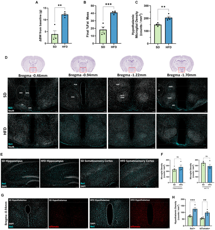
Microglia, the resident macrophages of the central nervous system, are recognized for their heterogeneity and integral role in brain function and diseases. In the context of high fat diet (HFD) feeding and obesity, microglia become overactive, acquiring a prevailing lipid associated microglial phenotype (also known as LAM). Yet, how microgliosis is induced and regulated remains unclear. Here we report a key role for the Complement 3a Receptor (C3aR), on HFD-induced hypothalamic gliosis and weight gain in mice. HFD consumption leads to elevated microglial expression of C3aR, which parallels widespread accumulation of reactive microglia, selectively in the hypothalamus. Conditional microglial C3aR deletion protects mice from HFD-induced hypothalamic reactive microgliosis. C3aR deletion or pharmacological antagonism opposes HFD-induced weight gain in male but not female mice. Mechanistically, we demonstrated that C3aR is essential for lipid-induced lipid droplet formation, and acquisition of a LAM molecular signature. In summary, we uncovered a previously unknown role for C3aR in the acquisition of a LAM signature driving diet-induced gliosis, identifying this receptor as a new viable therapeutic candidate for conditions associated with hypothalamic neuroinflammation.
Overview
CoChat Summary
TL;DR
In mice, microglial C3aR is selectively expressed by microglia, marks lipid-associated microglia (LAM), and is required for high-fat-diet (HFD)-induced hypothalamic microgliosis/astrogliosis and lipid droplet formation. Conditional microglial C3aR deletion or intranasal C3aR antagonism protects against HFD-induced hypothalamic gliosis and, in male mice on 45% HFD, weight/fat gain.
Key findings
- C3aR expression was selective to microglia in mouse and human CNS; scRNAseq identified hypothalamic microglial subclusters with foamy/LAM features (FoamDEX > 0.50), with subclusters 4, 7, and 10 showing highest C3ar1 and LAM scores.
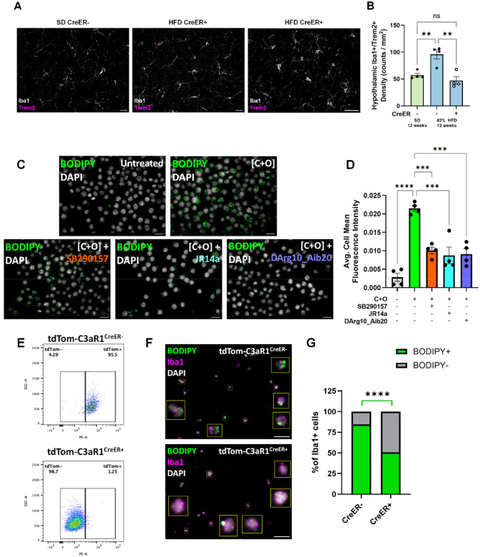
- HFD feeding increased hypothalamic microgliosis and astrogliosis independent of sex and body weight, and increased microglial C3aR; conditional microglial C3aR deletion normalized HFD-induced microgliosis, astrogliosis, amoeboid microglial morphology, CD68, and Trem2+ microglia in both sexes.
- C3aR deletion protected male mice from 45% HFD-induced obesity but not 60% HFD, and did not protect females; intranasal DArg10_Aib20 in 45% HFD-fed males limited body weight gain and decreased fat mass gain without changing food intake, and protected against hypothalamic microgliosis.
- In vitro, cholesterol + oleate induced lipid droplets and LAM/inflammatory genes; C3aR antagonism blocked lipid droplet accumulation, and primary microglia from C3aR-deleted mice showed <50% lipid-loaded cells versus >80% in controls.
Methods
- Mouse genetics and diets: tdTom-C3aR1lox/lox reporter mice and CX3CR1creERT2-mediated conditional microglial C3aR deletion; tamoxifen 100 mg/kg for 5 days; standard diet, 45% HFD, or 60% HFD for up to 12 weeks; n = 10–12/group/sex for SD/45% HFD and n = 5/group/sex for 60% HFD; body weight, EchoMRI, CLAMS indirect calorimetry, and glucose tolerance tests.
- Pharmacological and imaging experiments: intranasal DArg10_Aib20 (8.33 μg/μl; 12 μl/day; 100 μg daily) or saline for 12 weeks in 45% HFD-fed male mice; Cy5-TLQP-21 in vivo imaging via IVIS.
- In vitro and transcriptomic analysis: BV2 or primary microglia lipid-loaded with 20 μg/ml cholesterol + 400 μM oleic acid; BODIPY staining; antagonists SB290157 (20 μM), JR14a (1 μM), or DArg10_Aib20 (20 μM); public scRNAseq datasets analyzed with Seurat/Harmony, FoamDEX, and LAM scores.
Limitations
- Stated: the mechanism of sex-dependent vulnerability needs further study; further sequencing is needed to determine how hypothalamic microglial subclusters shift in diet-induced obesity; the functional implication of a slight reduction in cumulative energy expenditure in male C3aR1lox/lox-CreER+ mice on 45% HFD is currently unclear.
- Stated/evident: CX3CR1creERT2 can target some peripheral macrophages; the authors found transient C3aR depletion in the small intestine at 2 weeks post-tamoxifen that was later replenished, and state this may have limited metabolic effect.
- Evident: the protective metabolic effect was limited to male mice on 45% HFD, not 60% HFD or females, so generalizability across sexes and diet fat levels is constrained.
Model-written summary. Check it against the paper before relying on it.
Integrity signals
Registry
Cites
Statistics
Figures

![Figure 3. Conditional depletion of microglial C3aR protects against diet-induced obesity and hypothalamic microgliosis. A) Experimental timeline for in vivo studies. Change in body weight from baseline comparisons within male (B) or female (C) C3aR1lox/lox-CreER- or C3aR1lox/lox-CreER+ mice fed either SD, 45% HFD, or 60% HFD for 12 weeks. Male Cre+ animals on 45% HFD were protected against diet-induced weight gain meanwhile female mice regardless of genotype were resistant to 45% HFD-induced weight gain. Males 2-way ANOVA. Diet (main) effect: F(2,50)=18.58, p<0.0001; Genotype effect: F(1,49)=2.415, p=0.1265; Interaction: F(2,50)=1.422, p=0.2507). Females 2-way ANOVA. Diet (main) effect: F(2,47)=26.65, p<0.0001; Genotype effect: F(1,47)=0.02555, p=0.8737; Interaction: F(2,47)=0.3026, p=0.7403). Tukey’s post hoc]. Animals: SD & 45%HFD, N=10–12/genotype/sex; 60% N=5/genotype/sex. Representative 10x IF images (D) and quantification (E) showing HFD-induced hypothalamic microgliosis that was reduced in animals lacking microglial C3aR. Diet-induced hypothalamic microgliosis was protected when microglial C3aR was ablated in both male and female animals, and regardless of which HFD diet they were on. 2-way ANOVA. Diet (main) effect: F(2,45)=23.12, p<0.0001; Genotype effect: F(1,45)=23.41, p<0.0001; Interaction: F(2,45)=7.515, p=0.0015). F) Flow cytometry showing elevated whole brain microglial counts after 12 weeks of 45% HFD that was mitigated in animals with ablated microglial C3aR, consistent with the histology performed on replicate animals in C/D. Sexes were pooled although there appears to be clustering based on sex in HFD-fed CreER+ animals (Fig.S4C). Pooled 2-way ANOVA. Diet (main) effect: F(1,40)=52.62, p<0.0001; Genotype effect: F(1,40)=9.353, p=0.0040; Interaction: F(1,40)=5.290, p=0.0267). Representative IF images (G) and quantification (H) showing HFD-induced hypothalamic astrogliosis that was also reduced in animals lacking microglial C3aR, despite these astrocytes not expressing C3aR. Quantification shows an increase in percent area occupied by GFAP+ pixels in 10x magnification images (20x magnification images shown for representative images, G). Scale bars represent 30μm. 2-way ANOVA. Diet (main) effect: F(2,43)=15.79, p<0.0001; Genotype effect: F(1,43)=23.34, p<0.0001; Interaction: F(2,42)=13.16, p<0.0001). Error bars represent standard error of the mean (SEM).](https://figures.cochat.ai/paper-figures/8acb9c1087a647588b72aa62948d06fa/complement-3a-receptor-mediates-high-fat-diet-induced-hypothalamic-accumulation-figure-3.jpg)


Verification
Claims the paper makes
1 Valid · 2 Not valid · 33 Not checked · 83 Own results and interpretations
Some cited sources support this claim and some don’t — read each source’s verdict below.
In the paper
“Similarly, HFD resulted in increased CD68 staining as a proxy of phagocytic activity[73,75,85].”
§ Complement 3a Receptor mediates high fat diet induced hypothalamic accumulation of lipid associated microglia to regulate neuroinflammation and obesity > Discussion > An essential role for C3aR in HFD-induced hypothalamic gliosis
What the sources say
- Partly supportedcites [73] sourceExperimental — accuracy not yet measured
In the Preclinical models section (Results), our copy does report that HFD raises CD68 as a phagocytic marker — 'enhanced phagocytic activity (Iba1+/CD68+ cells)' — but bounds it to male microglia, noting that females showed no signs of HFD-mediated microglial phenotypic polarization (ref [188]). The citing sentence drops that sex-specific qualifier and states the increase as a general HFD effect. The only other CD68 mention in our copy is in a mechanistic context (reduced microglial phagocytic capacity/CD68 expression in lipoprotein-lipase knockdown mice on HFD, ref [190]), which does not add unconditional support to the claim as worded. (LLM, conf=0.75)
- Disputedcites [75] sourceExperimental — accuracy not yet measured
Our copy of Hendrickx et al. (2017) contains no high-fat-diet experiment at all: it is an immunohistochemical study of HLA-DR, Iba1 and CD68 in human post-mortem microglia/MS tissue (Introduction, §1; Results/Discussion 'Nimmerjahn' section), so the attributed HFD-induced increase in CD68 staining is not this paper's finding. More decisively, the paper contradicts the framing of CD68 staining as a proxy of phagocytic activity: in the Discussion it states that 'studies are inconclusive about cellular CD68 regulation during phagocytosis' (unaltered after zymosan or myelin phagocytosis, upregulated only in THP-1 and BV-2 systems) and concludes 'a convincing correlation between CD68 expression and phagocytic activity has yet to be found.' It does note CD68 intensity is highest in foamy macrophages and that CD68 is functionally involved in phagocytosis of oxLDL and apoptotic cells, but explicitly stops short of endorsing CD68 as a readout of phagocytic activity. (LLM, conf=0.78)
Checked by a reasoning model — may be wrong.
- Supportedcites [85] source
The Introduction (p. 1) states that CD68-ir 'is associated with microglial phagocytosis' and that 'a high-calorie diet not only increases the number of iba1-ir and CD68-ir positive microglia' in the ARC. The Results section 'Microglial Activation Is Mainly Induced by HFD Diet but Not by Increased Fat Mass' reports that HFD exposure raised CD68-ir in ob/ob and db/db mice versus SC-fed controls, and that microglial activation is mainly a consequence of HFD rather than obesity per se. Thus the source supports both the direction of the effect (HFD increases CD68 staining) and the interpretation of CD68 as a phagocytic-activity marker. (LLM, conf=0.90)
plainstates a cause- Asserted: passes — The authors put it forward as true.
- Checkable: passes — It can be checked against evidence.
- Load-bearing: unclear — It is unclear whether the paper’s conclusions depend on it.
- Based: passes — The paper gives a reason why it holds.
- Partly supported
The cited sources that could be checked couldn’t confirm or dispute this claim.
In the paper
“Analysis of single-cell RNA sequencing datasets[13,54,56] confirms that microglia are the predominant cell type in the CNS expressing C3ar1 in mice (Fig.S2A–C).”
§ Complement 3a Receptor mediates high fat diet induced hypothalamic accumulation of lipid associated microglia to regulate neuroinflammation and obesity > Results > Selective expression of C3aR by microglia in the central nervous system
What the sources say
- Could not verifycites [13] sourceExperimental — accuracy not yet measured
Our copy of Hajdarovic et al. (Trends in Neurosciences, 2022) is a review of single-cell 'omics of the aging hypothalamus; the only microglia passage states that 'a clear inflammatory signal emerged across age in microglia and macrophages[93]' (Machine learning applications in single cell analysis and aging research), and the single-cell sections discuss neuronal cluster definitions (Single cell ‘omics complements existing tools). No passage anywhere in our copy mentions C3ar1/C3aR1 or the relative expression of that receptor across CNS cell types, despite targeted searches ('C3ar1', 'C3aR1 complement component 3a receptor enriched in microglia', 'microglia marker gene expression central nervous system'). Absence of the point is not contradiction, so the claim can be neither confirmed nor disputed on this source. (LLM, conf=0.70)
- Could not verifycites [54] sourceExperimental — accuracy not yet measured
Our copy of HypoMap contains no passage mentioning C3ar1 (searched: 'C3ar1 microglia', 'complement 3a receptor gene expression marker', 'complement receptor C3aR1 expression cell type'); all returns were semantic neighbours about Glp1r/POMC validation and bacTRAP mapping, none exact-phrase hits. The paper does profile microglia — a large Ly86-expressing cluster subdivided into ten clusters (Results, 'Non-neuronal populations'; and 'The generation of HypoMap', where Ly86 marks the microglia node at C7) — but only for the murine hypothalamus, with no statement about C3ar1 or about which CNS cell type predominantly expresses it. Since the claim's decisive content (C3ar1 expression) is not addressed anywhere in our copy, and nothing contrary is stated, it can be neither confirmed nor disputed. (LLM, conf=0.72)
- Could not verifycites [56] sourceExperimental — accuracy not yet measured
Our copy of Kim et al. (Cell Reports 2025) is a hypothalamic/prethalamic scRNA-/scATAC-seq atlas and contains no mention of C3ar1 or complement receptor expression in any cell type. The only microglia references are the STAR★Methods note that immune cells were removed as contaminant populations during cluster annotation, and a RESULTS statement (Dlx1/2 section) that non-neuronal cells increased 'with no change in oligodendrocytes or microglia' in P21 Dlx1/2 mutants — neither addresses which CNS cell type predominantly expresses C3ar1. Searched: C3ar1/C3a receptor/complement, microglia/macrophage markers (Cx3cr1, P2ry12), and non-neuronal CNS cell types; nothing supports or contradicts the claim, so this citation cannot be verified from this source. (LLM, conf=0.70)
boostedstates an association- Asserted: passes — The authors put it forward as true.
- Checkable: passes — It can be checked against evidence.
- Load-bearing: passes — The paper’s conclusions depend on it.
- Based: passes — The paper gives a reason why it holds.
- Could not verify
The cited sources support only part of this claim — read each source’s verdict below.
In the paper
“Analysis of publicly available single-cell RNA sequencing datasets[13,54,56] revealed that even under healthy, physiological circumstances, there are unique sub-populations of hypothalamic microglia exhibiting a LAM signature.”
§ Complement 3a Receptor mediates high fat diet induced hypothalamic accumulation of lipid associated microglia to regulate neuroinflammation and obesity > Discussion > C3aR is a new LAM marker, necessary for lipid-induced lipid droplet formation
What the sources say
- Could not verifycites [13] sourceExperimental — accuracy not yet measured
Our copy of this review discusses single-cell transcriptomic discovery of neuronal (and some glial) subtypes in the hypothalamus, but nowhere mentions a lipid-associated macrophage (LAM) signature or LAM-like subpopulations of hypothalamic microglia under healthy, physiological conditions (searched: 'LAM/lipid-associated macrophage', 'microglia macrophages inflammatory', 'publicly available scRNA-seq datasets', 'glial cell types'). The closest passage, in 'Machine learning applications in single cell analysis and aging research', reports only that in the authors' own study 'a clear inflammatory signal emerged across age in microglia and macrophages[93]' — an age-related, not a healthy-state, observation, and it credits their own dataset rather than publicly available ones. Since our copy neither states nor contradicts the claim, attribution cannot be settled from this source. (LLM, conf=0.70)
- Partly supportedcites [54] sourceExperimental — accuracy not yet measured
Our copy does support the framing: HypoMap was built by integrating 17 publicly available datasets (Main; Methods, 'Dataset download'), and the Non-neuronal populations section reports that 'HypoMap also captures a large cluster of Ly86-expressing microglia ... which could be further divided into ten clusters' — i.e., distinct hypothalamic microglial sub-populations under physiological (ad libitum-fed/fasted) conditions. What our copy never states is the defining qualifier of the claim: no passage describes a LAM (lysosome-associated membrane / lipid-associated macrophage) signature in any microglial sub-cluster; the only state-labelled glial sub-population named is a reactive astrocyte cluster (C66-54: Lgals3.Astrocytes, Gfap/Lgals3). Searches for 'LAM', 'lysosome-associated membrane', 'lysosomal/autophagy microglia markers (Ctsd, Gpnmb, Apoe)' and 'microglia homeostatic markers (Cx3cr1, P2ry12)' returned no such characterization. So the sub-population half is supported and the LAM-signature half is not. (LLM, conf=0.68)
- Could not verifycites [56] sourceExperimental — accuracy not yet measured
Our copy of Kim et al. (Cell Reports 2025) is a developmental scRNA-seq/scATAC-seq atlas of the mouse hypothalamus and prethalamus (E11-P8) and contains no passage describing lipid-associated macrophage (LAM) signatures or LAM-like microglial subpopulations in healthy hypothalamus. Searching 'microglia', 'lipid-associated macrophage / Trem2 / Cd9 / Gpnmb / myeloid', and 'publicly available dataset reanalysis of microglia' returned only one mention of microglia, in the RESULTS section 'Dlx1/2 promote prethalamic identity and GABAergic differentiation while repressing SMN identity': 'A >3-fold increase in non-neuronal cells—primarily GFAP+ astrocytes—was observed in TRN and ZI, with no change in oligodendrocytes or microglia (Figures S13E and S13F)' — a mutant-comparison statement, not a claim about LAM subpopulations. The paper's own data are newly generated (GEO GSE284492; Zenodo processed Seurat objects), and its non-neuronal content covers radial glia/astrocyte markers (Slc1a3, Aldh1l1) rather than microglial subsets, so nothing in our copy either supports or contradicts the attributed claim. (LLM, conf=0.72)
plain- Asserted: passes — The authors put it forward as true.
- Checkable: passes — It can be checked against evidence.
- Load-bearing: passes — The paper’s conclusions depend on it.
- Based: passes — The paper gives a reason why it holds.
- Could not verify
A result the paper reports from its own data — it cites no other source for it, so there is none to check. The quote shows it is in the paper.
In the paper
“HFD consumption leads to elevated microglial expression of C3aR, which parallels widespread accumulation of reactive microglia, selectively in the hypothalamus.”
§ Complement 3a Receptor mediates high fat diet induced hypothalamic accumulation of lipid associated microglia to regulate neuroinflammation and obesity > Abstract
plainstates a cause- Asserted: passes — The authors put it forward as true.
- Checkable: passes — It can be checked against evidence.
- Load-bearing: passes — The paper’s conclusions depend on it.
- Based: passes — The paper gives a reason why it holds.
A result the paper reports from its own data — it cites no other source for it, so there is none to check. The quote shows it is in the paper.
In the paper
“C3aR deletion or pharmacological antagonism opposes HFD-induced weight gain in male but not female mice.”
- Also stated in: § Introduction · plain
“Finally, C3aR deletion or pharmacological antagonism opposes HFD-induced weight gain in a sex dependent-manner.”
§ Complement 3a Receptor mediates high fat diet induced hypothalamic accumulation of lipid associated microglia to regulate neuroinflammation and obesity > Abstract
plainstates a cause- Asserted: passes — The authors put it forward as true.
- Checkable: passes — It can be checked against evidence.
- Load-bearing: passes — The paper’s conclusions depend on it.
- Based: passes — The paper gives a reason why it holds.
- Rests on an assumption not shown here.
- Also stated in: § Introduction · plain
41 more claims
Create a free account to see all 119 claims and to verify the 121 references still open.
- It may state two separate claims as one. 59
- Probably not a substantial claim. 29
Self-check
Percentages, sums and reported differences re-computed from the paper’s own numbers.
Each contradiction between two passages listed here was confirmed by an independent check.
Rated minor: it would not touch a result — a value no result rests on, a p-value that stays on the same side of the significance level, or a conflict that is clerical, resolved elsewhere in the paper, or outside its own findings.
In the paper
§ Complement 3a Receptor mediates high fat diet induced hypothalamic accumulation of lipid associated microglia to regulate neuroinflammation and obesity > Results > Conditional C3aR deletion prevents high fat diet induced hypothalamic gliosis
“there was a slight reduction in cumulative energy expenditure only in male C3aR1lox/lox-CreER+ fed 45% HFD (Fig.S4A–B)”
§ Complement 3a Receptor mediates high fat diet induced hypothalamic accumulation of lipid associated microglia to regulate neuroinflammation and obesity > Discussion > C3aR exerts a sex dependent role in HFD-induced obesity.
“neither food intake measured during the entire study or energy expenditure assessed after 10 weeks on the diet were affected by gene deletion.”
Citations
Cites
Fast, sensitive and accurate integration of single-cell data with Harmony
Korsunsky, I., Millard, N., Fan, J., Slowikowski, K., Zhang, F., Wei, K., Baglaenko, Y., Brenner, M., Loh, P.-R., & Raychaudhuri, S. (2019) · Nature Methods · doi:10.1038/s41592-019-0619-0 · Cited by 10,488 · cited 1×
“4.6[60]).”
InfluentialMassively parallel digital transcriptional profiling of single cells
Zheng, G. X. Y., Terry, J. M., Belgrader, P., Ryvkin, P., Bent, Z. W., Wilson, R., Ziraldo, S. B., Wheeler, T. D., McDermott, G. P., Zhu, J., et al. (2017) · Nature Communications · doi:10.1038/ncomms14049 · Cited by 7,353
Dictionary learning for integrative, multimodal and scalable single-cell analysis
Hao, Y., Stuart, T., Kowalski, M. H., Choudhary, S., Hoffman, P., Hartman, A., Srivastava, A., Molla, G., Madad, S., Fernandez-Granda, C., et al. (2024) · Nature Biotechnology · doi:10.1038/s41587-023-01767-y · Cited by 5,631
Pfaffl, M. W., Tichopad, A., Prgomet, C., & Neuvians, T. P. (2004) · Biotechnology Letters · doi:10.1023/b:bile.0000019559.84305.47 · Cited by 5,065
Kopelman, P. G. (2000) · Nature · doi:10.1038/35007508 · Cited by 4,795
Fate Mapping Reveals Origins and Dynamics of Monocytes and Tissue Macrophages under Homeostasis
Yona, S., Kim, K.-W., Wolf, Y., Mildner, A., Varol, D., Breker, M., Strauss-Ayali, D., Viukov, S., Guilliams, M., Misharin, A., et al. (2013) · Immunity · doi:10.1016/j.immuni.2012.12.001 · Cited by 3,245
Krasemann, S., Madore, C., Cialic, R., Baufeld, C., Calcagno, N., El Fatimy, R., Beckers, L., O’Loughlin, E., Xu, Y., Fanek, Z., et al. (2017) · Immunity · doi:10.1016/j.immuni.2017.08.008 · Cited by 2,979
Microglia promote learning-dependent synapse formation through brain-derived neurotrophic factor
Parkhurst, C. N., Yang, G., Ninan, I., Savas, J. N., Yates, J. R., Lafaille, J. J., Hempstead, B. L., Littman, D. R., & Gan, W.-B. (2013) · Cell · doi:10.1016/j.cell.2013.11.030 · Cited by 2,599
Obesity is associated with hypothalamic injury in rodents and humans.
Thaler, J. P., Yi, C.-X., Schur, E. A., Guyenet, S. J., Hwang, B. H., Dietrich, M. O., Zhao, X., Sarruf, D. A., Izgur, V., Maravilla, K. R., et al. (2012) · Journal of Clinical Investigation · doi:10.1172/jci59660 · Cited by 1,857
Marschallinger, J., Iram, T., Zardeneta, M., Lee, S. E., Lehallier, B., Haney, M. S., Pluvinage, J. V., Mathur, V., Hahn, O., Morgens, D. W., et al. (2020) · Nature Neuroscience · doi:10.1038/s41593-019-0566-1 · Cited by 1,586
Microglia Biology: One Century of Evolving Concepts
Prinz, M., Jung, S., & Priller, J. (2019) · Cell · doi:10.1016/j.cell.2019.08.053 · Cited by 1,525
Disease-Associated Microglia: A Universal Immune Sensor of Neurodegeneration
Deczkowska, A., Keren-Shaul, H., Weiner, A., Colonna, M., Schwartz, M., & Amit, I. (2018) · Cell · doi:10.1016/j.cell.2018.05.003 · Cited by 1,474 · cited 2×
“…protection against it due to ablation of microglia C3aR, we took high-32 magnification images to profile the morphological complexity of microglia[74–76] in mice fed SD or 45% HFD. Sholl analysis revealed that under standard diet and normal C3aR expression (C3aR1 lox/lox -CreER- fed SD), the…”
Lipid-associated macrophages control metabolic homeostasis in a Trem2-dependent manner
Jaitin, D. A., Adlung, L., Thaiss, C. A., Weiner, A., Li, B., Descamps, H., Lundgren, P., Bleriot, C., Liu, Z., Deczkowska, A., et al. (2019) · Cell · doi:10.1016/j.cell.2019.05.054 · Cited by 1,377
High‐fat Diets: Modeling the Metabolic Disorders of Human Obesity in Rodents
Buettner, R., Schölmerich, J., & Bollheimer, L. C. (2007) · Obesity · doi:10.1038/oby.2007.608 · Cited by 1,231
Anti-obesity drug discovery: advances and challenges
Müller, T. D., Blüher, M., Tschöp, M. H., & DiMarchi, R. D. (2022) · Nature Reviews Drug Discovery · doi:10.1038/s41573-021-00337-8 · Cited by 1,021
Glia as architects of central nervous system formation and function
Allen, N. J., & Lyons, D. A. (2018) · Science · doi:10.1126/science.aat0473 · Cited by 1,001
TREM2 regulates microglial cholesterol metabolism upon chronic phagocytic challenge
Nugent, A. A., Lin, K., van Lengerich, B., Lianoglou, S., Przybyla, L., Davis, S. S., Llapashtica, C., Wang, J., Kim, D. J., Xia, D., et al. (2020) · Neuron · doi:10.1016/j.neuron.2019.12.007 · Cited by 798
A new type of microglia gene targeting shows TAK1 to be pivotal in CNS autoimmune inflammation
Goldmann, T., Wieghofer, P., Müller, P. F., Wolf, Y., Varol, D., Yona, S., Brendecke, S. M., Kierdorf, K., Staszewski, O., Datta, M., et al. (2013) · Nature Neuroscience · doi:10.1038/nn.3531 · Cited by 688
APOE4/4 is linked to damaging lipid droplets in Alzheimer’s disease microglia
Haney, M. S., Pálovics, R., Munson, C. N., Long, C., Johansson, P. K., Yip, O., Dong, W., Rawat, E., West, E., Schlachetzki, J. C. M., et al. (2024) · Nature · doi:10.1038/s41586-024-07185-7 · Cited by 667
Neuroinflammation in Alzheimer disease
Heneka, M. T., van der Flier, W. M., Jessen, F., Hoozemanns, J., Thal, D. R., Boche, D., Brosseron, F., Teunissen, C., Zetterberg, H., Jacobs, A. H., et al. (2025) · Nature Reviews Immunology · doi:10.1038/s41577-024-01104-7 · Cited by 638
Valdearcos, M., Robblee, M. M., Benjamin, D. I., Nomura, D. K., Xu, A. W., & Koliwad, S. K. (2014) · Cell Reports · doi:10.1016/j.celrep.2014.11.018 · Cited by 629
A new fate mapping system reveals context-dependent random or clonal expansion of microglia
Tay, T. L., Mai, D., Dautzenberg, J., Fernández-Klett, F., Lin, G., Sagar, Datta, M., Drougard, A., Stempfl, T., Ardura-Fabregat, A., et al. (2017) · Nature Neuroscience · doi:10.1038/nn.4547 · Cited by 597
Cognitive impairment following high fat diet consumption is associated with brain inflammation
Pistell, P. J., Morrison, C. D., Gupta, S., Knight, A. G., Keller, J. N., Ingram, D. K., & Bruce-Keller, A. J. (2010) · Journal of Neuroimmunology · doi:10.1016/j.jneuroim.2009.11.010 · Cited by 596
Complement C3 deficiency protects against neurodegeneration in aged plaque-rich APP/PS1 mice
Shi, Q., Chowdhury, S., Ma, R., Le, K. X., Hong, S., Caldarone, B. J., Stevens, B., & Lemere, C. A. (2017) · Science Translational Medicine · doi:10.1126/scitranslmed.aaf6295 · Cited by 593
Microglia in depression: an overview of microglia in the pathogenesis and treatment of depression
Wang, H., He, Y., Sun, Z., Ren, S., Liu, M., Wang, G., & Yang, J. (2022) · Journal of Neuroinflammation · doi:10.1186/s12974-022-02492-0 · Cited by 571
Self-citationAdamu, A., Li, S., Gao, F., & Xue, G. (2024) · Frontiers in Aging Neuroscience · doi:10.3389/fnagi.2024.1347987 · Cited by 564
Obesity-Induced Neuroinflammation: Beyond the Hypothalamus
Guillemot-Legris, O., & Muccioli, G. G. (2017) · Trends in Neurosciences · doi:10.1016/j.tins.2017.02.005 · Cited by 563
Olmos-Alonso, A., Schetters, S. T. T., Sri, S., Askew, K., Mancuso, R., Vargas-Caballero, M., Holscher, C., Perry, V. H., & Gomez-Nicola, D. (2016) · Brain · doi:10.1093/brain/awv379 · Cited by 532
Litvinchuk, A., Wan, Y.-W., Swartzlander, D. B., Chen, F., Cole, A., Propson, N. E., Wang, Q., Zhang, B., Liu, Z., & Zheng, H. (2018) · Neuron · doi:10.1016/j.neuron.2018.10.031 · Cited by 523
A clinical perspective of obesity, metabolic syndrome and cardiovascular disease
Han, T. S., & Lean, M. E. (2016) · JRSM Cardiovascular Disease · doi:10.1177/2048004016633371 · Cited by 500
Glial Cells and Their Function in the Adult Brain: A Journey through the History of Their Ablation
Jäkel, S., & Dimou, L. (2017) · Frontiers in Cellular Neuroscience · doi:10.3389/fncel.2017.00024 · Cited by 500
Inflammatory Links Between High Fat Diets and Diseases
Duan, Y., Zeng, L., Zheng, C., Song, B., Li, F., Kong, X., & Xu, K. (2018) · Frontiers in Immunology · doi:10.3389/fimmu.2018.02649 · Cited by 495
Valdearcos, M., Douglass, J. D., Robblee, M. M., Dorfman, M. D., Stifler, D. R., Bennett, M. L., Gerritse, I., Fasnacht, R., Barres, B. A., Thaler, J. P., et al. (2017) · Cell Metabolism · doi:10.1016/j.cmet.2017.05.015 · Cited by 477
SNT: a unifying toolbox for quantification of neuronal anatomy
Arshadi, C., Günther, U., Eddison, M., Harrington, K. I. S., & Ferreira, T. A. (2021) · Nature Methods · doi:10.1038/s41592-021-01105-7 · Cited by 470
Wu, T., Dejanovic, B., Gandham, V. D., Gogineni, A., Edmonds, R., Schauer, S., Srinivasan, K., Huntley, M. A., Wang, Y., Wang, T.-M., et al. (2019) · Cell Reports · doi:10.1016/j.celrep.2019.07.060 · Cited by 466
CalR: A Web-Based Analysis Tool for Indirect Calorimetry Experiments
Mina, A. I., LeClair, R. A., LeClair, K. B., Cohen, D. E., Lantier, L., & Banks, A. S. (2018) · Cell Metabolism · doi:10.1016/j.cmet.2018.06.019 · Cited by 458
Regulation of Microglial Proliferation during Chronic Neurodegeneration
Gómez-Nicola, D., Fransen, N. L., Suzzi, S., & Perry, V. H. (2013) · The Journal of Neuroscience · doi:10.1523/jneurosci.4440-12.2013 · Cited by 407
Emerging role of the brain in the homeostatic regulation of energy and glucose metabolism
Roh, E., Song, D. K., & Kim, M.-S. (2016) · Experimental & Molecular Medicine · doi:10.1038/emm.2016.4 · Cited by 396
High-Fat Diet Induced Anxiety and Anhedonia: Impact on Brain Homeostasis and Inflammation
Dutheil, S., Ota, K. T., Wohleb, E. S., Rasmussen, K., & Duman, R. S. (2016) · Neuropsychopharmacology · doi:10.1038/npp.2015.357 · Cited by 355
Role of astrocytes, microglia, and tanycytes in brain control of systemic metabolism
García-Cáceres, C., Balland, E., Prevot, V., Luquet, S., Woods, S. C., Koch, M., Horvath, T. L., Yi, C.-X., Chowen, J. A., Verkhratsky, A., et al. (2019) · Nature Neuroscience · doi:10.1038/s41593-018-0286-y · Cited by 329
Self-citationLipid accumulation induced by APOE4 impairs microglial surveillance of neuronal-network activity
Victor, M. B., Leary, N., Luna, X., Meharena, H. S., Scannail, A. N., Bozzelli, P. L., Samaan, G., Murdock, M. H., von Maydell, D., Effenberger, A. H., et al. (2022) · Cell Stem Cell · doi:10.1016/j.stem.2022.07.005 · Cited by 316
Hendrickx, D. A. E., van Eden, C. G., Schuurman, K. G., Hamann, J., & Huitinga, I. (2017) · Journal of Neuroimmunology · doi:10.1016/j.jneuroim.2017.04.007 · Cited by 313
Lipid Accumulation Induced by APOE4 Impairs Microglial Surveillance of Neuronal-Network Activity
Victor, M. B., Leary, N., Luna, X., Meharena, H. S., Bozzelli, P. L., Samaan, G., Murdock, M. H., von Maydell, D., Effenberger, A. H., Cerit, O., et al. (2022) · doi:10.1101/2022.03.21.484146 · Cited by 311
Dietary Fat, but Not Protein or Carbohydrate, Regulates Energy Intake and Causes Adiposity in Mice
Hu, S., Wang, L., Yang, D., Li, L., Togo, J., Wu, Y., Liu, Q., Li, B., Li, M., Wang, G., et al. (2018) · Cell Metabolism · doi:10.1016/j.cmet.2018.06.010 · Cited by 271
HypoMap—a unified single-cell gene expression atlas of the murine hypothalamus
Steuernagel, L., Lam, B. Y. H., Klemm, P., Dowsett, G. K. C., Bauder, C. A., Tadross, J. A., Hitschfeld, T. S., del Rio Martin, A., Chen, W., de Solis, A. J., et al. (2022) · Nature Metabolism · doi:10.1038/s42255-022-00657-y · Cited by 267
Hormones and diet, but not body weight, control hypothalamic microglial activity
Gao, Y., Ottaway, N., Schriever, S. C., Legutko, B., García‐Cáceres, C., de la Fuente, E., Mergen, C., Bour, S., Thaler, J. P., Seeley, R. J., et al. (2014) · Glia · doi:10.1002/glia.22580 · Cited by 263
Tissue-specific Role of CX3CR1 Expressing Immune Cells and Their Relationships with Human Disease
Lee, M., Lee, Y., Song, J., Lee, J., & Chang, S.-Y. (2018) · Immune Network · doi:10.4110/in.2018.18.e5 · Cited by 260 · cited 1×
“Nevertheless, because CX3CR1 is expressed by a subset of macrophages in other organs such as small intestine, liver, and adipose tissue[72], we extended our analysis to other organs as well to confirm specificity of C3aR deletion in microglia long-term.”
Morphology of Microglia Across Contexts of Health and Disease
Savage, J. C., Carrier, M., & Tremblay, M.-È. (2019) · Methods in Molecular Biology · doi:10.1007/978-1-4939-9658-2_2 · Cited by 232
Hypothalamic Inflammation: Marker or Mechanism of Obesity Pathogenesis?
Thaler, J. P., Guyenet, S. J., Dorfman, M. D., Wisse, B. E., & Schwartz, M. W. (2013) · Diabetes · doi:10.2337/db12-1605 · Cited by 209
Mamane, Y., Chung Chan, C., Lavallee, G., Morin, N., Xu, L.-J., Huang, J., Gordon, R., Thomas, W., Lamb, J., Schadt, E. E., et al. (2009) · Diabetes · doi:10.2337/db09-0323 · Cited by 188
Hypothalamic PGC-1α Protects Against High-Fat Diet Exposure by Regulating ERα
Morselli, E., Fuente-Martin, E., Finan, B., Kim, M., Frank, A., Garcia-Caceres, C., Navas, C. R., Gordillo, R., Neinast, M., Kalainayakan, S. P., et al. (2014) · Cell Reports · doi:10.1016/j.celrep.2014.09.025 · Cited by 187
Trem2 promotes foamy macrophage lipid uptake and survival in atherosclerosis
Patterson, M. T., Firulyova, M. M., Xu, Y., Hillman, H., Bishop, C., Zhu, A., Hickok, G. H., Schrank, P. R., Ronayne, C. E., Caillot, Z., et al. (2023) · Nature Cardiovascular Research · doi:10.1038/s44161-023-00354-3 · Cited by 180
Self-citationIntranasal Administration of CNS Therapeutics to Awake Mice
Hanson, L. R., Fine, J. M., Svitak, A. L., & Faltesek, K. A. (2013) · Journal of Visualized Experiments · doi:10.3791/4440 · Cited by 170
Glial cells more than support cells?
He, F., & Sun, Y. E. (2006) · The International Journal of Biochemistry & Cell Biology · doi:10.1016/j.biocel.2006.10.022 · Cited by 170
Beilharz, J. E., Maniam, J., & Morris, M. J. (2016) · Behavioural Brain Research · doi:10.1016/j.bbr.2016.03.018 · Cited by 168
Guillemot-Legris, O., Masquelier, J., Everard, A., Cani, P. D., Alhouayek, M., & Muccioli, G. G. (2016) · Journal of Neuroinflammation · doi:10.1186/s12974-016-0666-8 · Cited by 168
Tarantini, S., Valcarcel-Ares, M. N., Yabluchanskiy, A., Tucsek, Z., Hertelendy, P., Kiss, T., Gautam, T., Zhang, X. A., Sonntag, W. E., de Cabo, R., et al. (2018) · The Journals of Gerontology: Series A · doi:10.1093/gerona/glx177 · Cited by 164
de Paula, G. C., Brunetta, H. S., Engel, D. F., Gaspar, J. M., Velloso, L. A., Engblom, D., de Oliveira, J., & de Bem, A. F. (2021) · Frontiers in Neuroscience · doi:10.3389/fnins.2021.734158 · Cited by 162
Stress induced microglial activation contributes to depression
Li, B., Yang, W., Ge, T., Wang, Y., & Cui, R. (2022) · Pharmacological Research · doi:10.1016/j.phrs.2022.106145 · Cited by 151
Nose-to-brain drug delivery: from bench to bedside
Drath, I., Richter, F., & Feja, M. (2025) · Translational Neurodegeneration · doi:10.1186/s40035-025-00481-w · Cited by 150
Microglial control of neuronal activity
Béchade, C., Cantaut-Belarif, Y., & Bessis, A. (2013) · Frontiers in Cellular Neuroscience · doi:10.3389/fncel.2013.00032 · Cited by 147
Bartolomucci, A., La Corte, G., Possenti, R., Locatelli, V., Rigamonti, A. E., Torsello, A., Bresciani, E., Bulgarelli, I., Rizzi, R., Pavone, F., et al. (2006) · Proceedings of the National Academy of Sciences · doi:10.1073/pnas.0606102103 · Cited by 134
Self-citationComprehensive Review of Current and Upcoming Anti-Obesity Drugs
Son, J. W., & Kim, S. (2020) · Diabetes & Metabolism Journal · doi:10.4093/dmj.2020.0258 · Cited by 134
Lim, J., Iyer, A., Suen, J. Y., Seow, V., Reid, R. C., Brown, L., & Fairlie, D. P. (2013) · The FASEB Journal · doi:10.1096/fj.12-220582 · Cited by 129
Obesity-induced inflammation: connecting the periphery to the brain
Le Thuc, O., & García-Cáceres, C. (2024) · Nature Metabolism · doi:10.1038/s42255-024-01079-8 · Cited by 125
Lipoprotein lipase maintains microglial innate immunity in obesity.
Gao, Y., Vidal-Itriago, A., Kalsbeek, M. J., Layritz, C., García-Cáceres, C., Tom, R. Z., Eichmann, T. O., Vaz, F. M., Houtkooper, R. H., van der Wel, N., et al. (2017) · Cell Reports · doi:10.1016/j.celrep.2017.09.008 · Cited by 123 · cited 1×
“The notion that hypothalamic inflammation and gliosis are primarily driven by HFD consumption rather than body weight gain is consistent with previous data[14,30,85,94].”
Blanco-Carmona, E., & Kool, M. (2026) · Bioinformatics Advances · doi:10.1093/bioadv/vbag151 · Cited by 122
A comprehensive spatio-cellular map of the human hypothalamus
Tadross, J. A., Steuernagel, L., Dowsett, G. K. C., Kentistou, K. A., Lundh, S., Porniece, M., Klemm, P., Rainbow, K., Hvid, H., Kania, K., et al. (2025) · Nature · doi:10.1038/s41586-024-08504-8 · Cited by 112
Obesity-Induced Brain Neuroinflammatory and Mitochondrial Changes
Schmitt, L. O., & Gaspar, J. M. (2023) · Metabolites · doi:10.3390/metabo13010086 · Cited by 109
Neuroinflammation: A Modifiable Pathway Linking Obesity, Alzheimer's disease, and Depression
Ly, M., Yu, G. Z., Mian, A., Cramer, A., Meysami, S., Merrill, D. A., Samara, A., Eisenstein, S. A., Hershey, T., Babulal, G. M., et al. (2023) · The American Journal of Geriatric Psychiatry · doi:10.1016/j.jagp.2023.06.001 · Cited by 108
Generating publication ready visualizations for Single Cell transcriptomics using SCpubrPreprint
Blanco-Carmona, E. (2022) · bioRxiv (Cold Spring Harbor Laboratory) · doi:10.1101/2022.02.28.482303 · Cited by 107
CCR7 defines a precursor for murine iNKT cells in thymus and periphery
Wang, H., & Hogquist, K. A. (2018) · eLife · doi:10.7554/elife.34793 · Cited by 105
Self-citationVargas-Soria, M., García-Alloza, M., & Corraliza-Gómez, M. (2023) · Journal of Neuroinflammation · doi:10.1186/s12974-023-02740-x · Cited by 104
Neuroinflammation: The Pathogenic Mechanism of Neurological Disorders
Gorji, A. (2022) · International Journal of Molecular Sciences · doi:10.3390/ijms23105744 · Cited by 101
Dionysopoulou, S., Charmandari, E., Bargiota, Α., Vlahos, N., Mastorakos, G. P., & Valsamakis, G. (2021) · Nutrients · doi:10.3390/nu13020498 · Cited by 100
Targeting Microglia Using Cx3cr1-Cre Lines: Revisiting the Specificity
Zhao, X.-F., Alam, M. M., Liao, Y., Huang, T., Mathur, R., Zhu, X., & Huang, Y. (2019) · eneuro · doi:10.1523/eneuro.0114-19.2019 · Cited by 92
El Gaamouch, F., Audrain, M., Lin, W.-J., Beckmann, N., Jiang, C., Hariharan, S., Heeger, P. S., Schadt, E. E., Gandy, S., Ehrlich, M. E., et al. (2020) · Molecular Neurodegeneration · doi:10.1186/s13024-020-0357-x · Cited by 92 · cited 3×
“C3aR expression is high in the brain, and previous work suggested a prominent expression in microglia[41,42,64,65], but a detailed characterization is lacking.”
Self-citationSahasrabuddhe, V., & Ghosh, H. S. (2022) · Cell Reports · doi:10.1016/j.celrep.2021.110252 · Cited by 91
Monitoring C3aR Expression Using a Floxed tdTomato-C3aR Reporter Knock-in Mouse
Quell, K. M., Karsten, C. M., Kordowski, A., Almeida, L. N., Briukhovetska, D., Wiese, A. V., Sun, J., Ender, F., Antoniou, K., Schröder, T., et al. (2017) · The Journal of Immunology · doi:10.4049/jimmunol.1700318 · Cited by 89
Self-citationRole of glucagon-like peptide-1 receptor agonists in Alzheimer’s disease and Parkinson’s disease
Hong, C.-T., Chen, J.-H., & Hu, C.-J. (2024) · Journal of Biomedical Science · doi:10.1186/s12929-024-01090-x · Cited by 78
Gedam, M., Comerota, M. M., Propson, N. E., Chen, T., Jin, F., Wang, M. C., & Zheng, H. (2023) · Journal of Clinical Investigation · doi:10.1172/jci167501 · Cited by 76
The Impact of Obesity on Microglial Function: Immune, Metabolic and Endocrine Perspectives
Alexaki, V. I. (2021) · Cells · doi:10.3390/cells10071584 · Cited by 71
Underweight, Overweight and Obesity among Reproductive Bangladeshi Women: A Nationwide Survey
Khanam, M., Osuagwu, U. L., Sanin, K. I., Haque, M. A., Rita, R. S., Agho, K. E., & Ahmed, T. (2021) · Nutrients · doi:10.3390/nu13124408 · Cited by 67
da Silva, H. B., Wang, H., Qian, L. J., Hogquist, K. A., & Jameson, S. C. (2019) · The Journal of Immunology · doi:10.4049/jimmunol.1801613 · Cited by 66
Self-citationThe complement system in neurodegenerative and inflammatory diseases of the central nervous system
Negro‐Demontel, M. L., Maleki, A. F., Reich, D. S., & Kemper, C. (2024) · Frontiers in Neurology · doi:10.3389/fneur.2024.1396520 · Cited by 63
Hypothalamic mitochondrial abnormalities occur downstream of inflammation in diet-induced obesity
Carraro, R. S., Souza, G. F., Solon, C., Razolli, D. S., Chausse, B., Barbizan, R., Victorio, S. C., & Velloso, L. A. (2018) · Molecular and Cellular Endocrinology · doi:10.1016/j.mce.2017.07.029 · Cited by 54
Myeloid cells activate iNKT cells to produce IL-4 in the thymic medulla
Wang, H., Breed, E. R., Lee, Y. J., Qian, L. J., Jameson, S. C., & Hogquist, K. A. (2019) · Proceedings of the National Academy of Sciences · doi:10.1073/pnas.1910412116 · Cited by 52
Self-citationMicroglial Lipid Biology in the Hypothalamic Regulation of Metabolic Homeostasis
Folick, A., Koliwad, S. K., & Valdearcos, M. (2021) · Frontiers in Endocrinology · doi:10.3389/fendo.2021.668396 · Cited by 52
Deng, G., Morselli, L. L., Wagner, V. A., Balapattabi, K., Sapouckey, S. A., Knudtson, K. L., Rahmouni, K., Cui, H., Sigmund, C. D., Kwitek, A. E., et al. (2020) · Hypertension · doi:10.1161/hypertensionaha.120.15137 · Cited by 49
Bhatia, K., Ahmad, S., Kindelin, A., & Ducruet, A. F. (2021) · Journal of Clinical Investigation · doi:10.1172/jci144348 · Cited by 44
Cero, C., Razzoli, M., Han, R., Sahu, B. S., Patricelli, J., Guo, Z., Zaidman, N. A., Miles, J. M., O'Grady, S. M., & Bartolomucci, A. (2017) · Molecular Metabolism · doi:10.1016/j.molmet.2016.10.005 · Cited by 43
Self-citationDietary fatty acid composition drives neuroinflammation and impaired behavior in obesity
Sanchez, C., Colson, C., Gautier, N., Noser, P., Salvi, J., Villet, M., Fleuriot, L., Peltier, C., Schlich, P., Brau, F., et al. (2024) · Brain, Behavior, and Immunity · doi:10.1016/j.bbi.2024.01.216 · Cited by 41
Emerging roles for hypothalamic microglia as regulators of physiological homeostasis
Rosin, J. M., & Kurrasch, D. M. (2019) · Frontiers in Neuroendocrinology · doi:10.1016/j.yfrne.2019.100748 · Cited by 40
Xu, Y., Hillman, H., Chang, M., Barrow, F., Ivanov, S., Revelo, X. S., & Williams, J. W. (2025) · Communications Biology · doi:10.1038/s42003-025-08387-z · Cited by 39
Self-citationComplement 3a receptor in dorsal horn microglia mediates pronociceptive neuropeptide signaling
Doolen, S., Cook, J., Riedl, M., Kitto, K., Kohsaka, S., Honda, C. N., Fairbanks, C. A., Taylor, B. K., & Vulchanova, L. (2017) · Glia · doi:10.1002/glia.23208 · Cited by 38
Yao, X., Yang, C., Jia, X., Yu, Z., Wang, C., Zhao, J., Chen, Y., Xie, B., Zhuang, H., Sun, C., et al. (2024) · Brain, Behavior, and Immunity · doi:10.1016/j.bbi.2024.04.005 · Cited by 38
Potent Thiophene Antagonists of Human Complement C3a Receptor with Anti-Inflammatory Activity
Rowley, J. A., Reid, R. C., Poon, E. K. Y., Wu, K., Lim, J., Lohman, R., Hamidon, J. K., Yau, M., Halili, M. A., Durek, T., et al. (2020) · Journal of Medicinal Chemistry · doi:10.1021/acs.jmedchem.9b00927 · Cited by 37
Understanding the aging hypothalamus, one cell at a time
Hajdarovic, K. H., Yu, D., & Webb, A. E. (2022) · Trends in Neurosciences · doi:10.1016/j.tins.2022.10.004 · Cited by 37
Microglia Sing the Prelude of Neuroinflammation-Associated Depression
Li, Q., Xie, Y., Lin, J., Li, M., Gu, Z., Xin, T., Zhang, Y., Lu, Q., Guo, Y., Xing, Y., et al. (2024) · Molecular Neurobiology · doi:10.1007/s12035-024-04575-w · Cited by 32
Elmadany, N., de Almeida Sassi, F., Wendt, S., Logiacco, F., Visser, J., Haage, V., Hernandez, D. P., Mertins, P., Hambardzumyan, D., Wolf, S., et al. (2020) · The Journal of Neuroscience · doi:10.1523/jneurosci.1458-19.2020 · Cited by 30
Functional profiling of the G protein-coupled receptor C3aR1 reveals ligand-mediated biased agonism
Rodriguez, P., Laskowski, L. J., Pallais, J. P., Bock, H. A., Cavalco, N. G., Anderson, E. I., Calkins, M. M., Razzoli, M., Sham, Y. Y., McCorvy, J. D., et al. (2024) · Journal of Biological Chemistry · doi:10.1016/j.jbc.2023.105549 · Cited by 30
Self-citationBasic principles of neuroimmunology
Yoshida, T. M., Wang, A., & Hafler, D. A. (2022) · Seminars in Immunopathology · doi:10.1007/s00281-022-00951-7 · Cited by 29
Complement C3aR signaling: Immune and metabolic modulation and its impact on Alzheimer's disease
Gedam, M., & Zheng, H. (2024) · European Journal of Immunology · doi:10.1002/eji.202350815 · Cited by 27
Role of Non-Neuronal Cells in Body Weight and Appetite Control
Argente-Arizón, P., Freire-Regatillo, A., Argente, J., & Chowen, J. A. (2015) · Frontiers in Endocrinology · doi:10.3389/fendo.2015.00042 · Cited by 26
Peptide/Receptor Co-evolution Explains the Lipolytic Function of the Neuropeptide TLQP-21
Sahu, B. S., Rodriguez, P., Nguyen, M. E., Han, R., Cero, C., Razzoli, M., Piaggi, P., Laskowski, L. J., Pavlicev, M., Muglia, L., et al. (2019) · Cell Reports · doi:10.1016/j.celrep.2019.07.101 · Cited by 26
Self-citationCellular source of hypothalamic macrophage accumulation in diet-induced obesity
Lee, C. H., Shin, S. H., Kang, G. M., Kim, S., Kim, J., Yu, R., & Kim, M. (2019) · Journal of Neuroinflammation · doi:10.1186/s12974-019-1607-0 · Cited by 26
Karczewski, J., Zielińska, A., Staszewski, R., Eder, P., Dobrowolska, A., & Souto, E. B. (2022) · International Journal of Molecular Sciences · doi:10.3390/ijms23116145 · Cited by 26
Decoding gene networks controlling hypothalamic and prethalamic neuron development
Kim, D. W., Duncan, L. H., Xu, Z., Chang, M., Sejer, S., Terrillion, C. E., Kanold, P. O., Place, E., & Blackshaw, S. (2025) · Cell Reports · doi:10.1016/j.celrep.2025.115858 · Cited by 25
APOE4/4 is linked to damaging lipid droplets in Alzheimer’s microglia
Haney, M. S., Pálovics, R., Munson, C. N., Long, C., Johansson, P., Yip, O., Dong, W., Rawat, E., West, E., Schlachetzki, J. C., et al. (2023) · doi:10.1101/2023.07.21.549930 · Cited by 20
Changes in VGF and C3aR1 gene expression in human adipose tissue in obesity
Koc, G., Soyocak, A., Alis, H., Kankaya, B., & Kanigur, G. (2021) · Molecular Biology Reports · doi:10.1007/s11033-020-06043-9 · Cited by 19
ANGPTL4-mediated microglial lipid droplet accumulation: Bridging Alzheimer's disease and obesity
Li, N., Wang, X., Lin, R., Yang, F., Chang, H.-C., Gu, X., Shu, J., Liu, G., Yu, Y., Wei, W., et al. (2024) · Neurobiology of Disease · doi:10.1016/j.nbd.2024.106741 · Cited by 16
Chen, Y.-W., Ahn, I. S., Wang, S. S.-M., Majid, S., Diamante, G., Cely, I., Zhang, G., Cabanayan, A., Komzyuk, S., Bonnett, J., et al. (2025) · Cell Reports · doi:10.1016/j.celrep.2025.115690 · Cited by 16
GLP-1, GIP, and Glucagon Agonists for Obesity Treatment: A Hunger Perspective
d’Ávila, M., Hall, S., & Horváth, T. L. (2024) · Endocrinology · doi:10.1210/endocr/bqae128 · Cited by 14
ATP11B Modulates Microglial Lipid Metabolism and Alleviates Alzheimer's Disease Pathology
Zhang, Y., Zhang, S., Zhao, X., Wu, P., Ying, Y., Wu, L., Zhuang, J., Chen, Z., Chao, Y., Dong, X., et al. (2025) · MedComm · doi:10.1002/mco2.70139 · Cited by 13
Ninni, A., Zaccaria, F., Verteramo, L., Sciarretta, F., Silveira, L. S., Rosa-Neto, J. C., Carotti, S., Nevi, L., Grumati, P., Patel, S., et al. (2025) · Nature Communications · doi:10.1038/s41467-025-60295-2 · Cited by 11
Self-citationShifting Microglial Phenotypes: Targeting Disease-Associated Microglia in Neurodegeneration
Sasidharan, A., Somayaji, Y., & Fernandes, R. (2025) · ACS Chemical Neuroscience · doi:10.1021/acschemneuro.5c00159 · Cited by 11
The molecular identity of the TLQP-21 peptide receptor
Sahu, B. S., Nguyen, M. E., Rodriguez, P., Pallais, J. P., Ghosh, V., Razzoli, M., Sham, Y. Y., Salton, S. R., & Bartolomucci, A. (2021) · Cellular and Molecular Life Sciences · doi:10.1007/s00018-021-03944-1 · Cited by 10
Self-citationRecent Advances in Nose-to-Brain Gene Delivery for Central Nervous System Disorders
Fachel, F. N. S., Salatino‐Oliveira, A., Carniel, W. D. S., Zimmermann, R., Matte, Ú. D. S., Teixeira, H. F., Baldo, G., & Schuh, R. S. (2025) · Pharmaceutics · doi:10.3390/pharmaceutics17091177 · Cited by 9
Identification of inflammatory lipid-associated macrophages in human carotid atherosclerosis
Lavine, K. (2023) · Nature Cardiovascular Research · doi:10.1038/s44161-023-00299-7 · Cited by 8
Evolving insights into the identity and function of dark microglia
Flury, A., St-Pierre, M.-K., Ayata, P., & Tremblay, M.-È. (2026) · Trends in Neurosciences · doi:10.1016/j.tins.2025.11.002 · Cited by 8
Dietary wheat gluten induces astro‐ and microgliosis in the hypothalamus of male mice
Rizwan, M. Z., Kerbus, R., Kamstra, K., Keerthisinghe, P., & Tups, A. (2023) · Journal of Neuroendocrinology · doi:10.1111/jne.13326 · Cited by 7
A Novel Class of Complement 3a Receptor Agonists and Antagonists Derived from the TLQP-21 Peptide
Rodriguez, P., Pallais, J. P., He, R., Razzoli, M., McKee, J. L., Bock, H. A., McCorvy, J. D., Sham, Y. Y., DiMarchi, R., & Bartolomucci, A. (2025) · Journal of Medicinal Chemistry · doi:10.1021/acs.jmedchem.5c01177 · Cited by 5
Self-citationXu, Y., Hillman, H., Chang, M., Ivanov, S., & Williams, J. W. (2024) · doi:10.1101/2024.09.24.614807 · Cited by 3
Self-citationGragoll, C., Rawish, E., Rusack, T., Schneider, M., Kurz, K., Aherrahrou, Z., Köhl, J., & Eitel, I. (2025) · Immunobiology · doi:10.1016/j.imbio.2025.152990 · Cited by 2
Self-citation
Research works printed but not linked: 5
- 10.1016/j.tins.2017.02.005/attachment/54e28e50-9278-4d91-b850-bcb32105363e/mmc1.docx · doi:10.1016/j.tins.2017.02.005/attachment/54e28e50-9278-4d91-b850-bcb32105363e/mmc1.docx
- 10.3389/fneur.2024.1396520/bibtex · doi:10.3389/fneur.2024.1396520/bibtex
- 10.1126/scitranslmed.aaf6295/suppl_file/aaf6295_sm.pdf · doi:10.1126/scitranslmed.aaf6295/suppl_file/aaf6295_sm.pdf
- 10.3389/fnins.2021.734158/full · doi:10.3389/fnins.2021.734158/full
- 10.1186/s12974-016-0666-8/figures/5 · doi:10.1186/s12974-016-0666-8/figures/5
Cited by
Vastrad, B. M., Patil, S., & Vastrad, C. M. (2026) · Research Square · doi:10.21203/rs.3.rs-10369268/v1
Metadata
- Authors
- Pallais, J. P., Razzoli, M., Rodriguez, P., McGonigle, S., Daugherty, A., Hillman, H., Verteramo, L., Schrank, P. R., Parthiban, P., Chang, X., Wang, H., Veglia, G., Köhl, J., Bose, M., Ehrlich, M. E., Salton, S. R. J., Araque, A., Barbato, D. L., Revelo, X. S., Ruan, H.-B., Williams, J. W., & Bartolomucci, A.
- Affiliations
- University of Minnesota (US)
- University of Rome Tor Vergata (IT)
- University of Lübeck (DE)
- Icahn School of Medicine at Mount Sinai (US)
- Department of Biology, University of Rome Tor Vergata, Rome, Italy
- Corresponding author
- Bartolomucci, Alessandro
- ORCID
- Venue
- Journal of Neuroinflammation
- Publisher
- openRxiv
- Published
- 2026-04-22
- ISSN
- 1742-2094 (ISSN-L)
- PMC
- PMC13131461
- OpenAlex
- W7215059476
- Cited by
- 1
- References
- 120
- Citations by year
- 2026: 1
- FWCI
- 2.06
- Type
- journal article
- Language
- en
- Open access
- gold
- doi.org
- Licence
- Locations
- Journal of Neuroinflammation (publishedVersion, cc-by-nc-nd)
- bioRxiv (Cold Spring Harbor Laboratory) (submittedVersion, cc-by-nc-nd)
- PubMed Central (publishedVersion, cc-by-nc-nd)
- PubMed (submittedVersion)
- Funders
- National Institute of Allergy and Infectious Diseases (R01AI165553)
- Institute for Diabetes Obesity and Metabolism University of Minnesota (2022 Pilot and Feasibility Program)
- Minnesota Partnership for Biotechnology and Medical Genomics (24.01)
- American Heart Association (25PRE1361476)
- Cure Alzheimer’s Fund
- MN Dept. of Education Award (214654)
- Topics
- Life Sciences › Neuroscience › Neurology › Neuroinflammation and Neurodegeneration Mechanisms
- Health Sciences › Medicine › Endocrinology, Diabetes and Metabolism › Adipokines, Inflammation, and Metabolic Diseases
- Life Sciences › Neuroscience › Endocrine and Autonomic Systems › Regulation of Appetite and Obesity
- Keywords
- C3aR, microglial activation, hypothalamus, high-fat diet, obesity, neuroinflammation, lipid droplet formation, transcriptional remodeling
About these checks
Registry, retraction, Cites and statistics results are computed, not judged, with one exception: a printed reference that no index lists is read once by a model before it counts as a citation error. Claims are extracted by a model and kept only when their quote appears verbatim in the text; each cited source is then checked, with a model’s help, for whether it says what the claim relies on. Those verdicts can be wrong — read the claim’s quote and the cited source before relying on one.
The claim verdicts this report counts are experimental: their accuracy is not yet measured.
A language model drew the claims from the paper on 2026-11-17, under version 3 of the claim definition (model deepseek-v4.1-flash, prompt extract-v3).
A second model labelled each claim on 2026-11-17 (model jev-1.13-20260917, definition revision gold-guideline-v0.2).
Cited sources are checked with a language model’s help (qwen3.8-omni-flash).
Statistics and arithmetic are recomputed, not judged.
Confidence bands are shown only for fields calibrated on our annotated test set. None is calibrated yet, so no band is shown.
What counts as a claim ›How we measure confidence ›How we check this ›
Are you an author, or do you see a problem with this page? Request a review
Data sources
Bibliographic data: Semantic Scholar, OpenAlex, Crossref.
Contains information from the Semantic Scholar Academic Graph Datasets, made available under the ODC Attribution License (ODC-BY 1.0).
Semantic Scholar’s data is described in: Kinney, R., et al. (2023). The Semantic Scholar Open Data Platform. arXiv:2301.10140.
