

The DOI matches its Crossref record; 88 of the 88 references it cites were found in a registry or an index.
Global reorganisation of cis-regulatory units upon lineage commitment of human embryonic stem cells
Open accessAbstract
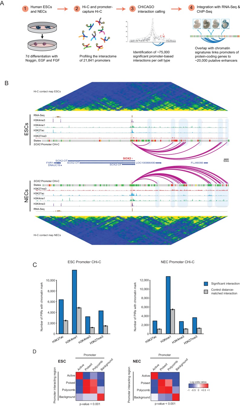
Long-range cis-regulatory elements such as enhancers coordinate cell-specific transcriptional programmes by engaging in DNA looping interactions with target promoters. Deciphering the interplay between the promoter connectivity and activity of cis-regulatory elements during lineage commitment is crucial for understanding developmental transcriptional control. Here, we use Promoter Capture Hi-C to generate a high-resolution atlas of chromosomal interactions involving ~22,000 gene promoters in human pluripotent and lineage-committed cells, identifying putative target genes for known and predicted enhancer elements. We reveal extensive dynamics of cis-regulatory contacts upon lineage commitment, including the acquisition and loss of promoter interactions. This spatial rewiring occurs preferentially with predicted changes in the activity of cis-regulatory elements and is associated with changes in target gene expression. Our results provide a global and integrated view of promoter interactome dynamics during lineage commitment of human pluripotent cells.
Overview
The claims and statistics of this paper haven’t been checked yet.
Run the check — free, no account needed. It reads the open-access text and checks the sources it cites.
Integrity signals
Registry
Cites
Statistics
Figures
Verification
Claims the paper makes
The claims and statistics of this paper haven’t been checked yet.
Run the check — free, no account needed. It reads the open-access text and checks the sources it cites.
Claims other papers make about this one
Each is checked against this paper’s own text: does it say what the citing paper says it does?
The source backs part of the claim, not all of it — or backs it for a different population, condition or comparison.
In the citing paper
“Notably, some contacts between PcG-associated regions, including poised promoters and enhancers in ESCs, are retained after these regions are activated during cell differentiation [141, 143].”
From Transcriptional enhancers and their communication with gene promoters (2021) · cites this paper as [141]
Experimental — accuracy not yet measuredThe source backs part of the claim, not all of it — or backs it for a different population, condition or comparison.
In the citing paper
“the chromatin state of the promoter at dual-state CRUs typically corresponded to the majority state of its enhancers, consistent with a competition of opposing regulatory effects at the promoters.”
From Transcriptional enhancers and their communication with gene promoters (2021) · cites this paper as [141]
Experimental — accuracy not yet measuredThe source backs part of the claim, not all of it — or backs it for a different population, condition or comparison.
In the citing paper
“Upon early neural differentiation, about a third of single-state CRUs in human ESCs became dual-state, while around half of all dual-state CRUs became single-state [141].”
From Transcriptional enhancers and their communication with gene promoters (2021) · cites this paper as [141]
Experimental — accuracy not yet measuredThe cited source says what the claim attributes to it.
In the citing paper
“Finally, some enhancers are marked with H3K27me3 alone and are considered to be “Polycomb-repressed” [139–141].”
From Transcriptional enhancers and their communication with gene promoters (2021) · cites this paper as [141]
The cited source says what the claim attributes to it.
In the citing paper
“In mammals, PcG-associated promoters and enhancers also engage in multitudes of looping contacts, particularly in early development [139, 141, 276, 357].”
From Transcriptional enhancers and their communication with gene promoters (2021) · cites this paper as [141]
The cited source says what the claim attributes to it.
In the citing paper
“In mammalian differentiation models, in contrast, extensive remodelling of enhancer–promoter contacts is typically observed consistently with enhancer activation [141, 394–397].”
From Transcriptional enhancers and their communication with gene promoters (2021) · cites this paper as [141]
The cited source says what the claim attributes to it.
In the citing paper
“Nonetheless, a significant minority of enhancers in human ESCs also show pre-existing “permissive” promoter contacts prior to activation [141, 394].”
From Transcriptional enhancers and their communication with gene promoters (2021) · cites this paper as [141]
The cited source says what the claim attributes to it.
In the citing paper
“Capture Hi-C analyses in multiple cell types consistently detect enhancer–promoter contacts that cross TAD boundaries [141, 243].”
From Transcriptional enhancers and their communication with gene promoters (2021) · cites this paper as [141]
The cited source says what the claim attributes to it.
In the citing paper
“with 20% of CRUs detected in these cells being “dual-state”, i.e. containing a mixture of active and poised or Polycomb-repressed enhancers [141].”
From Transcriptional enhancers and their communication with gene promoters (2021) · cites this paper as [141]
The cited source says what the claim attributes to it.
In the citing paper
“Changes in CRU state were predominantly driven by coordinated alterations in the chromatin state of enhancers and their promoter contacts, and were associated with significant changes in gene expression [141].”
From Transcriptional enhancers and their communication with gene promoters (2021) · cites this paper as [141]
The cited source says what the claim attributes to it.
In the citing paper
“Enhancers vastly outnumber promoters in human and mouse genomes [131], and consistently promoters often contact more than one enhancer in the same cell type [141, 243, 278, 279].”
From Transcriptional enhancers and their communication with gene promoters (2021) · cites this paper as [141]
The cited source says what the claim attributes to it.
In the citing paper
“Analyses in human ESCs and early ES-derived neural progenitors found that, as expected, enhancers within a CRU tended to have the same activity state in a given cell type [141].”
From Transcriptional enhancers and their communication with gene promoters (2021) · cites this paper as [141]
Self-check
Percentages, sums and reported differences re-computed from the paper’s own numbers.
The self-check needs the paper’s full text.
Citations
Cites
Moderated estimation of fold change and dispersion for RNA-seq data with DESeq2
Love, M. I., Huber, W., & Anders, S. (2014) · Genome Biology · doi:10.1186/s13059-014-0550-8 · Cited by 95,106 · cited 4×
“Genes differentially expressed between ESCs and NECs were identified using DESeq2 (FDR < 0.05 and a log2 fold change of >1.5).”
InfluentialAn integrated encyclopedia of DNA elements in the human genome
The ENCODE Project Consortium (2012) · Nature · doi:10.1038/nature11247 · Cited by 18,072
Heinz, S., Benner, C., Spann, N., Bertolino, E., Lin, Y. C., Laslo, P., Cheng, J. X., Murre, C., Singh, H., & Glass, C. K. (2010) · Molecular Cell · doi:10.1016/j.molcel.2010.05.004 · Cited by 13,382 · cited 1×
“7 (http://homer.salk.edu/homer/) (Heinz et al., 2010).”
TopHat: discovering splice junctions with RNA-Seq
Trapnell, C., Pachter, L. S., & Salzberg, S. L. (2009) · Bioinformatics · doi:10.1093/bioinformatics/btp120 · Cited by 11,682
The Human Genome Browser at UCSC
Kent, W. J., Sugnet, C. W., Furey, T. S., Roskin, K. M., Pringle, T. H., Zahler, A. M., & Haussler, A. D. (2002) · Genome Research · doi:10.1101/gr.229102 · Cited by 10,400 · cited 1×
“Data were converted to GRCh37 using liftOver (Kent et al., 2002).”
MethodComprehensive Mapping of Long-Range Interactions Reveals Folding Principles of the Human Genome
Lieberman-Aiden, E., van Berkum, N. L., Williams, L., Imakaev, M., Ragoczy, T., Telling, A., Amit, I., Lajoie, B. R., Sabo, P. J., Dorschner, M. O., et al. (2009) · Science · doi:10.1126/science.1181369 · Cited by 8,898 · cited 1×
“A/B compartments were called by computing the principal components of the distance- and coverage-corrected interaction profile correlation matrix at 250 kb resolution (Lieberman-Aiden et al., 2009).”
MethodTopological domains in mammalian genomes identified by analysis of chromatin interactions
Dixon, J. R., Selvaraj, S., Yue, F., Kim, A., Li, Y., Shen, Y., Hu, M., Liu, J. S., & Ren, B. (2012) · Nature · doi:10.1038/nature11082 · Cited by 6,829 · cited 2×
“TADs were identified based on directionality indices (Dixon et al., 2012) of Hi-C interactions 1 Mb upstream and downstream from a 25 kb sliding window every 5 kb steps, which were then smoothed using a running average over a ± 25 kb window.”
MethodIntegrative analysis of 111 reference human epigenomes
Roadmap Epigenomics Consortium, Kundaje, A., Meuleman, W., Ernst, J., Bilenky, M., Yen, A., Heravi-Moussavi, A., Kheradpour, P., Zhang, Z., Wang, J., et al. (2015) · Nature · doi:10.1038/nature14248 · Cited by 6,368 · cited 1×
“The definition of chromatin signatures has enabled the genome-wide identification of enhancer elements across multiple human cell types (ENCODE Project Consortium, 2012; Heintzman et al., 2007, 2009; Pennacchio et al., 2006; Rada-Iglesias et al., 2011; Kundaje et al., 2015).”
BackgroundHistone H3K27ac separates active from poised enhancers and predicts developmental state
Creyghton, M. P., Cheng, A. W., Welstead, G. G., Kooistra, T., Carey, B. W., Steine, E. J., Hanna, J., Lodato, M. A., Frampton, G. M., Sharp, P. A., et al. (2010) · Proceedings of the National Academy of Sciences · doi:10.1073/pnas.1016071107 · Cited by 4,302 · cited 1×
“…activity associated with poised and active enhancers and have been used to show that widespread changes in enhancer position and activity occur upon cell fate decisions such as the lineage commitment of pluripotent cells (Creyghton et al., 2010; Rada-Iglesias et al., 2011; Zentner et al., 2011).”
BackgroundHeintzman, N. D., Stuart, R. K., Hon, G., Fu, Y., Ching, C. W., Hawkins, R. D., Barrera, L. O., Van Calcar, S., Qu, C., Ching, K. A., et al. (2007) · Nature Genetics · doi:10.1038/ng1966 · Cited by 3,398 · cited 1×
“The definition of chromatin signatures has enabled the genome-wide identification of enhancer elements across multiple human cell types (ENCODE Project Consortium, 2012; Heintzman et al., 2007, 2009; Pennacchio et al., 2006; Rada-Iglesias et al., 2011; Kundaje et al., 2015).”
Background
Cited by
Sorted by citations
Long-range enhancer–promoter contacts in gene expression control
Schoenfelder, S., & Fraser, P. (2019) · Nature Reviews Genetics · doi:10.1038/s41576-019-0128-0 · Cited by 1,102
Self-citationThe role of 3D genome organization in development and cell differentiation
Zheng, H., & Xie, W. (2019) · Nature Reviews Molecular Cell Biology · doi:10.1038/s41580-019-0132-4 · Cited by 742
A compendium of promoter-centered long-range chromatin interactions in the human genome
Jung, I., Schmitt, A., Diao, Y., Lee, A. J., Liu, T., Yang, D., Tan, C., Eom, J., Chan, M., Chee, S., et al. (2019) · Nature Genetics · doi:10.1038/s41588-019-0494-8 · Cited by 362
The relationship between genome structure and function
Oudelaar, A. M., & Higgs, D. R. (2021) · Nature Reviews Genetics · doi:10.1038/s41576-020-00303-x · Cited by 304
Rubin, A. J., Barajas, B. C., Furlan-Magaril, M., Lopez-Pajares, V., Mumbach, M. R., Howard, I., Kim, D. S., Boxer, L. D., Cairns, J., Spivakov, M., et al. (2017) · Nature Genetics · doi:10.1038/ng.3935 · Cited by 293
Self-citationEvaluating Enhancer Function and Transcription
Field, A. R., & Adelman, K. (2020) · Annual Review of Biochemistry · doi:10.1146/annurev-biochem-011420-095916 · Cited by 227
“Consistent with this, studies using chromatin conformation capture (3C)-based methods have given a wide range of estimates, from 7% (22) to 46% (28), of enhancer elements that contact the closest gene.”
ResultChromatin three-dimensional interactions mediate genetic effects on gene expression
Delaneau, O., Zazhytska, M., Borel, C., Giannuzzi, G., Rey, G., Howald, C., Kumar, S., Ongen, H., Popadin, K. Y., Marbach, D., et al. (2019) · Science · doi:10.1126/science.aat8266 · Cited by 212
“This hypothesis is in line with recent models of trans interactions (28, 35), phase separation (36, 37), and gene activation (38).”
ResultThe Bivalent Genome: Characterization, Structure, and Regulation
Blanco, E., González-Ramírez, M., Alcaine-Colet, A., Aranda, S., & Croce, L. D. (2019) · Trends in Genetics · doi:10.1016/j.tig.2019.11.004 · Cited by 226
Dynamic 3D chromatin architecture contributes to enhancer specificity and limb morphogenesis
Kragesteen, B. K., Spielmann, M., Paliou, C., Heinrich, V., Schöpflin, R., Esposito, A., Annunziatella, C., Bianco, S., Chiariello, A. M., Jerković, I., et al. (2018) · Nature Genetics · doi:10.1038/s41588-018-0221-x · Cited by 190
Candidate silencer elements for the human and mouse genomes
Jayavelu, N. D., Jajodia, A., Mishra, A., & Hawkins, R. D. (2020) · Nature Communications · doi:10.1038/s41467-020-14853-5 · Cited by 193
Preformed chromatin topology assists transcriptional robustness of Shh during limb development
Paliou, C., Guckelberger, P., Schöpflin, R., Heinrich, V., Esposito, A., Chiariello, A. M., Bianco, S., Annunziatella, C., Helmuth, J., Haas, S., et al. (2019) · Proceedings of the National Academy of Sciences · doi:10.1073/pnas.1900672116 · Cited by 163
Thiecke, M. J., Wutz, G., Muhar, M., Tang, W., Bevan, S., Malysheva, V., Stocsits, R., Neumann, T., Zuber, J., Fraser, P., et al. (2020) · Cell Reports · doi:10.1016/j.celrep.2020.107929 · Cited by 177
Self-citationRinzema, N. J., Sofiadis, K., Tjalsma, S. J. D., Verstegen, M. J. A. M., Oz, Y., Valdes-Quezada, C., Felder, A.-K., Filipovska, T., van der Elst, S., de Andrade dos Ramos, Z., et al. (2022) · Nature Structural & Molecular Biology · doi:10.1038/s41594-022-00787-7 · Cited by 166
Promoter bivalency favors an open chromatin architecture in embryonic stem cells
Martín, G. M., Blanco, E., Ballaré, C., Sansó, M., Spill, Y. G., Hu, D., Aoi, Y., Dily, F. L., Shilatifard, A., Martı́-Renom, M. A., et al. (2018) · Nature Genetics · doi:10.1038/s41588-018-0218-5 · Cited by 151
Greenwald, W. W. Y., Li, H., Benaglio, P., Jakubosky, D. A., Matsui, H., Schmitt, A. D., Selvaraj, S., D’Antonio, M., D’Antonio‐Chronowska, A., Smith, E. N., et al. (2019) · Nature Communications · doi:10.1038/s41467-019-08940-5 · Cited by 144
A promoter interaction map for cardiovascular disease genetics
Montefiori, L. E., Sobreira, D. R., Sakabe, N. J., Aneas, I., Joslin, A. C., Hansen, G. T., Bozek, G., Moskowitz, I. P., McNally, E. M., & Nóbrega, M. A. (2018) · eLife · doi:10.7554/elife.35788 · Cited by 145
The three-dimensional genome: regulating gene expression during pluripotency and development
Andrey, G., & Mundlos, S. (2017) · Development · doi:10.1242/dev.148304 · Cited by 134
“…in a large-scale study of the promoter interactomes of PCs and neuroectodermal cells, around 50% of promoter-interacting regions were re-wired and 50% were retained between both cell types, arguing for extensive chromatin dynamics upon cell differentiation (Freire-Pritchett et al., 2017).”
BackgroundMethodSingle-cell measurement of higher-order 3D genome organization with scSPRITE
Arrastia, M. V., Jachowicz, J. W., Ollikainen, N., Curtis, M. S., Lai, C., Quinodoz, S. A., Selck, D. A., Ismagilov, R. F., & Guttman, M. (2021) · Nature Biotechnology · doi:10.1038/s41587-021-00998-1 · Cited by 127
G-quadruplex DNA structures in human stem cells and differentiation
Zyner, K. G., Simeone, A., Flynn, S. M., Doyle, C., Marsico, G., Adhikari, S., Portella, G., Tannahill, D., & Balasubramanian, S. (2022) · Nature Communications · doi:10.1038/s41467-021-27719-1 · Cited by 124
Dynamics of the 4D Genome during in Vivo Lineage Specification and Differentiation
Oudelaar, A. M., Beagrie, R. A., Gosden, M., de Ornellas, S., Georgiades, E., Kerry, J., Hidalgo, D., Carrelha, J., Shivalingam, A., El-Sagheer, A. H., et al. (2020) · Nature Communications · doi:10.1038/s41467-020-16598-7 · Cited by 119
Novo, C. L., Javierre, B.-M., Cairns, J., Segonds-Pichon, A., Wingett, S. W., Freire-Pritchett, P., Furlan-Magaril, M., Schoenfelder, S., Fraser, P., & Rugg-Gunn, P. J. (2018) · Cell Reports · doi:10.1016/j.celrep.2018.02.040 · Cited by 119
“In contrast, intra-TAD DNA loops are typically cell-type specific and are frequently rewired upon cell state changes (Smith et al., 2016; Denholtz et al., 2013; Schoenfelder et al., 2015b; Freire-Pritchett et al., 2017).”
BackgroundSelf-citationPromoter Capture Hi-C: High-resolution, Genome-wide Profiling of Promoter Interactions
Schoenfelder, S., Javierre, B. M., Furlan-Magaril, M., Wingett, S., & Fraser, P. (2018) · Journal of Visualized Experiments · doi:10.3791/57320 · Cited by 121
Self-citationMadsen, J. G. S., Madsen, M. S., Rauch, A., Traynor, S., Hauwaert, E. L. V., Haakonsson, A. K., Javierre, B. M., Hyldahl, M., Fraser, P., & Mandrup, S. (2020) · Nature Genetics · doi:10.1038/s41588-020-0709-z · Cited by 97
Self-citationNucleome Dynamics during Retinal Development
Norrie, J. L., Lupo, M. S., Xu, B., Aldiri, I., Valentine, M. B., Putnam, D. K., Griffiths, L. M., Zhang, J., Johnson, D. A., Easton, J. B., et al. (2019) · Neuron · doi:10.1016/j.neuron.2019.08.002 · Cited by 87
Increased enhancer–promoter interactions during developmental enhancer activation in mammals
Chen, Z., Snetkova, V., Bower, G., Jacinto, S. H., Clock, B., Dizehchi, A., Barozzi, I., Mannion, B. J., Alcaina‐Caro, A., López-Ríos, J., et al. (2024) · Nature Genetics · doi:10.1038/s41588-024-01681-2 · Cited by 98
Transcriptional enhancers and their communication with gene promoters
Ray-Jones, H., & Spivakov, M. (2021) · Cellular and Molecular Life Sciences · doi:10.1007/s00018-021-03903-w · Cited by 81
Self-citationHiCAR is a robust and sensitive method to analyze open-chromatin-associated genome organization
Wei, X., Yu, X., Peters, D., Marius, C., Sun, T., Shan, R., Ou, J., Lin, X., Yue, F., Li, W., et al. (2022) · Molecular Cell · doi:10.1016/j.molcel.2022.01.023 · Cited by 84
Three-dimensional genome architecture and emerging technologies: looping in disease
Mishra, A., & Hawkins, R. D. (2017) · Genome Medicine · doi:10.1186/s13073-017-0477-2 · Cited by 79
“Integrating 3D genomic data with genetic data and applying polymer modeling approaches with Hi-C catalogues may recapitulate architectural effects of disease variants [8, 43, 44] and can serve as reference 3D genome maps for clinically relevant samples—useful for generating testable hypotheses toward therapeutic oppor…”
BackgroundChoy, M., Javierre, B. M., Williams, S. G., Baross, S. L., Liu, Y., Wingett, S., Akbarov, A., Wallace, C., Freire-Pritchett, P., Rugg‐Gunn, P. J., et al. (2018) · Nature Communications · doi:10.1038/s41467-018-04931-0 · Cited by 70
Self-citationThe interdependence of gene-regulatory elements and the 3D genome
Vermunt, M. W., Zhang, D., & Blobel, G. A. (2018) · The Journal of Cell Biology · doi:10.1083/jcb.201809040 · Cited by 72
Interpreting the impact of noncoding structural variation in neurodevelopmental disorders
D’haene, E., & Vergult, S. (2020) · Genetics in Medicine · doi:10.1038/s41436-020-00974-1 · Cited by 63
Zhang, S., Bell, E., Zhi, H., Brown, S., Imran, S. A. M., Azuara, V., & Cui, W. (2019) · Stem Cell Research & Therapy · doi:10.1186/s13287-019-1228-7 · Cited by 59
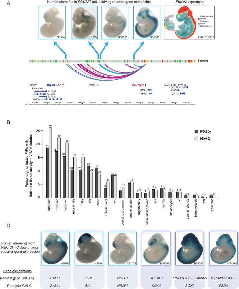
“moter Capture Hi-C analysis as an enhancer region of POU3F2, which exhibited regulation on reporter gene expression in the CNS of mouse embryos [48].”
BackgroundSchoenfelder, S., Mifsud, B., Senner, C. E., Todd, C. D., Chrysanthou, S., Darbo, E., Hemberger, M., & Branco, M. R. (2018) · Nature Communications · doi:10.1038/s41467-018-06666-4 · Cited by 54
Self-citationWatt, S., Vasquez, L., Walter, K., Mann, A. L., Kundu, K., Chen, L., Sims, Y., Ecker, S., Burden, F., Farrow, S., et al. (2021) · Nature Communications · doi:10.1038/s41467-021-22548-8 · Cited by 63
Self-citationChromatin architecture reorganization during neuronal cell differentiation in Drosophila genomePreprint
Chathoth, K. T., & Zabet, N. R. (2019) · Genome Research · doi:10.1101/gr.246710.118 · Cited by 56
“Similar observations have been 3D genome after differentiation in Drosophila Genome Research 621 www.genome.org reported in human cells (Bonev et al. 2017; Freire-Pritchett et al. 2017),whichsuggeststhatthereorganizationduringdevelopment is conserved during evolution.”
Three-dimensional organization and dynamics of the genome
Szałaj, P., & Plewczyński, D. (2018) · Cell Biology and Toxicology · doi:10.1007/s10565-018-9428-y · Cited by 59
Integrative single-cell RNA-seq and ATAC-seq analysis of myogenic differentiation in pig
Cai, S., Hu, B., Wang, X., Liu, T., Lin, Z., Tong, X., Xu, R., Chen, M., Duo, T., Zhu, Q., et al. (2023) · BMC Biology · doi:10.1186/s12915-023-01519-z · Cited by 60
“The cis-regulatory contact upon lineage commitment is a dynamic process that includes acquiring and losing specific promoter interactions [61].”
BackgroundRegulation of Chromatin Structure During Neural Development
Kishi, Y., & Gotoh, Y. (2018) · Frontiers in Neuroscience · doi:10.3389/fnins.2018.00874 · Cited by 51
Chovanec, P., Collier, A. J., Krueger, C., Várnai, C., Semprich, C. I., Schoenfelder, S., Corcoran, A. E., & Rugg‐Gunn, P. J. (2021) · Nature Communications · doi:10.1038/s41467-021-22201-4 · Cited by 48
Self-citationRaviram, R., Rocha, P. P., Luo, V. M., Swanzey, E., Miraldi, E. R., Chuong, E. B., Feschotte, C., Bonneau, R., & Skok, J. A. (2018) · Genome biology · doi:10.1186/s13059-018-1598-7 · Cited by 50
Thompson, J. J., Lee, D. J., Mitra, A., Frail, S., Dale, R., & Rocha, P. P. (2022) · Nature Communications · doi:10.1038/s41467-022-31938-5 · Cited by 48
Enhancers in the Peril lincRNA locus regulate distant but not local genes
Groff, A. F., Barutcu, A. R., Lewandowski, J. P., & Rinn, J. L. (2018) · Genome biology · doi:10.1186/s13059-018-1589-8 · Cited by 42
Pahl, M. C., Doege, C. A., Hodge, K. M., Littleton, S. H., Leonard, M. E., Lu, S., Rausch, R., Pippin, J. A., Rosa, M. C. D., Basak, A., et al. (2021) · Nature Communications · doi:10.1038/s41467-021-27001-4 · Cited by 46
Koohy, H., Bolland, D. J., Matheson, L. S., Schoenfelder, S., Stellato, C., Dimond, A., Várnai, C., Chovanec, P., Chessa, T. A. M., Denizot, J., et al. (2018) · Genome biology · doi:10.1186/s13059-018-1489-y · Cited by 41
“7e), in contrast to findings in other systems such as ES cell differentiation [34].”
ResultSelf-citationSu, C., Argenziano, M., Lu, S., Pippin, J. A., Pahl, M. C., Leonard, M. E., Cousminer, D. L., Johnson, M. E., Lasconi, C., Wells, A. D., et al. (2021) · Progress in Neurobiology · doi:10.1016/j.pneurobio.2021.102000 · Cited by 41
Enhancer–gene maps in the human and zebrafish genomes using evolutionary linkage conservation
Clément, Y., Torbey, P., Gilardi‐Hebenstreit, P., & Crollius, H. R. (2019) · Nucleic Acids Research · doi:10.1093/nar/gkz1199 · Cited by 43
Comoglio, F., Park, H. J., Schoenfelder, S., Barozzi, I., Bode, D., Fraser, P., & Green, A. R. (2018) · Genome Research · doi:10.1101/gr.227272.117 · Cited by 41
Self-citationCollier, A. J., & Rugg‐Gunn, P. J. (2018) · BioEssays · doi:10.1002/bies.201700239 · Cited by 40
Self-citationAnalysis of the structural variability of topologically associated domains as revealed by Hi-C
Sauerwald, N., Singhal, A., & Kingsford, C. (2019) · NAR Genomics and Bioinformatics · doi:10.1093/nargab/lqz008 · Cited by 38
“hESC (H9) Human embryonic stem cell 1 HindIII Dilution GSM2309023 (41) Adrenal gland Tissue 1 HindIII Dilution SRX2179246 (31) Bladder Tissue 2 HindIII Dilution SRX2179247, SRX2179248 (31) DPC Dorsolateral prefrontal cortex tissue 1 HindIII Dilution SRX2179249 (31)”
Balasubramanian, D., Pinto, P. B., Grasso, A., Vincent, S., Tarayre, H., Lajoignie, D., & Ghavi-Helm, Y. (2023) · Nucleic Acids Research · doi:10.1093/nar/gkad1183 · Cited by 45
“…nature of these interactions and the elements involved have not been studied in detail, promoter-interacting regions were found to be enriched in histone marks associated with active enhancers, active promoters, and transcribed regions, highly suggestive of non-coding regulatory elements ( 29 ).”
BackgroundInfluentialYun, H., Narayan, N., Vohra, S., Giotopoulos, G., Mupo, A., Madrigal, P., Sasca, D., Lara‐Astiaso, D., Horton, S. J., Singh, S. A., et al. (2021) · Nature Genetics · doi:10.1038/s41588-021-00925-9 · Cited by 38
“…11% of all high-confidence interactions were significantly differential (gained or lost) in SM or DM HSPC, based on stringent alterations of the ranking of CHiCAGO scores 29,30 (Figure 4g), with more interactions lost than newly formed in the mutant states (Figure 4h and Supplementary Table 8).”
Self-citationDetecting chromosomal interactions in Capture Hi-C data with CHiCAGO and companion tools
Freire-Pritchett, P., Ray-Jones, H., Rosa, M. D., Eijsbouts, C., Orchard, W. R., Wingett, S., Wallace, C., Cairns, J. M., Spivakov, M., & Malysheva, V. (2021) · Nature Protocols · doi:10.1038/s41596-021-00567-5 · Cited by 34
Self-citationChromatin modules and their implication in genomic organization and gene regulation
van Mierlo, G., Pushkarev, O., Kribelbauer, J. F., & Deplancke, B. (2022) · Trends in Genetics · doi:10.1016/j.tig.2022.11.003 · Cited by 35
Navigating the non-coding genome in heart development and Congenital Heart Disease
Chahal, G., Tyagi, S., & Ramialison, M. (2019) · Differentiation · doi:10.1016/j.diff.2019.05.001 · Cited by 31
Laverré, A., Tannier, É., & Necşulea, A. (2021) · Genome Research · doi:10.1101/gr.275901.121 · Cited by 30
A complex regulatory landscape involved in the development of mammalian external genitals
Amândio, A. R., Lopez‐Delisle, L., Bolt, C. C., Mascrez, B., & Duboule, D. (2020) · eLife · doi:10.7554/elife.52962 · Cited by 26
Wolfe, J. C., Mikheeva, L. A., Hagras, H., & Zabet, N. R. (2021) · Genome biology · doi:10.1186/s13059-021-02532-7 · Cited by 28
“To achieve regulation of distal target genes, enhancers must make 3D contacts with the promoters of genes that they control [9].”
3D genome organization links non-coding disease-associated variants to genes
Orozco, G., Schoenfelder, S., Walker, N. J., Eyre, S., & Fraser, P. (2022) · Frontiers in Cell and Developmental Biology · doi:10.3389/fcell.2022.995388 · Cited by 30
“Furthermore, although cell differentiation is often characterized by dynamic rewiring of promoter-enhancer loops (Freire-Pritchett et al., 2017; Siersbæk et al., 2017), some specific interactions do not contemporaneously drive gene expression.”
BackgroundSelf-citationBlank spots on the map: some current questions on nuclear organization and genome architecture
Adriaens, C., Serebryannyy, L., Feric, M., Schibler, A., Meaburn, K. J., Kubben, N., Trzaskoma, P., Shachar, S., Vidak, S., Finn, E. H., et al. (2018) · Histochemistry and Cell Biology · doi:10.1007/s00418-018-1726-1 · Cited by 30
Middelkamp, S., Vlaar, J. M., Giltay, J. C., Korzelius, J., Besselink, N. J. M., Boymans, S., Janssen, R., de la Fonteijne, L., van Binsbergen, E., van Roosmalen, M. J., et al. (2019) · Genome Medicine · doi:10.1186/s13073-019-0692-0 · Cited by 27
“The bottom track displays the PCHiC 795 interactions of the HOXD13 gene in neuroectodermal cells [42].”
BackgroundContributions of 3D chromatin structure to cell-type-specific gene regulation
Schaeffer, M., & Nöllmann, M. (2023) · Current Opinion in Genetics & Development · doi:10.1016/j.gde.2023.102032 · Cited by 26
FoxA1/2-dependent epigenomic reprogramming drives lineage switching in lung adenocarcinoma
Gillis, K., Orellana, W. A., Wilson, E., Parnell, T. J., Fort, G., Fang, P., Dadzie, H. E., Murphy, B., Zhang, X., & Snyder, E. L. (2024) · Developmental Cell · doi:10.1016/j.devcel.2024.10.009 · Cited by 23
Su, C., Pahl, M. C., Grant, S. F. A., & Wells, A. D. (2021) · Human Genetics · doi:10.1007/s00439-021-02326-8 · Cited by 24
Decoding the organization, dynamics, and function of the 4D genome
Aboelnour, E., & Bonev, B. (2021) · Developmental Cell · doi:10.1016/j.devcel.2021.04.023 · Cited by 24
Vijayabaskar, M. S., Goode, D. K., Obier, N., Lichtinger, M., Emmett, A., Abidin, F. N. Z., Shar, N. A., Hannah, R., Assi, S. A., Lie‐A‐Ling, M., et al. (2019) · PLoS Computational Biology · doi:10.1371/journal.pcbi.1007337 · Cited by 19
“This coCRE comprises of the promoter and a -17kB upstream region, which has been shown to be involved in Spi1 expression control [28].”
BackgroundWelzenbach, J., Hammond, N. L., Nikolić, M., Thieme, F., Ishorst, N., Leslie, E. J., Weinberg, S. M., Beaty, T. H., Marazita, M. L., Mangold, E., et al. (2021) · Human Genetics and Genomics Advances · doi:10.1016/j.xhgg.2021.100038 · Cited by 21
“, DNA-DNA interactions of enhancers and TSS) at specific risk loci, we accessed pCHi-C cis-interaction data collected in hESCs (GEO: GSE86821).(41)”
MethodVisualizing Structure and Transitions for Biological Data ExplorationPreprint
Moon, K. R., van Dijk, D., Wang, Z., Burkhardt, D. B., Chen, W. S., Yim, K., van den Elzen, A. M. G., Hirn, M., Coifman, R. R., Ivanova, N. B., et al. (2018) · SSRN Electronic Journal · doi:10.2139/ssrn.3155891 · Cited by 22
“To show that the PHATE embedding agrees with known biology, we compared the scRNA-seq data with bulk RNA-seq data available from the literature including datasets from ESCs [64]; ESC-derived neuroectodermal cells (NEC) [65]; ESC differentiating into neural progenitor cells (NPC) at days 0, 8, and 16 [66]; neural crest…”
MethodApplication of Hi-C and other omics data analysis in human cancer and cell differentiation research
Gong, H., Yang, Y., Zhang, S., Li, M., & Zhang, X. (2021) · Computational and Structural Biotechnology Journal · doi:10.1016/j.csbj.2021.04.016 · Cited by 20
Rosa, M. D., & Spivakov, M. (2020) · Nature Genetics · doi:10.1038/s41588-020-0583-8 · Cited by 19
Self-citationSignaling‐to‐chromatin pathways in the immune system
de Paz, A. M., & Josefowicz, S. Z. (2021) · Immunological Reviews · doi:10.1111/imr.12955 · Cited by 18
preciseTAD: a transfer learning framework for 3D domain boundary prediction at base-pair resolution
Stilianoudakis, S. C., Marshall, M. A., & Dozmorov, M. G. (2021) · Bioinformatics · doi:10.1093/bioinformatics/btab743 · Cited by 19
Enhancer-Promoter Communication: It’s Not Just About Contact
Wurmser, A., & Basu, S. (2022) · Frontiers in Molecular Biosciences · doi:10.3389/fmolb.2022.867303 · Cited by 19
Huang, H., Liu, J., Li, M., Guo, H., Zhu, J., Zhu, L., Wu, S., Mo, K., Huang, Y., Tan, J., et al. (2022) · Science Advances · doi:10.1126/sciadv.abo5668 · Cited by 17
“These chromatin interactions identified promoter interaction regions with active enhancer that are enriched for epigenomic features, such as H3K4me1 and H3K27ac, which was consistent with the results of several recent elegant studies, demonstrating that forced looping of enhancers to promoters induces gene activation…”
ResultGenetic coupling of enhancer activity and connectivity in gene expression control
Ray-Jones, H. F., Sung, C. K., Chan, L. T., Haglund, A., Artemov, P., Rosa, M. D., Ruje, L., Burden, F., Kreuzhuber, R., Litovskikh, A., et al. (2025) · Nature Communications · doi:10.1038/s41467-025-55900-3 · Cited by 17
Self-citationInterplay between regulatory elements and chromatin topology in cellular lineage determination
Shukla, V., Cetnarowska, A., Hyldahl, M., & Mandrup, S. (2022) · Trends in Genetics · doi:10.1016/j.tig.2022.05.011 · Cited by 15
Feldmann, A., Dimitrova, E., Kenney, A., Lastuvkova, A., & Klose, R. J. (2020) · Nucleic Acids Research · doi:10.1093/nar/gkaa064 · Cited by 15
“Firstly, they are not maintained upon differentiation-induced gene activation as is the case for other stimuli-induced interactions (46), developmental genes (25), or interactions described in other developmental model systems (2,45).”
BackgroundArrastia, M. V., Jachowicz, J. W., Ollikainen, N., Curtis, M. S., Lai, C., Quinodoz, S. A., Selck, D. A., Guttman, M., & Ismagilov, R. F. (2020) · bioRxiv (Cold Spring Harbor Laboratory) · doi:10.1101/2020.08.11.242081 · Cited by 15
González-Ramírez, M., Ballaré, C., Mugianesi, F., Beringer, M., Santanach, A., Blanco, E., & Croce, L. D. (2021) · PLoS Computational Biology · doi:10.1371/journal.pcbi.1009368 · Cited by 15
Molecular Basis of the Function of Transcriptional Enhancers
Ibragimov, A. N., Bylino, O. V., & Shidlovskii, Y. V. (2020) · Cells · doi:10.3390/cells9071620 · Cited by 15
Wei, X., Xu, Y.-Y., Yang, D.-C., Kim, K., Yi, L., Luo, W.-Z., Lin, X., Xiang, Y., Williams, A. B., Wang, X.-T., et al. (2026) · Nature Biotechnology · doi:10.1038/s41587-026-03013-7 · Cited by 16
Salviato, E., Djordjilović, V., Hariprakash, J. M., Tagliaferri, I., Pal, K., & Ferrari, F. (2021) · Nucleic Acids Research · doi:10.1093/nar/gkab547 · Cited by 13
Making connections: enhancers in cellular differentiation
Herrmann, J. C., Beagrie, R. A., & Hughes, J. R. (2021) · Trends in Genetics · doi:10.1016/j.tig.2021.10.008 · Cited by 14
Pavlaki, I., Shapiro, M., Pisignano, G., Jones, S. M. E., Telenius, J. M., Descalzo, S. M., Williams, R. J., Hughes, J. R., & Vance, K. W. (2022) · PLoS Genetics · doi:10.1371/journal.pgen.1010230 · Cited by 13
The Role of Chromatin Density in Cell Population Heterogeneity during Stem Cell Differentiation
Golkaram, M., Jang, J., Hellander, S., Kosik, K. S., & Petzold, L. R. (2017) · Scientific Reports · doi:10.1038/s41598-017-13731-3 · Cited by 14
Epigenetic priming of mammalian embryonic enhancer elements coordinates developmental gene networks
Todd, C. D., Ijaz, J., Torabi, F., Dovgusha, O., Bevan, S., Cracknell, O., Lohoff, T., Clark, S. J., Argelaguet, R., Pearce, J., et al. (2025) · Genome biology · doi:10.1186/s13059-025-03658-8 · Cited by 12
“…tissues we are investigating, we utilised a combination of genomic proximity models and Promoter Capture Hi-C data of mEpiLCs/hESCs [21, 24] to assign enhancers to promoters they likely interact with, as previous studies have shown that enhancers can interact with promoters prior to…”
Self-citationEpigenome rewiring in human pluripotent stem cells
Yan, J., & Huangfu, D. (2021) · Trends in Cell Biology · doi:10.1016/j.tcb.2021.12.001 · Cited by 11
Fernández-Valverde, S. L., Aguilera, F., & Ramos-Díaz, R. A. (2018) · Integrative and Comparative Biology · doi:10.1093/icb/icy061 · Cited by 12
Dynamic chromatin organization and regulatory interactions in human endothelial cell differentiation
Alavattam, K. G., Mitzelfelt, K. A., Bonora, G., Fields, P., Yang, X., Chiu, H. S., Pabon, L., Bertero, A., Palpant, N. J., Noble, W. S., et al. (2022) · Stem Cell Reports · doi:10.1016/j.stemcr.2022.11.003 · Cited by 10
“While work remains to understand how TADs strengthen and PPIs arise in endothelial cells, our findings indicate that these features are influenced by stable and dynamic compartments to regulate transcription necessary for differentiation: strengthened TAD boundaries are accompanied by increased numbers of intra-TAD co…”
Multi-layered transcriptional control of cranial neural crest development
Candido-Ferreira, I., Lukoseviciute, M., & Sauka‐Spengler, T. (2022) · Seminars in Cell and Developmental Biology · doi:10.1016/j.semcdb.2022.07.010 · Cited by 8
Comoglio, F., Park, H. J., Schoenfelder, S., Barozzi, I., Bode, D., Fraser, P., & Green, A. R. (2017) · bioRxiv (Cold Spring Harbor Laboratory) · doi:10.1101/163113 · Cited by 9
Self-citationGolov, A. K., Gavrilov, A. A., Kaplan, N., & Razin, S. V. (2024) · eLife · doi:10.7554/elife.91596.3 · Cited by 10
Rinzema, N. J., Sofiadis, K., Tjalsma, S. J. D., Verstegen, M. J. A. M., Oz, Y., Valdes-Quezada, C., Felder, A.-K., Filipovska, T., van der Elst, S., de Andrade dos Ramos, Z., et al. (2021) · doi:10.1101/2021.10.05.463209 · Cited by 8
Transient promoter interactions modulate developmental gene activation
Mahara, S., Prüssing, S., Smialkovska, V., Krall, S., Holliman, S., Blum, B., Dachtler, V., Borgers, H., Sollier, E., Plass, C., et al. (2024) · Molecular Cell · doi:10.1016/j.molcel.2024.10.005 · Cited by 9
Zibetti, C. (2022) · Cells · doi:10.3390/cells11050806 · Cited by 6
Regulatory de novo mutations underlying intellectual disability
Vas, M. G. D., Boulet, F., Joshi, S. S., Garstang, M. G., Khan, T. N., Atla, G., Parry, D., Moore, D., Cebola, I., Zhang, S., et al. (2023) · Life Science Alliance · doi:10.26508/lsa.202201843 · Cited by 6
“The significant interactions between promoters and enhancers identified using PCHi-C in neuronal progenitor cells (21) were used to assign target genes to the DNM-containing enhancers.”
Cellular reprogramming is driven by widespread rewiring of promoter-enhancer interactions
Wang, M., He, B., Hao, Y., Srinivasan, D., Shrinet, J., & Fraser, P. (2023) · BMC Biology · doi:10.1186/s12915-023-01766-0 · Cited by 5
“PCHi-C studies have shown dynamic rewiring of enhancer-promoter interactions during human ESC differentiation to neuroectodermal cells [22], during mouse adipocyte differentiation [7], and during keratinogenesis [23] suggesting that promoter interaction changes regulate gene expression associated…”
BackgroundSelf-citationAlgorithmic considerations when analysing capture Hi-C data
Disney–Hogg, L., Kinnersley, B., & Houlston, R. S. (2020) · Wellcome Open Research · doi:10.12688/wellcomeopenres.16394.1 · Cited by 6
Malysheva, V., Mendoza-Parra, M. A., Blum, M., Spivakov, M., & Gronemeyer, H. (2018) · bioRxiv (Cold Spring Harbor Laboratory) · doi:10.1101/303842 · Cited by 7
Self-citationGolov, A. K., Gavrilov, A. A., Kaplan, N., & Razin, S. V. (2023) · eLife · doi:10.7554/elife.91596 · Cited by 5
Epigenetic regulation of cis-regulatory elements and transcription factors during development
Khan, S., Mishra, R. K., & Srivastava, S. (2023) · Elsevier eBooks · doi:10.1016/b978-0-12-821785-6.00004-9 · Cited by 5
Metadata
- Authors
- Freire-Pritchett, P., Schoenfelder, S., Várnai, C., Wingett, S. W., Cairns, J., Collier, A. J., García-Vílchez, R., Furlan-Magaril, M., Osborne, C. S., Fraser, P., Rugg-Gunn, P. J., & Spivakov, M.
- Affiliations
- Babraham Institute (GB)
- King's College London (GB)
- Epigenetics Programme, Babraham Institute, Cambridge, United Kingdom
- Corresponding author
- Spivakov, Mikhail
- ORCID
- Venue
- eLife
- Publisher
- eLife Sciences Publications, Ltd
- Published
- 2017-03-23
- Volume
- 6
- ISSN
- 2050-084X (ISSN-L)
- PMC
- PMC5407860
- OpenAlex
- W2602710346
- Cited by
- 161
- References
- 88
- Citations by year
- 2026: 9 · 2025: 12 · 2024: 13 · 2023: 16 · 2022: 20 · 2021: 25 · 2020: 17 · 2019: 19 · 2018: 24 · 2017: 6
- FWCI
- 7.37
- Type
- journal article
- Language
- en
- Open access
- gold
- doi.org
- Licence
- Locations
- eLife (publishedVersion, cc-by)
- Europe PMC (PubMed Central) (publishedVersion, cc-by)
- PubMed Central (publishedVersion, cc-by)
- PubMed (publishedVersion)
- Research Portal (King's College London) (submittedVersion, cc-by)
- DOAJ (DOAJ: Directory of Open Access Journals) (submittedVersion, cc-by-sa)
- UCL Discovery (University College London) (submittedVersion)
- University of Birmingham Research Portal (University of Birmingham) (submittedVersion, cc-by)
- Funders
- Wellcome (WT093736)
- Biotechnology and Biological Sciences Research Council (BB/J004480/1)
- Medical Research Council (MR/J003808/1)
- Topics
- Life Sciences › Biochemistry, Genetics and Molecular Biology › Molecular Biology › Genomics and Chromatin Dynamics
- Life Sciences › Biochemistry, Genetics and Molecular Biology › Molecular Biology › Pluripotent Stem Cells Research
- Life Sciences › Biochemistry, Genetics and Molecular Biology › Molecular Biology › Epigenetics and DNA Methylation
- Keywords
- cis-regulatory elements, enhancers, lineage commitment, human embryonic stem cells, chromatin interactions, promoter capture Hi-C, enhancer target genes, pluripotency
- MeSH terms
- Human Embryonic Stem Cells, Cell Differentiation, Gene Expression Regulation, Humans, Promoter Regions, Genetic, Transcription, Genetic, Regulatory Elements, Transcriptional
About these checks
Registry, retraction, Cites and statistics results are computed, not judged, with one exception: a printed reference that no index lists is read once by a model before it counts as a citation error. Claims are extracted by a model and kept only when their quote appears verbatim in the text; each cited source is then checked, with a model’s help, for whether it says what the claim relies on. Those verdicts can be wrong — read the claim’s quote and the cited source before relying on one.
Statistics and arithmetic are recomputed, not judged.
Confidence bands are shown only for fields calibrated on our annotated test set. None is calibrated yet, so no band is shown.
What counts as a claim ›How we measure confidence ›How we check this ›
Are you an author, or do you see a problem with this page? Request a review
