

The DOI matches its Crossref record; 91 of the 91 references it cites were found in a registry or an index.
NPTX2 and cognitive dysfunction in Alzheimer’s Disease
Open accessAbstract
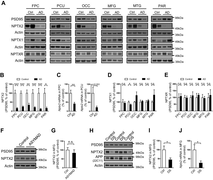
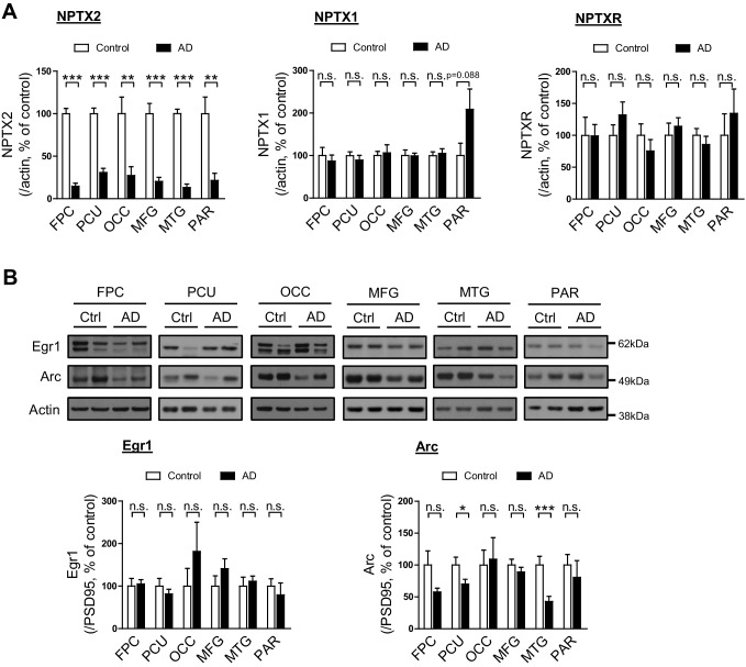
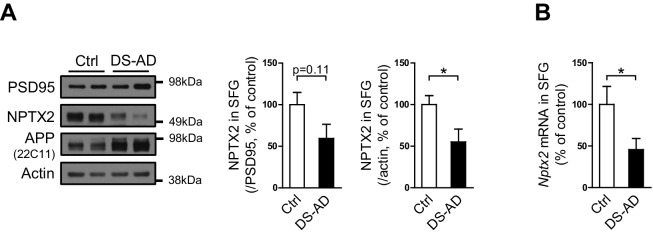
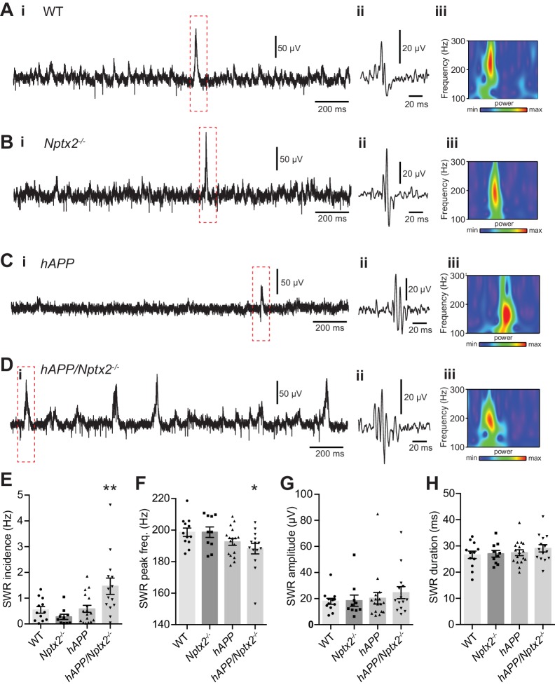
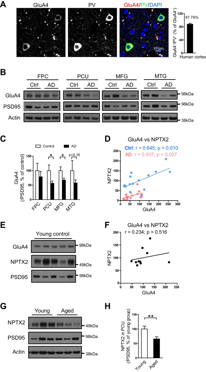
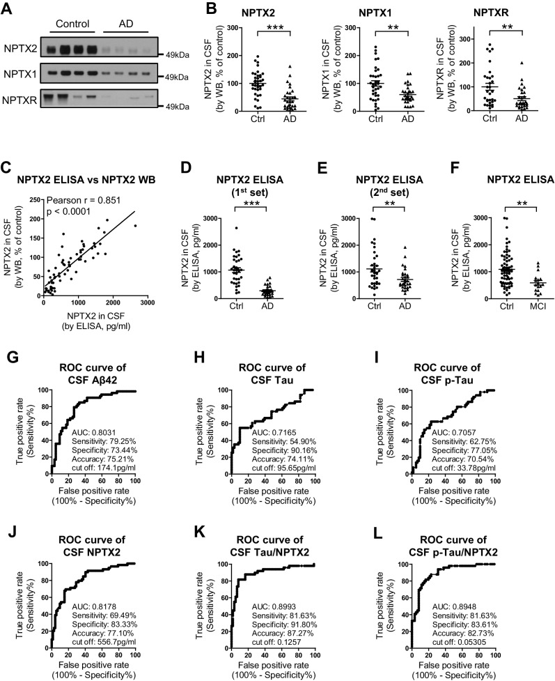
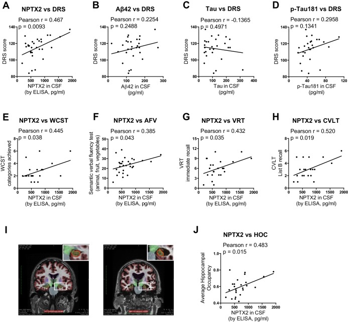
Memory loss in Alzheimer’s disease (AD) is attributed to pervasive weakening and loss of synapses. Here, we present findings supporting a special role for excitatory synapses connecting pyramidal neurons of the hippocampus and cortex with fast-spiking parvalbumin (PV) interneurons that control network excitability and rhythmicity. Excitatory synapses on PV interneurons are dependent on the AMPA receptor subunit GluA4, which is regulated by presynaptic expression of the synaptogenic immediate early gene NPTX2 by pyramidal neurons. In a mouse model of AD amyloidosis, Nptx2-/- results in reduced GluA4 expression, disrupted rhythmicity, and increased pyramidal neuron excitability. Postmortem human AD cortex shows profound reductions of NPTX2 and coordinate reductions of GluA4. NPTX2 in human CSF is reduced in subjects with AD and shows robust correlations with cognitive performance and hippocampal volume. These findings implicate failure of adaptive control of pyramidal neuron-PV circuits as a pathophysiological mechanism contributing to cognitive failure in AD.
Overview
The claims and statistics of this paper haven’t been checked yet.
Run the check — free, no account needed. It reads the open-access text and checks the sources it cites.
Integrity signals
Registry
Cites
Statistics
Figures
Verification
Claims the paper makes
The claims and statistics of this paper haven’t been checked yet.
Run the check — free, no account needed. It reads the open-access text and checks the sources it cites.
Claims other papers make about this one
Each is checked against this paper’s own text: does it say what the citing paper says it does?
The cited source says what the claim attributes to it.
In the citing paper
“Thus, they might be a good candidate biomarker for disease staging and cognitive prognosis (Galasko et al. 2019; Libiger et al. 2021; Nilsson et al. 2021; Swanson et al. 2016; Xiao et al. 2017).”
From Neuronal pentraxins as biomarkers of synaptic activity: from physiological functions to pathological changes in neurodegeneration (2022) · cites this paper as Xiao et al. 2017
In the citing paper
“NPTXs might be a selective marker for the imbalance of pyramidal neuron-interneuron microcircuits, thought to underlie cognitive impairment in AD (Xiao et al. 2017).”
From Neuronal pentraxins as biomarkers of synaptic activity: from physiological functions to pathological changes in neurodegeneration (2022) · cites this paper as Xiao et al. 2017
In the citing paper
“Deregulation of neuronal activity could explain changes in NPTXs and drive neurogenerative processes (Xiao et al. 2017).”
From Neuronal pentraxins as biomarkers of synaptic activity: from physiological functions to pathological changes in neurodegeneration (2022) · cites this paper as Xiao et al. 2017
In the citing paper
“The receiver operating characteristic (ROC) analysis concluded a similar test performance to distinguish AD from controls than that of the classical AD biomarkers (Xiao et al. 2017).”
From Neuronal pentraxins as biomarkers of synaptic activity: from physiological functions to pathological changes in neurodegeneration (2022) · cites this paper as Xiao et al. 2017
In the citing paper
“The subunit of AMPAR, GluA4, was also reduced in this study (both in the precuneus and medial frontal gyri) and correlated with NPTX2 in both control and AD group (Xiao et al. 2017).”
From Neuronal pentraxins as biomarkers of synaptic activity: from physiological functions to pathological changes in neurodegeneration (2022) · cites this paper as Xiao et al. 2017
In the citing paper
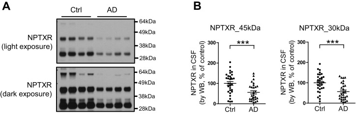
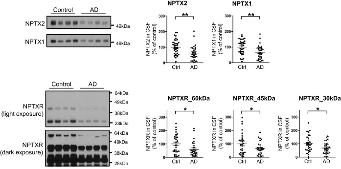
“CSF NPTXR levels, both the full length protein and the peptides resulting from TACE cleavage, were reduced in AD in comparison with controls (Xiao et al. 2017).”
From Neuronal pentraxins as biomarkers of synaptic activity: from physiological functions to pathological changes in neurodegeneration (2022) · cites this paper as Xiao et al. 2017
In the citing paper
“This was further sustained by studies that found NPTX2 correlating with medial temporal atrophy, hippocampal volume and cognitive impairment (Nilsson et al. 2021; Swanson et al. 2016; Xiao et al. 2017).”
From Neuronal pentraxins as biomarkers of synaptic activity: from physiological functions to pathological changes in neurodegeneration (2022) · cites this paper as Xiao et al. 2017
In the citing paper
“Consequently, the reduction of NPTX2 could be linked to the disruption of pyramidal neuron-PV interneuron microcircuit, contributing to cognitive failure in AD (Xiao et al. 2017).”
From Neuronal pentraxins as biomarkers of synaptic activity: from physiological functions to pathological changes in neurodegeneration (2022) · cites this paper as Xiao et al. 2017
In the citing paper
“NPTX2 was also decreased in post-mortem cortex from AD patients (hippocampus not analysed) (Xiao et al. 2017).”
From Neuronal pentraxins as biomarkers of synaptic activity: from physiological functions to pathological changes in neurodegeneration (2022) · cites this paper as Xiao et al. 2017
In the citing paper
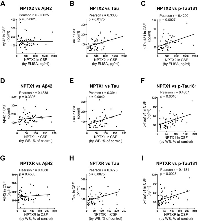
“NPTX2 exhibited a weak correlation with p-tau181 and total tau (Xiao et al. 2017).”
From Neuronal pentraxins as biomarkers of synaptic activity: from physiological functions to pathological changes in neurodegeneration (2022) · cites this paper as Xiao et al. 2017
In the citing paper
“NPTX1 and NPTXR did not correlate with Aβ42, but did weakly correlate with total tau and p-tau181 (Xiao et al. 2017).”
From Neuronal pentraxins as biomarkers of synaptic activity: from physiological functions to pathological changes in neurodegeneration (2022) · cites this paper as Xiao et al. 2017
In the citing paper
“NPTX1 and NPTXR did not correlate with Aβ42, but did weakly correlate with total tau and p-tau181 (Xiao et al. 2017).”
From Neuronal pentraxins as biomarkers of synaptic activity: from physiological functions to pathological changes in neurodegeneration (2022) · cites this paper as Xiao et al. 2017
In the citing paper
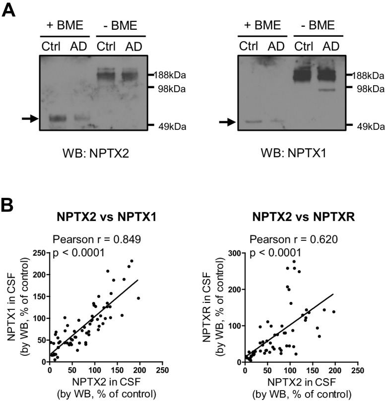
“Finally, CSF levels of the different NPTXs strongly correlated with each other in both AD and genetic FTLD, indicating an overall reduction of these proteins in CSF (Nilsson et al. 2021; van der Ende et al. 2020; Xiao et al. 2017).”
From Neuronal pentraxins as biomarkers of synaptic activity: from physiological functions to pathological changes in neurodegeneration (2022) · cites this paper as Xiao et al. 2017
In the citing paper
“Some authors described a weak correlation with Aβ42, mainly in MCI (Galasko et al. 2019; Nilsson et al. 2021), whereas others found no correlation (Xiao et al. 2017).”
From Neuronal pentraxins as biomarkers of synaptic activity: from physiological functions to pathological changes in neurodegeneration (2022) · cites this paper as Xiao et al. 2017
In the citing paper
“NPTXs may contribute to the cognitive decline in neurodegeneration and other diseases of the CNS by modifying the excitatory synaptic drive onto fast-spiking PV interneurons which leads to E/I imbalance.”
From Neuronal pentraxins as biomarkers of synaptic activity: from physiological functions to pathological changes in neurodegeneration (2022) · cites this paper as Xiao et al. 2017
In the citing paper
“This might promote or accelerate disease progression by increasing the demand in other compensatory mechanisms (Hu et al. 2010; Shepherd et al. 2006; Xiao et al. 2017).”
From Neuronal pentraxins as biomarkers of synaptic activity: from physiological functions to pathological changes in neurodegeneration (2022) · cites this paper as Xiao et al. 2017
In the citing paper
“NPTXs also showed an added role when combined with core AD biomarkers in predicting the progression from MCI to AD dementia and in discriminating AD and healthy controls, e.g., NPTX2 in ratio with either total tau or p-tau (Galasko et al. 2019; Xiao et al. 2017).”
From Neuronal pentraxins as biomarkers of synaptic activity: from physiological functions to pathological changes in neurodegeneration (2022) · cites this paper as Xiao et al. 2017
In the citing paper
“Unlike core biomarkers such as Aβ (Williams et al. 2011), CSF NPTXs levels correlated with cognitive scores in AD patients, even in the absence of brain pathology.”
From Neuronal pentraxins as biomarkers of synaptic activity: from physiological functions to pathological changes in neurodegeneration (2022) · cites this paper as Xiao et al. 2017
In the citing paper
“In the pre-symptomatic stage, NPTXs may act as a resilience factor keeping the integrity of the synapses, as previously hypothesised (Xiao et al. 2017).”
From Neuronal pentraxins as biomarkers of synaptic activity: from physiological functions to pathological changes in neurodegeneration (2022) · cites this paper as Xiao et al. 2017
Self-check
Percentages, sums and reported differences re-computed from the paper’s own numbers.
The self-check needs the paper’s full text.
Citations
Cites
Folstein, M. F., Folstein, S. E., & McHugh, P. R. (1975) · Journal of Psychiatric Research · doi:10.1016/0022-3956(75)90026-6 · Cited by 88,282
Hirono, N., Mori, E., Ikejiri, Y., Imamura, T., Shimomura, T., Hashimoto, M., Yamashita, H., & Ikeda, M. (1997) · PubMed · Cited by 21,012
Kok, J. S., Berg, I. J., & Scherder, E. J. A. (2013) · Dementia and Geriatric Cognitive Disorders Extra · doi:10.1159/000353441 · Cited by 20,589
Older Adults' Reasons for Using Technology while Aging in Place
Peek, S. T. M., Luijkx, K. G., Rijnaard, M. D., Nieboer, M. E., van der Voort, C. S., Aarts, S., van Hoof, J., Vrijhoef, H. J., & Wouters, E. (2015) · Gerontology · doi:10.1159/000430949 · Cited by 19,852
Sensitivity and False Alarm Rate of a Fall Sensor in Long-Term Fall Detection in the Elderly
Kangas, M., Korpelainen, R., Vikman, I., Nyberg, L., & Jämsä, T. J. (2014) · Gerontology · doi:10.1159/000362720 · Cited by 19,007
The Brain's Default Network: Anatomy, Function, and Relevance to Disease
Buckner, R. L., Andrews‐Hanna, J. R., & Schacter, D. L. (2008) · Annals of the New York Academy of Sciences · doi:10.1196/annals.1440.011 · Cited by 9,761
Basic principles of ROC analysis
Metz, C. E. (1978) · Seminars in Nuclear Medicine · doi:10.1016/s0001-2998(78)80014-2 · Cited by 6,122
Hypothetical model of dynamic biomarkers of the Alzheimer's pathological cascade
Jack, C. R., Knopman, D. S., Jagust, W. J., Shaw, L. M., Aisen, P. S., Weiner, M. W., Petersen, R. C., & Trojanowski, J. Q. (2010) · The Lancet Neurology · doi:10.1016/s1474-4422(09)70299-6 · Cited by 4,806
Kaplan, E., Goodglass, H., Weıntraub, S., & Segal, O. (2001) · Medical Entomology and Zoology · Cited by 4,378
The Alzheimer’s disease neuroimaging initiative (ADNI): MRI methods
Jack, C. R., Bernstein, M. A., Fox, N. C., Thompson, P., Alexander, G., Harvey, D., Borowski, B., Britson, P. J., L. Whitwell, J., Ward, C., et al. (2008) · Journal of Magnetic Resonance Imaging · doi:10.1002/jmri.21049 · Cited by 4,347
Cited by
Sorted by citations
Antonarakis, S. E., Skotko, B. G., Rafii, M. S., Strydom, A., Pape, S. E., Bianchi, D. W., Sherman, S. L., & Reeves, R. H. (2020) · Nature Reviews Disease Primers · doi:10.1038/s41572-019-0143-7 · Cited by 964
Self-citationLópez, J. A., González, H. M., & Léger, G. C. (2019) · Handbook of clinical neurology · doi:10.1016/b978-0-12-804766-8.00013-3 · Cited by 887
Deep Multilayer Brain Proteomics Identifies Molecular Networks in Alzheimer’s Disease Progression
Bai, B., Wang, X., Li, Y., Chen, P.-C., Yu, K., Dey, K. K., Yarbro, J. M., Han, X., Lutz, B. M., Rao, S., et al. (2020) · Neuron · doi:10.1016/j.neuron.2019.12.015 · Cited by 565
“(F) Consistent protein changes of 8 proteins in AD brain tissue and CSF.”
BackgroundInfluentialSynaptic degeneration in Alzheimer disease
Tzioras, M., McGeachan, R. I., Durrant, C. S., & Spires-Jones, T. L. (2023) · Nature Reviews Neurology · doi:10.1038/s41582-022-00749-z · Cited by 504
Mathys, H., Peng, Z., Boix, C. A., Victor, M. B., Leary, N., Babu, S., Abdelhady, G., Jiang, X., Ng, A. P., Ghafari, K., et al. (2023) · Cell · doi:10.1016/j.cell.2023.08.039 · Cited by 495
Higginbotham, L., Ping, L., Dammer, E. B., Duong, D. M., Zhou, M., Gearing, M., Hurst, C., Glass, J. D., Factor, S. A., Johnson, E. C. B., et al. (2020) · Science Advances · doi:10.1126/sciadv.aaz9360 · Cited by 367
Higginbotham, L., Ping, L., Dammer, E. B., Duong, D. M., Zhou, M., Gearing, M., Johnson, E. C. B., Hajjar, I., Lah, J. J., Levey, A. I., et al. (2019) · doi:10.1101/806752 · Cited by 367
Proteomic landscape of Alzheimer’s Disease: novel insights into pathogenesis and biomarker discovery
Bai, B., Vanderwall, D., Li, Y., Wang, X., Poudel, S., Wang, H., Dey, K. K., Chen, P., Yang, K., & Peng, J. (2021) · Molecular Neurodegeneration · doi:10.1186/s13024-021-00474-z · Cited by 267
Optogenetic gamma stimulation rescues memory impairments in an Alzheimer’s disease mouse model
Etter, G., van der Veldt, S., Manseau, F., Zarrinkoub, I., Trillaud-Doppia, É., & Williams, S. (2019) · Nature Communications · doi:10.1038/s41467-019-13260-9 · Cited by 248
Goetzl, E. J., Abner, E. L., Jicha, G. A., Kapogiannis, D., & Schwartz, J. B. (2018) · The FASEB Journal · doi:10.1096/fj.201700731r · Cited by 219
Turk, B. R., Theda, C., Fatemi, A. S., & Moser, A. B. (2020) · International Journal of Developmental Neuroscience · doi:10.1002/jdn.10003 · Cited by 212
Farhy-Tselnicker, I., van Casteren, A. C. M., Lee, A., Chang, V. T., Aricescu, A. R., & Allen, N. J. (2017) · Neuron · doi:10.1016/j.neuron.2017.09.053 · Cited by 199
Fluid Biomarkers for Synaptic Dysfunction and Loss
Camporesi, E., Nilsson, J., Brinkmalm, A., Becker, B., Ashton, N. J., Blennow, K., & Zetterberg, H. (2020) · Biomarker Insights · doi:10.1177/1177271920950319 · Cited by 165
Shaping Synapses by the Neural Extracellular Matrix
Ferrer-Ferrer, M., & Dityatev, A. (2018) · Frontiers in Neuroanatomy · doi:10.3389/fnana.2018.00040 · Cited by 169
Immediate and deferred epigenomic signatures of in vivo neuronal activation in mouse hippocampus
Fernandez-Albert, J., Lipinski, M., Lopez-Cascales, M. T., Rowley, M. J., Martin-Gonzalez, A. M., del Blanco, B., Corces, V. G., & Barco, A. (2019) · Nature Neuroscience · doi:10.1038/s41593-019-0476-2 · Cited by 166
Pros and Cons of Clinically Relevant Methods to Assess Pain in Rodents
Tappe‐Theodor, A., King, T. E., & Morgan, M. M. (2019) · Neuroscience & Biobehavioral Reviews · doi:10.1016/j.neubiorev.2019.03.009 · Cited by 165
Targeting synapse function and loss for treatment of neurodegenerative diseases
Dejanovic, B., Sheng, M., & Hanson, J. E. (2023) · Nature Reviews Drug Discovery · doi:10.1038/s41573-023-00823-1 · Cited by 177
Stimulus-dependent representational drift in primary visual cortex
Marks, T. D., & Goard, M. J. (2021) · Nature Communications · doi:10.1038/s41467-021-25436-3 · Cited by 185
The visual word form area (VWFA) is part of both language and attention circuitry
Chen, L., Wassermann, D., Abrams, D. A., Kochalka, J., Gallardo, G., & Menon, V. (2019) · Nature Communications · doi:10.1038/s41467-019-13634-z · Cited by 165
Zhou, J., Wade, S. D., Graykowski, D., Xiao, M.-F., Zhao, B., Giannini, L. A. A., Hanson, J. E., van Swieten, J. C., Sheng, M., Worley, P. F., et al. (2023) · Science Translational Medicine · doi:10.1126/scitranslmed.adf0141 · Cited by 152
Self-citationConnolly, K., Lehoux, M., O’Rourke, R., Assetta, B., Erdemir, G. A., Elias, J. A., Lee, C. G., & Huang, Y. A. (2022) · Alzheimer s & Dementia · doi:10.1002/alz.12612 · Cited by 149
Fast-spiking parvalbumin-positive interneurons in brain physiology and Alzheimer’s disease
Hijazi, S., Smit, A. B., & van Kesteren, R. E. (2023) · Molecular Psychiatry · doi:10.1038/s41380-023-02168-y · Cited by 157
Galasko, D., Xiao, M., Xu, D., Smirnov, D., Salmon, D. P., Dewit, N., Vanbrabant, J., Jacobs, D., Vanderstichele, H., Vanmechelen, E., et al. (2019) · Alzheimer's & Dementia: Translational Research & Clinical Interventions · doi:10.1016/j.trci.2019.11.002 · Cited by 133
Self-citationLongitudinal CSF proteomics identifies NPTX2 as a prognostic biomarker of Alzheimer's disease
Libiger, O., Shaw, L. M., Watson, M. H., Nairn, A. C., Umaña, K. L., Biarnes, M. C., Canet‐Avilés, R. M., Jack, C. R., Breton, Y., Cortes, L., et al. (2021) · Alzheimer's & Dementia · doi:10.1002/alz.12353 · Cited by 108
Sathe, G., Na, C. H., Renuse, S., Madugundu, A. K., Albert, M., Moghekar, A., & Pandey, A. (2019) · PROTEOMICS – Clinical Applications · doi:10.1002/prca.201800105 · Cited by 126
Input–output organization of the mouse claustrum
Zingg, B., Dong, H., Tao, H. W., & Zhang, L. I. (2018) · The Journal of Comparative Neurology · doi:10.1002/cne.24502 · Cited by 129
The functional organization of cortical feedback inputs to primary visual cortex
Marques, T., Nguyen, J. K., Fioreze, G. T., & Petreanu, L. (2018) · Nature Neuroscience · doi:10.1038/s41593-018-0135-z · Cited by 122
Mozafari, M., Ganjtabesh, ., Nowzari-Dalini, A., & Masquelier, T. (2019) · Frontiers in Neuroscience · doi:10.3389/fnins.2019.00625 · Cited by 125
Sub-second Dopamine and Serotonin Signaling in Human Striatum during Perceptual Decision-Making
Bang, D., Kishida, K. T., Lohrenz, T. M., White, J. P., Laxton, A. W., Tatter, S. B., Fleming, S. M., & Montague, P. R. (2020) · Neuron · doi:10.1016/j.neuron.2020.09.015 · Cited by 130
Excitatory-inhibitory imbalance in Alzheimer's disease and therapeutic significance
Varela, E. V., Etter, G., & Williams, S. (2019) · Neurobiology of Disease · doi:10.1016/j.nbd.2019.04.010 · Cited by 122
Mossink, B., Negwer, M., Schubert, D., & Nadif Kasri, N. (2021) · Cellular and Molecular Life Sciences · doi:10.1007/s00018-020-03714-5 · Cited by 114
Gómez de San José, N., Massa, F., Halbgebauer, S., Oeckl, P., Steinacker, P., & Otto, M. (2022) · Journal of Neural Transmission · doi:10.1007/s00702-021-02411-2 · Cited by 112
An update on fluid biomarkers for neurodegenerative diseases: recent success and challenges ahead
Simrén, J., Ashton, N. J., Blennow, K., & Zetterberg, H. (2019) · Current Opinion in Neurobiology · doi:10.1016/j.conb.2019.11.019 · Cited by 105
ATN profiles among cognitively normal individuals and longitudinal cognitive outcomes
Soldan, A., Pettigrew, C. A., Fagan, A. M., Schindler, S. E., Moghekar, A., Fowler, C. J., Li, Q.-X., Collins, S. J., Carlsson, C. M., Asthana, S., et al. (2019) · Neurology · doi:10.1212/wnl.0000000000007248 · Cited by 107
N-myristoylation: from cell biology to translational medicine
Meng, Y. G., Song, Z., Ying, M., Zhu, H., He, Q., Yang, B., & Cao, J. (2020) · Acta Pharmacologica Sinica · doi:10.1038/s41401-020-0388-4 · Cited by 101
Krishnaswamy, V. R., Benbenishty, A., Blinder, P., & Sagi, I. (2019) · Cellular and Molecular Life Sciences · doi:10.1007/s00018-019-03182-6 · Cited by 96
Neuronal pentraxin 2: a synapse-derived CSF biomarker in genetic frontotemporal dementia
van der Ende, E. L., Xiao, M., Xu, D., Poos, J. M., Panman, J. L., Jiskoot, L. C., Meeter, L. H., Dopper, E. G., Papma, J. M., Heller, C., et al. (2020) · Journal of Neurology, Neurosurgery & Psychiatry · doi:10.1136/jnnp-2019-322493 · Cited by 94
Self-citationCurrent advances in plasma and cerebrospinal fluid biomarkers in Alzheimer's disease
Leuzy, A., Cullen, N. C., Mattsson, N., & Hansson, O. (2021) · Current Opinion in Neurology · doi:10.1097/wco.0000000000000904 · Cited by 91
Delling, J. P., & Boeckers, T. M. (2021) · Journal of Neurodevelopmental Disorders · doi:10.1186/s11689-021-09397-8 · Cited by 104
Chen, J., Zhao, X., Cui, L., He, G., Wang, X., Wang, F., Duan, S., He, L., Li, Q., Yu, X., et al. (2020) · Aging · doi:10.18632/aging.103150 · Cited by 91
Caccavano, A., Bozzelli, P. L., Forcelli, P. A., Pak, D. T. S., Wu, J., Conant, K. E., & Vicini, S. (2020) · Journal of Neuroscience · doi:10.1523/jneurosci.0425-20.2020 · Cited by 94
Vermeiren, S., Bellefroid, E., & Desiderio, S. (2020) · Frontiers in Cell and Developmental Biology · doi:10.3389/fcell.2020.587699 · Cited by 82
Fermie, J., Liv, N., ten Brink, C., van Donselaar, E. G., Müller, W. H., Schieber, N. L., Schwab, Y., Gerritsen, H. C., & Klumperman, J. (2018) · Traffic · doi:10.1111/tra.12557 · Cited by 84
Panchal, V. V., Griffiths, C., Mosaei, H., Bilyk, B. L., Sutton, J. A. F., Carnell, O. T., HORNBY, D. P., Green, J. D., Hobbs, J. K., Kelley, W. L., et al. (2020) · PLoS Pathogens · doi:10.1371/journal.ppat.1008672 · Cited by 85
van Steenoven, I., Koel-Simmelink, M. J. A., Vergouw, L. J. M., Tijms, B. M., Piersma, S. R., Pham, T. V., Bridel, C., Ferri, G.-L., Cocco, C., Noli, B., et al. (2020) · Molecular Neurodegeneration · doi:10.1186/s13024-020-00388-2 · Cited by 83
Self-citationMitofusins: Disease Gatekeepers and Hubs in Mitochondrial Quality Control by E3 Ligases
Escobar‐Henriques, M., & Joaquim, M. (2019) · Frontiers in Physiology · doi:10.3389/fphys.2019.00517 · Cited by 85
Cerebrospinal fluid biomarker panel for synaptic dysfunction in Alzheimer's disease
Nilsson, J., Gobom, J., Sjödin, S., Brinkmalm, G., Ashton, N. J., Svensson, J., Johansson, P., Portelius, E., Zetterberg, H., Blennow, K., et al. (2021) · Alzheimer's & Dementia: Diagnosis, Assessment & Disease Monitoring · doi:10.1002/dad2.12179 · Cited by 82
Nilsson, J., Binette, A. P., Palmqvist, S., Brum, W. S., Janelidze, S., Ashton, N. J., Spotorno, N., Stomrud, E., Gobom, J., Zetterberg, H., et al. (2024) · Brain · doi:10.1093/brain/awae032 · Cited by 90
Latest advances in cerebrospinal fluid and blood biomarkers of Alzheimer’s disease
Milà‐Alomà, M., Suárez‐Calvet, M., & Molinuevo, J. L. (2019) · Therapeutic Advances in Neurological Disorders · doi:10.1177/1756286419888819 · Cited by 81
Arroyo‐García, L. E., Isla, A. G., Andrade‐Talavera, Y., Balleza‐Tapia, H., Loera‐Valencia, R., Alvarez-Jimenez, L., Pizzirusso, G., Tambaro, S., Nilsson, P., & Fisahn, A. (2021) · Molecular Psychiatry · doi:10.1038/s41380-021-01257-0 · Cited by 75
Aryal, S., Bonanno, K., Song, B. J., Mani, D. R., Keshishian, H., Carr, S. A., Sheng, M., & Dejanovic, B. (2023) · Cell Reports · doi:10.1016/j.celrep.2023.112497 · Cited by 77
14-3-3 modulation of the inflammatory response
Munier, C. C., Ottmann, C., & Perry, M. W. D. (2020) · Pharmacological Research · doi:10.1016/j.phrs.2020.105236 · Cited by 79
Hippocampal network oscillations at the interplay between innate anxiety and learned fear
Çalışkan, G., & Stork, O. (2019) · Psychopharmacology · doi:10.1007/s00213-018-5109-z · Cited by 72
Neuroinflammation in Alzheimer’s disease: Pleiotropic roles for cytokines and neuronal pentraxins
Swanson, A., Wolf, T., Sitzmann, A., & Willette, A. A. (2018) · Behavioural Brain Research · doi:10.1016/j.bbr.2018.02.015 · Cited by 72
Kluever, V., Russo, B., Mandad, S., Kumar, N. H., Alevra, M., Ori, A., Rizzoli, S. O., Urlaub, H., Schneider, A., & Fornasiero, E. F. (2022) · Science Advances · doi:10.1126/sciadv.abn4437 · Cited by 73
Burghardt, L. T. (2019) · New Phytologist · doi:10.1111/nph.16045 · Cited by 70
Eat, sleep, repeat – endocrine regulation of behavioural circadian rhythms
Koop, S., & Oster, H. (2021) · FEBS Journal · doi:10.1111/febs.16109 · Cited by 75
Novel CSF biomarkers in genetic frontotemporal dementia identified by proteomics
van der Ende, E. L., Meeter, L. H., Stingl, C., van Rooij, J. G. J., Stoop, M. P., Nijholt, D. A. T., Sanchez‐Valle, R., Graff, C., Öijerstedt, L., Grossman, M., et al. (2019) · Annals of Clinical and Translational Neurology · doi:10.1002/acn3.745 · Cited by 65
Modeste, E. S., Ping, L., Watson, C. M., Duong, D. M., Dammer, E. B., Johnson, E. C. B., Roberts, B. R., Lah, J. J., Levey, A. I., & Seyfried, N. T. (2023) · Molecular Neurodegeneration · doi:10.1186/s13024-023-00638-z · Cited by 66
Quantitative proteomic analysis of the frontal cortex in Alzheimer’s disease
Sathe, G. J., Albert, M., Darrow, J. A., Saito, A., Troncoso, J. C., Pandey, A., & Moghekar, A. (2020) · Journal of Neurochemistry · doi:10.1111/jnc.15116 · Cited by 65
Self-citationHaque, R., Watson, C. M., Liu, J., Carter, E. K., Duong, D. M., Lah, J. J., Wingo, A. P., Roberts, B. R., Johnson, E. C. B., Saykin, A. J., et al. (2023) · Science Translational Medicine · doi:10.1126/scitranslmed.adg4122 · Cited by 64
Gyengési, E., Rangel, A., Ullah, F., Liang, H., Niedermayer, G., Asgarov, R., Venigalla, M., Gunawardena, D., Karl, T., & Münch, G. W. (2019) · Frontiers in Neuroscience · doi:10.3389/fnins.2019.00303 · Cited by 64
HIV-1 Tat-Induced Astrocytic Extracellular Vesicle miR-7 Impairs Synaptic Architecture
Hu, G., Niu, F., Liao, K., Periyasamy, P., Sil, S., Liu, J., Dravid, S. M., & Buch, S. J. (2019) · Journal of Neuroimmune Pharmacology · doi:10.1007/s11481-019-09869-8 · Cited by 64
Defining the Teratoma as a Model for Multi-lineage Human Development
McDonald, D., Wu, Y., Dailamy, A., Tat, J., Parekh, U., Zhao, D., Hu, M. Y., Tipps, A. M. P., Zhang, K., & Mali, P. (2020) · Cell · doi:10.1016/j.cell.2020.10.018 · Cited by 60
Ca2+-Triggered Synaptic Vesicle Fusion Initiated by Release of Inhibition
Brünger, A. T., Leitz, J., Zhou, Q., Choi, U. B., & Lai, Y. (2018) · Trends in Cell Biology · doi:10.1016/j.tcb.2018.03.004 · Cited by 61
Xu, C., Tian, G.-A., Jiang, C., Xue, H., Kuerbanjiang, M., Sun, L., Gu, L., Zhou, H., Liu, Y., Zhang, Z., et al. (2019) · Cell Death and Disease · doi:10.1038/s41419-019-1467-7 · Cited by 58
Fluid biomarkers in Alzheimer's disease
Simrén, J., Elmgren, A., Blennow, K., & Zetterberg, H. (2023) · Advances in Clinical Chemistry · doi:10.1016/bs.acc.2022.09.006 · Cited by 58
Singer, R. A., Arnes, L., Cui, Y., Wang, J., Gao, Y., Guney, M. A., Burnum-Johnson, K., Rabadán, R., Ansong, C. K., Orr, G., et al. (2019) · Cell Metabolism · doi:10.1016/j.cmet.2019.09.013 · Cited by 58
A data-driven disease progression model of fluid biomarkers in genetic frontotemporal dementia
van der Ende, E. L., Bron, E. E., Poos, J. M., Jiskoot, L. C., Panman, J. L., Papma, J. M., Meeter, L. H. H., Dopper, E. G. P., Wilke, C., Synofzik, M., et al. (2021) · Brain · doi:10.1093/brain/awab382 · Cited by 61
Harada, Y., Yamada, M., Imayoshi, I., Kageyama, R., Suzuki, Y., Kuniya, T., Furutachi, S., Kawaguchi, D., & Gotoh, Y. (2021) · Nature Communications · doi:10.1038/s41467-021-26605-0 · Cited by 56
Nilsson, J., Constantinescu, J., Nellgård, B., Jakobsson, P., Brum, W. S., Gobom, J., Forsgren, L., Dalla, K., Constantinescu, R., Zetterberg, H., et al. (2022) · Movement Disorders · doi:10.1002/mds.29287 · Cited by 59
A model of human neural networks reveals NPTX2 pathology in ALS and FTLD
Hruška-Plocháň, M., Wiersma, V. I., Betz, K. M., Mallona, I., Ronchi, S., Maniecka, Z., Hock, E., Tantardini, E., Laferrière, F., Sahadevan, S., et al. (2024) · Nature · doi:10.1038/s41586-024-07042-7 · Cited by 73
Invasive Insects: Management Methods Explored
McLaughlin, G. M., & Dearden, P. K. (2019) · Journal of Insect Science · doi:10.1093/jisesa/iez085 · Cited by 58
Structural basis for the tryptophan sensitivity of TnaC-mediated ribosome stalling
van der Stel, A., Gordon, E. R., Sengupta, A., Martínez, A. K., Klepacki, D., Perry, T. N., del Valle, A. H., Vázquez‐Laslop, N., Sachs, M. S., Cruz-Vera, L. R., et al. (2021) · Nature Communications · doi:10.1038/s41467-021-25663-8 · Cited by 55
Precision Aging: Applying Precision Medicine to the Field of Cognitive Aging
Ryan, L., Hay, M., Huentelman, M. J., Duarte, A., Rundek, T., Levin, B. E., Soldan, A., Pettigrew, C. A., Mehl, M. R., & Barnes, C. A. (2019) · Frontiers in Aging Neuroscience · doi:10.3389/fnagi.2019.00128 · Cited by 55
Moreno‐Rodríguez, M., Perez, S. E., Nadeem, M., Malek‐Ahmadi, M. H., & Mufson, E. J. (2020) · Journal of Neuroinflammation · doi:10.1186/s12974-020-1723-x · Cited by 53
Lang, Y., Li, Y., Yu, H., Lin, L., Chen, X., Wang, S., & Zhang, H. (2020) · Aging · doi:10.18632/aging.103028 · Cited by 50
Soldan, A., Oh, S., Ryu, T., Pettigrew, C. A., Zhu, Y., Moghekar, A., Xiao, M., Pontone, G. M., Albert, M., Na, C. H., et al. (2023) · Annals of Neurology · doi:10.1002/ana.26725 · Cited by 49
Self-citationXie, H., Heier, C., Kien, B., Vesely, P. W., Tang, Z., Sexl, V., Schoiswohl, G., Strießnig-Bina, I., Höefler, G., Zechner, R., et al. (2020) · Biochimica et Biophysica Acta (BBA) - Molecular and Cell Biology of Lipids · doi:10.1016/j.bbalip.2020.158737 · Cited by 48
The Role of Neuronal Pentraxin 2 (NP2) in Regulating Glutamatergic Signaling and Neuropathology
Chapman, G., Shanmugalingam, U., & Smith, P. D. (2020) · Frontiers in Cellular Neuroscience · doi:10.3389/fncel.2019.00575 · Cited by 48
Ma, Q., Zhu, C., Morselli, M., Su, T., Pelligrini, M., Lu, Z., Jones, M. R., Denver, P., Castro, D., Gu, X., et al. (2020) · Frontiers in Immunology · doi:10.3389/fimmu.2020.558036 · Cited by 48
A biomarker-authenticated model of schizophrenia implicating NPTX2 loss of function
Xiao, M.-F., Roh, S., Zhou, J., Chien, D. C.-C., Lucey, B. P., Craig, M. T., Hayes, L. N., Coughlin, J. M., Leweke, F. M., Jia, M., et al. (2021) · Science Advances · doi:10.1126/sciadv.abf6935 · Cited by 47
Self-citationPrecision Medicine and cardiac channelopathies: when dreams meet reality
Gnecchi, M., Sala, L., & Schwartz, P. J. (2021) · European Heart Journal · doi:10.1093/eurheartj/ehab007 · Cited by 45
Spatiotemporal transcriptomic maps of mouse intracerebral hemorrhage at single-cell resolution
Xiang, R., Wang, J., Chen, Z., Tao, J. Z., Peng, Q., Ding, R., Zhou, T., Tu, Z., Yang, T., Yang, T., et al. (2025) · Neuron · doi:10.1016/j.neuron.2025.04.026 · Cited by 51
LEAFY activity is post‐transcriptionally regulated by BLADE ON PETIOLE 2 and CULLIN 3 in Arabidopsis
Chahtane, H., Zhang, B., Norberg, M., Masson, M. L., Thévenon, E., Bakó, L., Benlloch, R., Holmlund, M., Parcy, F., Nilsson, O. R., et al. (2018) · New Phytologist · doi:10.1111/nph.15329 · Cited by 49
Soldan, A., Moghekar, A., Walker, K. A., Pettigrew, C. A., Hou, X., Lu, H., Miller, M. I., Alfini, A. J., Albert, M., Xu, D., et al. (2019) · Frontiers in Aging Neuroscience · doi:10.3389/fnagi.2019.00132 · Cited by 45
Self-citationCysteine oxidation and disulfide formation in the ribosomal exit tunnel
Schulte, L., Mao, J., Reitz, J., Sreeramulu, S., Kudlinzki, D., Hodirnau, V.-V., Meier‐Credo, J., Saxena, K., Buhr, F., Langer, J. D., et al. (2020) · Nature Communications · doi:10.1038/s41467-020-19372-x · Cited by 47
Katsuki, F., Gerashchenko, D., & Brown, R. E. (2022) · Brain Research Bulletin · doi:10.1016/j.brainresbull.2022.07.002 · Cited by 50
Data-Driven vs Consensus Diagnosis of MCI
Edmonds, E. C., Smirnov, D. S., Thomas, K. R., Graves, L. V., Bangen, K. J., Delano‐Wood, L., Galasko, D. R., Salmon, D. P., & Bondi, M. W. (2021) · Neurology · doi:10.1212/wnl.0000000000012600 · Cited by 45
Self-citationAPOE 𝜀4‐related blood–brain barrier breakdown is associated with microstructural abnormalities
Reas, E. T., Solders, S. K., Tsiknia, A. A., Triebswetter, C., Shen, Q., Rivera, C. S., Andrews, M., Alderson‐Myers, A., & Brewer, J. B. (2024) · Alzheimer s & Dementia · doi:10.1002/alz.14302 · Cited by 45
Self-citationKovács, R. Á., Vadászi, H., Bulyáki, É., Török, G., Tóth, V., Mátyás, D., Kun, J., Hunyadi-Gulyás, É., Fedor, F. Z., Csincsi, Á., et al. (2021) · Frontiers in Immunology · doi:10.3389/fimmu.2020.599771 · Cited by 43
Higginbotham, L. A., Carter, E. K., Dammer, E. B., Haque, R. U., Johnson, E. C. B., Duong, D. M., Yin, L., Jager, P. L. D., Bennett, D. A., Felsky, D., et al. (2023) · Neurobiology of Disease · doi:10.1016/j.nbd.2023.106286 · Cited by 41
Guanosine monophosphate reductase 1 is a potential therapeutic target for Alzheimer’s disease
Liu, H., Luo, K., & Luo, D. (2018) · Scientific Reports · doi:10.1038/s41598-018-21256-6 · Cited by 42
Boiten, W., van Steenoven, I., Xiao, M.-F., Worley, P., Noli, B., Cocco, C., Ferri, G.-L., Lemstra, A., & Teunissen, C. (2020) · Cells · doi:10.3390/cells10010038 · Cited by 42
Self-citationDendritic processing of spontaneous neuronal sequences for single-trial learning
Haga, T., & Fukai, T. (2018) · Scientific Reports · doi:10.1038/s41598-018-33513-9 · Cited by 43
Tang, C., Zhang, D., Yang, J., Liu, Q.-H., Wang, Y., & Wang, W. (2019) · Cell Death and Disease · doi:10.1038/s41419-019-1522-4 · Cited by 44
Welcome, M. O. (2020) · PharmaNutrition · doi:10.1016/j.phanu.2020.100176 · Cited by 41
Belbin, O., Xiao, M.-F., Xu, D., Carmona-Iragui, M., Pegueroles, J., Benejam, B., Videla, L., Fernández, S., Barroeta, I., Nuñez-Llaves, R., et al. (2020) · Molecular Neurodegeneration · doi:10.1186/s13024-020-00398-0 · Cited by 39
Self-citationLista, S., Santos‐Lozano, A., Emanuele, E., Mercuri, N. B., Gabelle, A., López‐Ortiz, S., Martín-Hernández, J., Maisto, N., Imbimbo, C., Caraci, F., et al. (2024) · Molecular Psychiatry · doi:10.1038/s41380-023-02376-6 · Cited by 44
Structure of Myosin VI/Tom1 complex reveals a cargo recognition mode of Myosin VI for tethering
Hu, S., Guo, Y., Wang, Y., Li, Y., Fu, T., Zhou, Z., Wang, Y., Liu, J., & Pan, L. (2019) · Nature Communications · doi:10.1038/s41467-019-11481-6 · Cited by 40
Metadata
- Authors
- Xiao, M.-F., Xu, D., Craig, M. T., Pelkey, K. A., Chien, C.-C., Shi, Y., Zhang, J., Resnick, S., Pletnikova, O., Salmon, D., Brewer, J., Edland, S., Wegiel, J., Tycko, B., Savonenko, A., Reeves, R. H., Troncoso, J. C., McBain, C. J., Galasko, D., & Worley, P. F.
- Affiliations
- Johns Hopkins University (US)
- Eunice Kennedy Shriver National Institute of Child Health and Human Development (US)
- National Institute on Aging (US)
- Department of Pathology, Johns Hopkins University School of Medicine, Baltimore, United States
- University of California San Diego (US)
- Institute for Basic Research, New York City, United States
- Columbia University (US)
- Corresponding author
- Worley, Paul F
- ORCID
- Venue
- eLife
- Publisher
- eLife Sciences Publications, Ltd
- Published
- 2017-03-23
- Volume
- 6
- ISSN
- 2050-084X (ISSN-L)
- PMC
- PMC5404919
- OpenAlex
- W2606988847
- Cited by
- 316
- References
- 91
- Citations by year
- 2026: 26 · 2025: 35 · 2024: 43 · 2023: 28 · 2022: 27 · 2021: 49 · 2020: 49 · 2019: 37 · 2018: 18 · 2017: 4
- FWCI
- 14.45
- Type
- journal article
- Language
- en
- Open access
- gold
- doi.org
- Licence
- Locations
- eLife (publishedVersion, cc-by)
- PubMed Central (publishedVersion, cc-by)
- PubMed (publishedVersion)
- ENLIGHTEN (Jurnal Bimbingan dan Konseling Islam) (acceptedVersion)
- DOAJ (DOAJ: Directory of Open Access Journals) (submittedVersion, cc-by-sa)
- eScholarship (California Digital Library) (submittedVersion)
- eScholarship (California Digital Library) (submittedVersion, cc-by)
- Figshare (submittedVersion, cc-by)
- Open Research Exeter (University of Exeter) (submittedVersion, cc-by)
- Funders
- LuMind Research Down Syndrome Foundation
- National Institute on Aging (AG05131)
- National Institute of Mental Health (P50 AG005146-27, MH100024, R35 NS-097966)
- Alzheimer’s drug discovery foundation (20021106)
- Topics
- Life Sciences › Neuroscience › Cellular and Molecular Neuroscience › Neuroscience and Neuropharmacology Research
- Life Sciences › Neuroscience › Cellular and Molecular Neuroscience › Alzheimer's disease research and treatments
- Health Sciences › Medicine › Psychiatry and Mental health › Dementia and Cognitive Impairment Research
- Keywords
- NPTX2, Alzheimer's disease, cognitive impairment, GluA4, AMPA receptors, parvalbumin interneurons, fast-spiking interneurons, pyramidal neurons, hippocampus, cortical excitability, neural oscillations, amyloidosis, memory loss, cerebrospinal fluid biomarkers, hippocampal volume, synaptogenesis
- MeSH terms
- Pentraxins, Alzheimer Disease, Animals, C-Reactive Protein, Cerebral Cortex, Disease Models, Animal, Hippocampus, Humans, Nerve Tissue Proteins, Mice, Knockout, Mice, Cognitive Dysfunction
About these checks
Registry, retraction, Cites and statistics results are computed, not judged, with one exception: a printed reference that no index lists is read once by a model before it counts as a citation error. Claims are extracted by a model and kept only when their quote appears verbatim in the text; each cited source is then checked, with a model’s help, for whether it says what the claim relies on. Those verdicts can be wrong — read the claim’s quote and the cited source before relying on one.
Statistics and arithmetic are recomputed, not judged.
Confidence bands are shown only for fields calibrated on our annotated test set. None is calibrated yet, so no band is shown.
What counts as a claim ›How we measure confidence ›How we check this ›
Are you an author, or do you see a problem with this page? Request a review
