

The DOI matches its Crossref record; 85 of the 85 references it cites were found in a registry or an index.
Environmental cystine drives glutamine anaplerosis and sensitizes cancer cells to glutaminase inhibition
Open accessPublished version of a preprint
Abstract
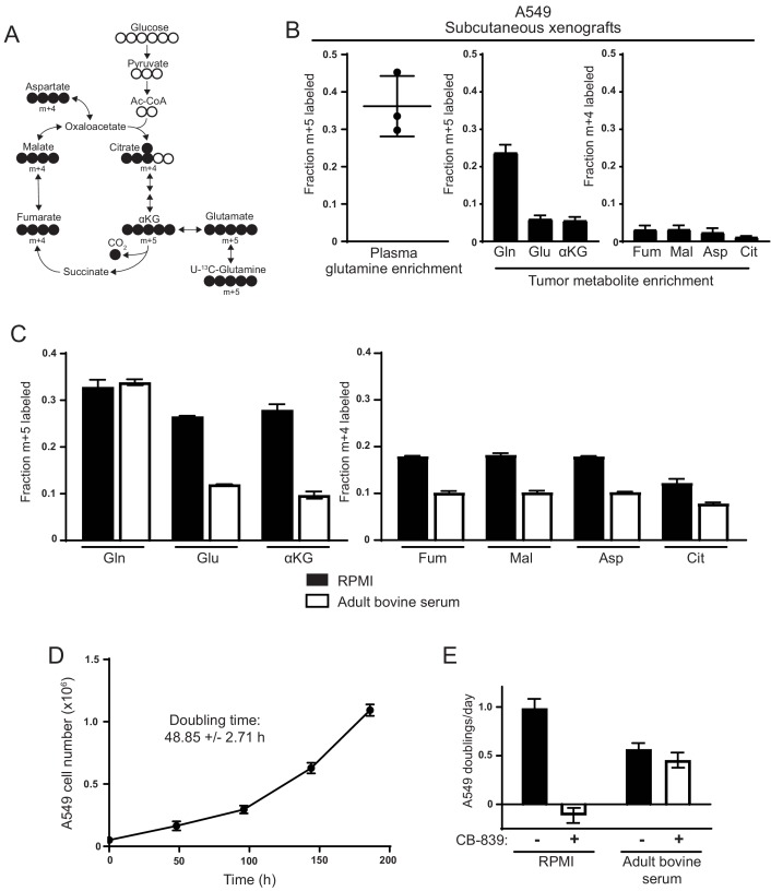
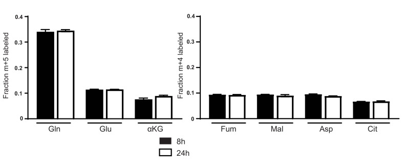
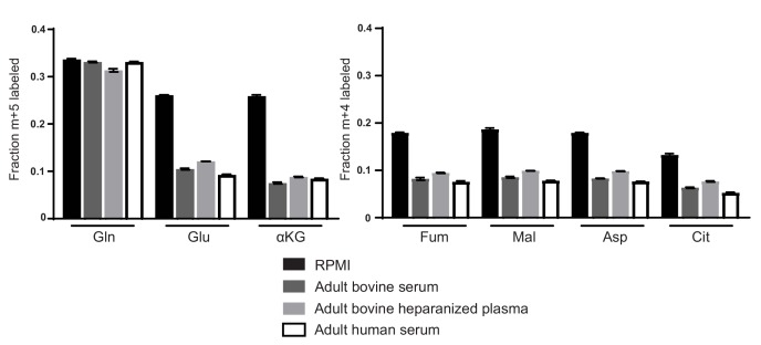
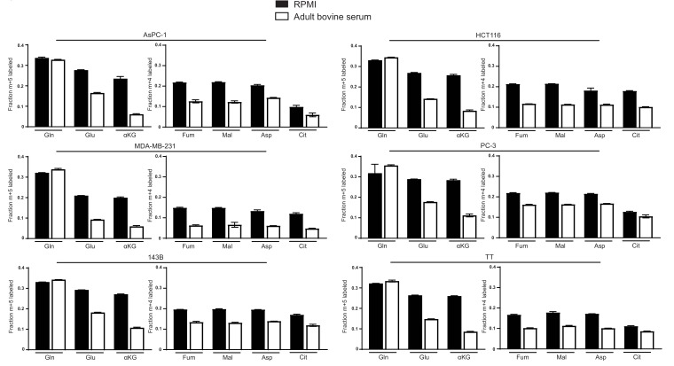
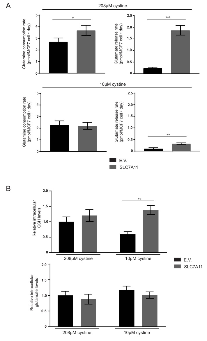
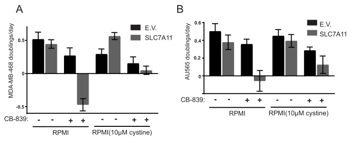
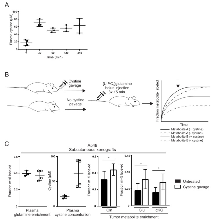
Many mammalian cancer cell lines depend on glutamine as a major tri-carboxylic acid (TCA) cycle anaplerotic substrate to support proliferation. However, some cell lines that depend on glutamine anaplerosis in culture rely less on glutamine catabolism to proliferate in vivo. We sought to understand the environmental differences that cause differential dependence on glutamine for anaplerosis. We find that cells cultured in adult bovine serum, which better reflects nutrients available to cells in vivo, exhibit decreased glutamine catabolism and reduced reliance on glutamine anaplerosis compared to cells cultured in standard tissue culture conditions. We find that levels of a single nutrient, cystine, accounts for the differential dependence on glutamine in these different environmental contexts. Further, we show that cystine levels dictate glutamine dependence via the cystine/glutamate antiporter xCT/SLC7A11. Thus, xCT/SLC7A11 expression, in conjunction with environmental cystine, is necessary and sufficient to increase glutamine catabolism, defining important determinants of glutamine anaplerosis and glutaminase dependence in cancer.
Overview
The claims and statistics of this paper haven’t been checked yet.
Run the check — free, no account needed. It reads the open-access text and checks the sources it cites.
Integrity signals
Registry
Cites
Statistics
Figures
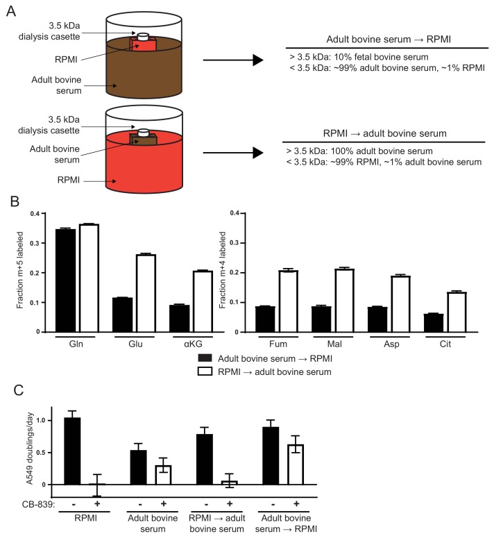
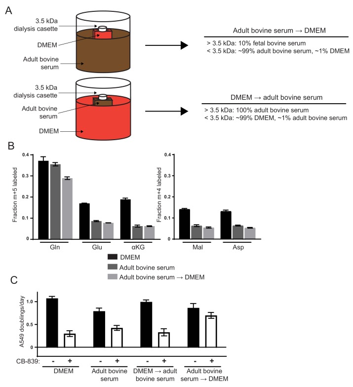
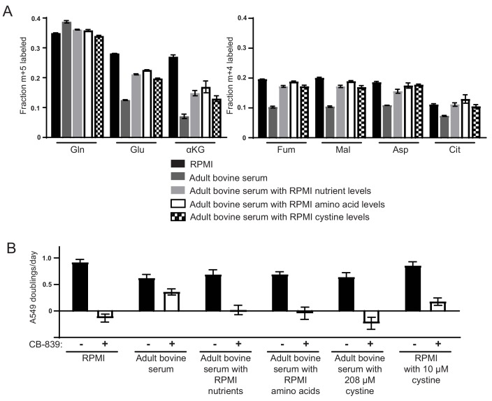
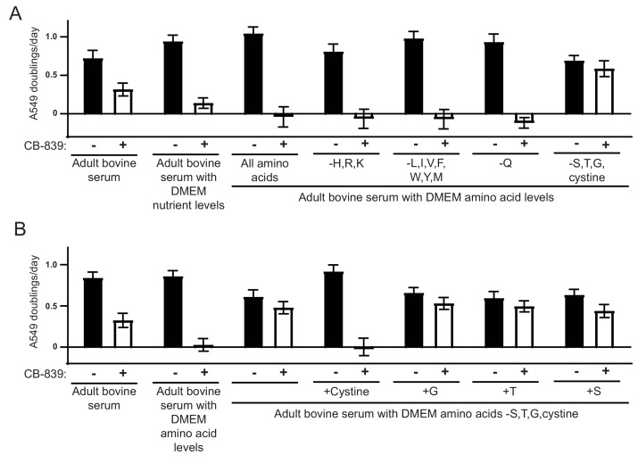
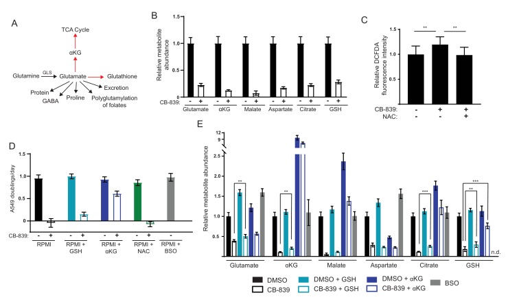
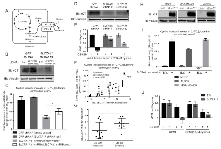
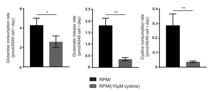
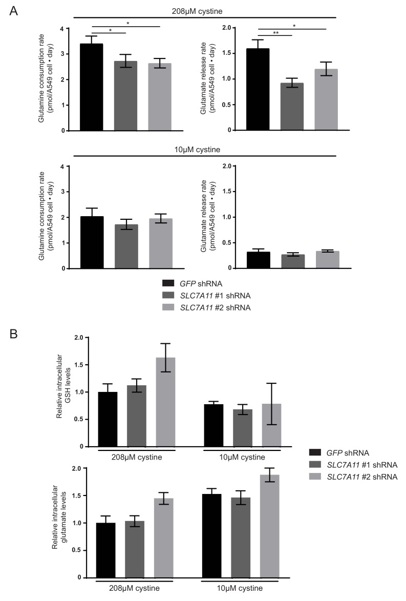
Verification
Claims the paper makes
The claims and statistics of this paper haven’t been checked yet.
Run the check — free, no account needed. It reads the open-access text and checks the sources it cites.
Claims other papers make about this one
Each is checked against this paper’s own text: does it say what the citing paper says it does?
In the citing paper
“implying that xc− may constitute a therapeutic target in this setting [553, 554].”
From Molecular mechanisms of cell death: recommendations of the nomenclature committee on cell death 2018. (2018) · cites this paper as [554]
In the citing paper
“the pronounced addiction of triple-negative breast carcinoma to glutamine relates (at least in part) to its ability to drive cystine uptake via xc−, implying that xc− may constitute a therapeutic target in this setting [553, 554].”
From Molecular mechanisms of cell death: recommendations of the nomenclature committee on cell death 2018. (2018) · cites this paper as [554]
In the citing paper
“However, environmental factors may also come into play, as for instance, the availability of extracellular as discussed below, can influence the dependence of cancer cells on glutamine metabolism73.”
From The metabolic landscape of RAS-driven cancers from biology to therapy (2021) · cites this paper as [73]
In the citing paper
“As shown in Fig. 3, glutamine is a direct fuel for the use of glutathione as a source of glutamate and is indirectly responsible for cystine uptake via the xCT antiporter system, which takes up cystine and simultaneously secretes glutamate61.”
From Glutamine reliance in cell metabolism (2020) · cites this paper as [61]
In the citing paper
“However, sensitivity to GLS inhibition in vitro is not always consistent in vivo, and is dependent on extracellular cystine levels.40”
From The multifaceted contributions of mitochondria to cellular metabolism (2018) · cites this paper as [40]
Self-check
Percentages, sums and reported differences re-computed from the paper’s own numbers.
The self-check needs the paper’s full text.
Citations
Cites
1 retracted
Li, J., Csibi, A., Yang, S., Hoffman, G. R., Li, C., Zhang, E. Y., Yu, J. J., & Blenis, J. (2014) · Proceedings of the National Academy of Sciences · doi:10.1073/pnas.1417015112 · Cited by 55
RetractedHallmarks of Cancer: The Next Generation
Hanahan, D., & Weinberg, R. A. (2011) · Cell · doi:10.1016/j.cell.2011.02.013 · Cited by 66,590
The Cancer Cell Line Encyclopedia enables predictive modelling of anticancer drug sensitivity
Barretina, J., Caponigro, G., Stransky, N., Venkatesan, K., Margolin, A. A., Kim, S., Wilson, C. J., Lehár, J., Kryukov, G. V., Sonkin, D., et al. (2012) · Nature · doi:10.1038/nature11003 · Cited by 7,950
Genome-scale CRISPR-Cas9 knockout screening in human cells
Shalem, O., Sanjana, N. E., Hartenian, E., Shi, X., Scott, D. A., Mikkelsen, T. S., Heckl, D., Ebert, B. L., Root, D. E., Doench, J. G., et al. (2014) · Science · doi:10.1126/science.1247005 · Cited by 5,234
The metabolism of tumors in the body
Warburg, O., Wind, F., & Negelein, E. (1927) · Journal of General Physiology · doi:10.1085/jgp.8.6.519 · Cited by 4,416
Fundamentals of Cancer Metabolism
DeBerardinis, R. J., & Chandel, N. S. (2016) · Science Advances · doi:10.1126/sciadv.1600200 · Cited by 3,013
DeBerardinis, R. J., Mancuso, A., Daikhin, E., Nissim, I., Yudkoff, M., Wehrli, S., & Thompson, C. B. (2007) · Proceedings of the National Academy of Sciences · doi:10.1073/pnas.0709747104 · Cited by 2,622
Understanding the Intersections between Metabolism and Cancer Biology
Heiden, M. G. V., & DeBerardinis, R. J. (2017) · Cell · doi:10.1016/j.cell.2016.12.039 · Cited by 2,369
Gao, P., Tchernyshyov, I., Chang, T.-C., Lee, Y.-S., Kita, K., Ochi, T., Zeller, K. I., De Marzo, A. M., Van Eyk, J. E., Mendell, J. T., et al. (2009) · Nature · doi:10.1038/nature07823 · Cited by 2,124
From Krebs to clinic: glutamine metabolism to cancer therapy
Altman, B. J., Stine, Z. E., & Dang, C. V. (2016) · Nature Reviews Cancer · doi:10.1038/nrc.2016.71 · Cited by 2,119
Cited by
Sorted by citations
Items 201–300 of 314
Wang, G., Chen, L., Qin, S., Zheng, Y., Xia, C., Yao, J., Wang, P., & Deng, L. (2022) · Molecular Nutrition & Food Research · doi:10.1002/mnfr.202200186 · Cited by 15
Mechanisms and therapeutic potential of disulphidptosis in cancer
Li, Y., Zhang, H., Yang, F., Zhu, D., Chen, S., Wang, Z., Wei, Z., Yang, Z., Jia, J., Zhang, Y., et al. (2024) · Cell Proliferation · doi:10.1111/cpr.13752 · Cited by 15
Koch, K., Hartmann, R., Suwala, A. K., Herrera-Rios, D., Kamp, M. A., Sabel, M., Steiger, H., Willbold, D., Sharma, A., Kahlert, U. D., et al. (2021) · Cancers · doi:10.3390/cancers13236001 · Cited by 15
Gene expression is a poor predictor of steady‐state metabolite abundance in cancer cells
Li, H., Barbour, J. A., Zhu, X., & Wong, J. W. H. (2022) · The FASEB Journal · doi:10.1096/fj.202101921rr · Cited by 17
Claiborne, M. D., & Leone, R. D. (2022) · Frontiers in Oncology · doi:10.3389/fonc.2022.1011191 · Cited by 16
Dietary thiamine influences l -asparaginase sensitivity in a subset of leukemia cells
Guarecuco, R., Williams, R. T., Baudrier, L., La, K., Passarelli, M. C., Ekizoglu, N., Mestanoğlu, M., Alwaseem, H., Rostandy, B., Fidelin, J., et al. (2020) · Science Advances · doi:10.1126/sciadv.abc7120 · Cited by 15
Metabolomic profiling of early inactive hepatic alveolar and cystic echinococcosis
Bai, Z., Ma, X., Yan, R., Lei, W., Zhang, Y., Ren, Y., & Liu, S. (2023) · Acta Tropica · doi:10.1016/j.actatropica.2023.106875 · Cited by 14
Cui, Y., Sun, Y., Li, D., Zhang, Y., Zhang, Y., Cao, D., & Cao, X. (2023) · Frontiers in Cell and Developmental Biology · doi:10.3389/fcell.2023.1275543 · Cited by 16
Re-imagining human cell culture media: Challenges, innovations, and future directions
Yapa, A. W., Isaac, K. S., Combe, M., Hume, S., & Sokolenko, S. (2025) · Biotechnology Advances · doi:10.1016/j.biotechadv.2025.108564 · Cited by 14
Metabolic Phenotypes, Dependencies, and Adaptation in Lung CancerPreprint
DeNicola, G. M., & Shackelford, D. B. (2021) · Cold Spring Harbor Perspectives in Medicine · doi:10.1101/cshperspect.a037838 · Cited by 13
Rethinking our approach to cancer metabolism to deliver patient benefit
Tardito, S., & MacKay, C. (2023) · British Journal of Cancer · doi:10.1038/s41416-023-02324-9 · Cited by 10
Daurio, N. A., Zhou, H., Chen, Y., Sheth, P. R., Imbriglio, J. E., McLaren, D. G., Tawa, P., Rachdaoui, N., Previs, M. J., Kasumov, T., et al. (2020) · ACS Chemical Biology · doi:10.1021/acschembio.0c00380 · Cited by 12
Zhang, H., Nabel, C. S., Li, D., O’Connor, R. Í., Crosby, C. R., Chang, S. M., Yuan, H., Stanley, R., Sahu, S., Levin, D. S., et al. (2023) · Journal of Thoracic Oncology · doi:10.1016/j.jtho.2023.03.014 · Cited by 11
Aspirin reprogrammes colorectal cancer cell metabolism and sensitises to glutaminase inhibition
Holt, A. K., Najumudeen, A. K., Collard, T. J., Li, H., Millett, L. M., Hoskin, A. J., Legge, D., Mortensson, E., Flanagan, D. J., Jones, N., et al. (2023) · Cancer & Metabolism · doi:10.1186/s40170-023-00318-y · Cited by 11
Exploring the role of ferroptosis in esophageal cancer: mechanisms and therapeutic implications
Zhao, D., Li, W., Han, Z., Wang, Z., Li, D., & Li, W. (2025) · Cell Death Discovery · doi:10.1038/s41420-025-02696-2 · Cited by 12
Excess cysteine drives conjugate formation and impairs proliferation of NRF2-activated cancer cells
Brain, J. A., Vigil, A.-L. B. G., Davidsen, K., Itokawa, A., Jurasin, A. C., Kerbyson, H. J., Kobiesa, M., Hart, M. L., Yoon, S. J., Bellotti, P., et al. (2026) · Nature Metabolism · doi:10.1038/s42255-026-01499-8 · Cited by 12
Rawat, V., DeLear, P. R., Prashanth, P., Özgürses, M. E., Tebeje, A. T., Burns, P. A., Conger, K. O., Solı́s, C., Hasnain, Y., Novikova, A., et al. (2024) · JCI Insight · doi:10.1172/jci.insight.174329 · Cited by 11
Besic, V., Habibolahi, F., Noël, B., Rupp, S., Genovesio, A., & Lebreton, A. (2020) · RNA Biology · doi:10.1080/15476286.2020.1777380 · Cited by 10
Scaglione, M., Knight, M., Trihemasava, K., Rome, K., Archambault, A., Oh, J., Tanaka, E., Hall, E., Le, T. N. V., Lines, C. L., et al. (2026) · Immunity · doi:10.1016/j.immuni.2026.04.004 · Cited by 10
Jiménez‐Alonso, J. J., Guillén-Mancina, E., Calderón‐Montaño, J. M., Jiménez-González, V., Díaz-Ortega, P., Burgos‐Morón, E., & López‐Lázaro, M. (2023) · International Journal of Molecular Sciences · doi:10.3390/ijms24054587 · Cited by 9
Loss of hexokinase 1 sensitizes ovarian cancer to high-dose metformin
Šimčíková, D., Gardáš, D., Hložková, K., Hruda, M., Žáček, P., Rob, L., & Heneberg, P. (2021) · Cancer & Metabolism · doi:10.1186/s40170-021-00277-2 · Cited by 10
Wang, K., Zhao, Q., Yan, T., Guo, D., Liu, J., Wang, G., & Du, J. (2022) · Frontiers in Surgery · doi:10.3389/fsurg.2022.830642 · Cited by 9
Cysteine metabolism at the crossroads of ferroptosis and cancer therapy
Lee, J., & Roh, J. (2025) · Critical Reviews in Oncology/Hematology · doi:10.1016/j.critrevonc.2025.104906 · Cited by 8
Ge, Z., Wallace, M., Turner, R., Yin, M., Rooney, M. F., & Porter, R. K. (2025) · Cell Death Discovery · doi:10.1038/s41420-025-02714-3 · Cited by 8
Microbial metabolites in tumor metabolic reprogramming and immunotherapy: new insights
Wang, Y., Huang, D., Wang, R., Yin, J.-J., Pei, J., & Zhang, C. (2025) · Frontiers in Cellular and Infection Microbiology · doi:10.3389/fcimb.2025.1706040 · Cited by 9
Aspartate availability drives differential engagement of the malate-aspartate shuttle
Brunner, J. S., Bridgeman, A. E., Jackson, B. T., Chakraborty, S., Fagoaga-Eugui, M., Paras, K. I., Xie, A., Arnold, P. K., Losner, J., & Finley, L. W. S. (2026) · Molecular Cell · doi:10.1016/j.molcel.2026.02.004 · Cited by 8
Kinslow, C. J., ll, M. B., Cai, Y., Yan, J., Lorkiewicz, P., Al‐Attar, A., Tan, J., Higashi, R. M., Lane, A. N., & Fan, T. W. ‐. (2024) · Metabolomics · doi:10.1007/s11306-024-02126-x · Cited by 10
Effect of glutaminase inhibition on cancer-induced bone pain
Fazzari, J., & Singh, G. (2019) · Breast Cancer Targets and Therapy · doi:10.2147/bctt.s215655 · Cited by 8
Xie, W., Dong, Y., Lv, J., & Wang, X. (2026) · Biochemical Pharmacology · doi:10.1016/j.bcp.2026.117720 · Cited by 8
Effect of glutamine metabolism on chemoresistance and its mechanism in tumors
Hu, X., Jin, H., & Zhu, L. (2021) · Journal of Zhejiang University (Medical Sciences) · doi:10.3724/zdxbyxb-2021-0040 · Cited by 10
Liu, Y., Zhang, L., Wang, J., Xu, J., Xu, J., Xie, M., & Wang, R. (2025) · Cancer Biology & Therapy · doi:10.1080/15384047.2025.2510041 · Cited by 8
Kanaan, M. N., Karam, C. Y., Kennedy, L. S., Pileggi, C. A., Hamilton, L., Čuperlović‐Culf, M., & Harper, M. (2025) · Redox Biology · doi:10.1016/j.redox.2025.103839 · Cited by 6
Pathway metabolite ratios reveal distinctive glutamine metabolism in a subset of proliferating cells
Santiappillai, N. T., Cao, Y., Hakeem-Sanni, M. F., Yang, J. Y. H., Quek, L., & Hoy, A. J. (2025) · Molecular Systems Biology · doi:10.1038/s44320-025-00099-0 · Cited by 6
Cysteine Metabolism in Cancer Progression and Therapy Resistance
Nin, D. S., Idres, S. B., & Deng, L. (2021) · Springer eBooks · doi:10.1007/978-981-16-0522-2_7 · Cited by 6
Site of breast cancer metastasis is independent of single nutrient levelsPreprint
Abbott, K. L., Subudhi, S., Ferreira, R., Gültekin, Y., Steinbuch, S. C., Munim, M. B., Honeder, S. E., Kumar, A. S., Ramesh, D. L., Wu, M., et al. (2024) · bioRxiv (Cold Spring Harbor Laboratory) · doi:10.1101/2024.10.24.616714 · Cited by 6
Prognostic Significance of the Comprehensive Biomarker Analysis in Colorectal Cancer
Potievskaya, V. I., Tyukanova, E. S., Sekacheva, M. I., Fashafsha, Z. Z. A., Fatyanova, A. S., Potievskiy, M. B., Kononova, E., Kholstinina, A., Polishchuk, E., Shegai, P., et al. (2025) · Life · doi:10.3390/life15071100 · Cited by 7
MYC dependency in GLS1 and NAMPT is a therapeutic vulnerability in multiple myeloma
Issa, L. H. B., Fléchon, L., Laine, W., Ouelkdite, A., Gaggero, S., Cozzani, A., Tilmont, R., Chauvet, P., Gower, N., Sklavenitis‐Pistofidis, R., et al. (2024) · iScience · doi:10.1016/j.isci.2024.109417 · Cited by 5
Liu, K., Wang, Z., Guo, X., Luo, J., Wu, X., Wang, F., & Mei, Y. (2025) · International Journal of Biological Macromolecules · doi:10.1016/j.ijbiomac.2025.142388 · Cited by 5
Glycome Profiling of Cancer Cell Lines Cultivated in Physiological and Commercial Media
Wang, J., Peng, W., Yu, A., Fokar, M., & Mechref, Y. S. (2022) · Biomolecules · doi:10.3390/biom12060743 · Cited by 5
Jariyal, H., Thakkar, H., Kumar, A. S., Bhattacharyya, M., Shah, R. P., & Srivastava, A. (2022) · International Journal of Biological Macromolecules · doi:10.1016/j.ijbiomac.2022.07.099 · Cited by 5
Caloric restriction alters lipid metabolism to contribute to tumor growth inhibitionPreprint
Lien, E. C., Westermark, A. M., Li, Z., Sapp, K. M., & Heiden, M. G. V. (2020) · bioRxiv (Cold Spring Harbor Laboratory) · doi:10.1101/2020.03.09.984302 · Cited by 5
Ensink, E., Jordan, T., Medeiros, H. C. D., Thurston, G., Pardal, A., Yu, L., & Lunt, S. Y. (2023) · bioRxiv (Cold Spring Harbor Laboratory) · doi:10.1101/2023.09.15.557984 · Cited by 5
Han, X., Kim, L. C., Lesner, N. P., Cai, X., Le, T. N. V., & Simon, M. C. (2025) · Cancer & Metabolism · doi:10.1186/s40170-025-00389-z · Cited by 4
Patient-Derived Xenografts to Study Cancer Metabolism: When Does X Mark the Spot?
Nabel, C. S., & Heiden, M. G. V. (2021) · Cancer Research · doi:10.1158/0008-5472.can-21-0770 · Cited by 4
Molecular mechanisms of cell death
Shanmugam, M. K., & Sethi, G. (2021) · Elsevier eBooks · doi:10.1016/b978-0-12-814208-0.00002-6 · Cited by 5
Santos-Jiménez, J. D. L., Campos-Sandoval, J. A., Rosales, T. I., Ko, B., Alonso, F. J., Márquez, J., DeBerardinis, R. J., & Matés, J. M. (2025) · International Journal of Molecular Sciences · doi:10.3390/ijms26010427 · Cited by 4
Fang, X. H., Ruan, Y., Yin, X., Wang, J., Chen, C., Hu, Y., Wang, H., Pi, J., & Xu, Y. (2025) · Environmental Pollution · doi:10.1016/j.envpol.2025.126381 · Cited by 4
Mitochondria and Tumor Metabolic Flexibility: Mechanisms and Therapeutic Perspectives
Rovini, A. M. F., Heslop, K. A., & Maldonado, E. N. (2021) · Elsevier eBooks · doi:10.1016/b978-0-12-820472-6.00090-6 · Cited by 5
Putting the K+ in K+aloric Restriction
Lien, E. C., Lau, A. N., & Heiden, M. G. V. (2019) · Immunity · doi:10.1016/j.immuni.2019.04.016 · Cited by 4
N-acetylaspartate improves cell survival when glucose is limitingPreprint
Alkan, H. F., Walter, K. E., Hackl, H., Heiden, M. G. V., Madl, T., & Bogner‐Strauß, J. G. (2020) · bioRxiv (Cold Spring Harbor Laboratory) · doi:10.1101/2020.05.28.114629 · Cited by 4
Wang, J., Peng, W., Yu, A., Fokar, M., & Mechref, Y. S. (2022) · Biomolecules · doi:10.3390/biom12111575 · Cited by 3
PI5P4Kα promotes glucose and iron acquisition to support metabolic fitness in pancreatic cancer
Arora, G. K., Loughran, R. M., Ly, K., Galapate, C. M., Lope, A. L., Anderson, T. R., Zhang, C. Y., Grenier, S. F., Li, L., Crabtree, S., et al. (2025) · Cell Reports · doi:10.1016/j.celrep.2025.116199 · Cited by 3
Lisci, M., Vericel, F., Liu, Y., Gallart‐Ayala, H., Ivanišević, J., Skinner, O. S., & Jourdain, A. A. (2026) · Molecular Cell · doi:10.1016/j.molcel.2026.02.002 · Cited by 4
Bile acids mediate fructose-associated liver tumour growth in mice
Hargett, S. R., Lahiri, S., Kowalski, G. M., Corley, S., Nelson, M. E., Lackner, C., Olzomer, E. M., Aleksovska, I., Hearn, B. A., Shrestha, R., et al. (2024) · Biochimica et Biophysica Acta (BBA) - Molecular Basis of Disease · doi:10.1016/j.bbadis.2024.167029 · Cited by 3
Repurposing Asparaginase Therapy to Target Cisplatin‐Resistant Cancer Cells
Wang, J. S., Pouryaghoub, N., Strauss, R., Bártek, J., Zhang, S. M., & Rudd, S. G. (2025) · Fundamental and Clinical Pharmacology · doi:10.1111/fcp.70044 · Cited by 3
Balancing the cellular budget: Lessons in metabolism from microbes to cancer
Vibishan, B., Jolly, M. K., & Goyal, A. (2025) · Biosystems · doi:10.1016/j.biosystems.2025.105571 · Cited by 3
Analysis of Leukemia Cell Metabolism through Stable Isotope Tracing in Mice
van Gastel, N., Spinelli, J. B., Haigis, M. C., & Scadden, D. T. (2021) · BIO-PROTOCOL · doi:10.21769/bioprotoc.4171 · Cited by 3
Moon, S. J., Chiappino-Pepe, A., Dong, W., Kelleher, J. K., Heiden, M. G. V., Stephanopoulos, G. N., & Sikes, H. D. (2024) · bioRxiv (Cold Spring Harbor Laboratory) · doi:10.1101/2024.04.27.591477 · Cited by 3
Catabolism of extracellular glutathione supplies amino acids to support tumor growthPreprint
Hecht, F., Zocchi, M., Tuttle, E. T., Ward, N. P., Smith, B., Kang, Y. P., Cazarin, J., Soares, Z. G., Özgürses, M. E., Zhao, H., et al. (2024) · bioRxiv (Cold Spring Harbor Laboratory) · doi:10.1101/2024.10.10.617667 · Cited by 3
Self-citationAbbott, K. L., Ali, A., Reinfeld, B. I., Deik, A., Subudhi, S., Landis, M. D., Hongo, R. A., Young, K. L., Kunchok, T., Nabel, C. S., et al. (2024) · eLife · doi:10.7554/elife.95652.3 · Cited by 3
Self-citationComplex I drives glutamine-dependent TCA cycle to support viability of MYChigh breast cancer cells
Anttila, J. M., Savelius, M., Id, L., Nicorici, D., Somani, J., Hiltunen, A. O., Munne, P., Peura, A., Peltonen, M., Aung, J., et al. (2026) · Cell Reports · doi:10.1016/j.celrep.2026.117629 · Cited by 2
FOXO1 links KRAS G12D and G12V alleles to glutamine and nitrogen metabolism in colorectal cancer
Ber, S., Yang, M. H., Sciacovelli, M., Samarajiwa, S., Patel, K., Nikitopoulou, E., Howitt, A., Cook, S. J., Venkitaraman, A. R., Frezza, C., et al. (2025) · EMBO Reports · doi:10.1038/s44319-025-00641-z · Cited by 2
Xiao, Y., Wang, W., Wang, G., Liu, Y., & Gong, J. (2026) · Cell Death Discovery · doi:10.1038/s41420-026-02987-2 · Cited by 2
Frank, A. R., Vandiver, F., & McFadden, D. G. (2024) · ACS Chemical Biology · doi:10.1021/acschembio.3c00663 · Cited by 2
CRL3 E3 ligase regulates glutamine and cystine metabolisms
Zhou, Q., Li, Z., & Sun, Y. (2024) · Protein & Cell · doi:10.1093/procel/pwae051 · Cited by 2
Metabolic pathway analysis of tumors using stable isotopes
Bi, Q., Zhao, J., Nie, J. H., & Huang, F. (2025) · Seminars in Cancer Biology · doi:10.1016/j.semcancer.2025.05.002 · Cited by 3
When Cancer Cells Are Given Lemo[NH3]s, They Make Lemo[NH3]ade
Shan, M., & Lyssiotis, C. A. (2017) · Cell Metabolism · doi:10.1016/j.cmet.2017.11.008 · Cited by 2
Radiosynthesis and Analysis of (S)-4-(3-[18F]Fluoropropyl)-L-Glutamic Acid
Brown, G. D., Soloviev, D., & Lewis, D. Y. (2022) · Molecular Imaging and Biology · doi:10.1007/s11307-022-01793-3 · Cited by 2
Induction of a Timed Metabolic Collapse to Overcome Cancer ChemoresistancePreprint
van Gastel, N., Spinelli, J., Sharda, A., Schajnovitz, A., Baryawno, N., Rhee, C., Oki, T., Grace, E., Soled, H. J., Milošević, J., et al. (2020) · Cell Metabolism · doi:10.2139/ssrn.3537794 · Cited by 123
Aging promotes lung cancer metastasis through epigenetic ATF4 inductionPreprint
Patel, A. A. H., Dzanan, J. J., Ali, K. X., Eklund, E. Ä., Alvarez, S., Altinönder, I., Raj, D., Zowalaty, A. E. E., Sayin, S. I., Dankis, M., et al. (2024) · bioRxiv (Cold Spring Harbor Laboratory) · doi:10.1101/2024.07.03.601209 · Cited by 2
Wang, J. S., Strauss, R., Bártek, J., & Rudd, S. G. (2024) · bioRxiv (Cold Spring Harbor Laboratory) · doi:10.1101/2024.07.19.604261 · Cited by 2
Metabolic Analysis of Tumor Cells Within Ameloblastoma at the Single‐Cell Level
Li, R., Zhao, Y. F., & Man, Q. (2024) · Oral Diseases · doi:10.1111/odi.15239 · Cited by 1
Why Targeting Tumor Acidity Fails: Translational Barriers and Emerging Solutions
Kim, K. D., & Yoo, B. C. (2026) · International Journal of Molecular Sciences · doi:10.3390/ijms27104623 · Cited by 1
Environmental factors shape methionine metabolism in p16/MTAP deleted cellsPreprint
Sanderson, S. M., Mikhael, P. G., Dai, Z., & Locasale, J. W. (2018) · bioRxiv (Cold Spring Harbor Laboratory) · doi:10.1101/313288 · Cited by 1
Limited Environmental Serine Confers Sensitivity to PHGDH Inhibition in Brain MetastasisPreprint
Ngo, B., Kim, E., Osorio-Vasquez, V., Doll, S., Bustraan, S., Luengo, A., Davidson, S. M., Ali, A., Ferraro, G. D., Kang, D. S., et al. (2020) · bioRxiv (Cold Spring Harbor Laboratory) · doi:10.1101/2020.03.03.974980 · Cited by 1
Complex I Drives Glutamine-Dependent TCA Cycle to Support Viability of MYC high Breast Cancer CellsPreprint
Anttila, J. M., Savelius, M., Somani, J., Nicorici, D., Munne, P., Id, L., Peura, A., Hiltunen, A. O., Aung, J., Awadhpersad, R., et al. (2025) · bioRxiv (Cold Spring Harbor Laboratory) · doi:10.1101/2025.05.27.656356 · Cited by 1
Comparative analysis of plasma and bone marrow nutrient levels in pediatric B-ALL patientsPreprint
Abbott, K. L., Ali, A., Munim, M. B., Bagchi, D. P., Kunchok, T., Waite, M., Wiggers, C. R. M., Dimitrov, B., Harris, M. H., Knoechel, B., et al. (2025) · bioRxiv (Cold Spring Harbor Laboratory) · doi:10.1101/2025.08.05.668707 · Cited by 1
Devenport, S. N. (2019) · Deep Blue (University of Michigan)
Rondon-Soto, D., & Vela-Anton, P. (2020) · Communications in computer and information science · doi:10.1007/978-3-030-46140-9_18
Wang, H., Chen, X., Schwalbe, M., Gorka, J. E., Mandel, J. A., Wang, J., Lu, J. L., Goetzman, E. S., Dobrowolski, S. F., & Prochownik, E. V. (2020) · bioRxiv (Cold Spring Harbor Laboratory) · doi:10.1101/2020.08.24.265421
Tumor metabolic reprogramming in therapeutic resistance
Siegel, P. M., & Ursini‐Siegel, J. (2021) · Elsevier eBooks · doi:10.1016/b978-0-12-821310-0.00005-x · Cited by 1
Tumor starvation by deprivation of glutamine and aspartate
Fried, R., Carlton, R. M., & Fried, D. A. (2021) · Elsevier eBooks · doi:10.1016/b978-0-12-824013-7.00008-0
Chirila, A. (2019) · TSpace (University of Toronto)
Kang, S., Liu, L., Wang, T., Cannon, M. V., Lin, P., Fan, T. W. ‐., Scott, D. A., Wu, H.-J. J., Lane, A. N., & Wang, R. (2022) · bioRxiv (Cold Spring Harbor Laboratory) · doi:10.1101/2022.02.17.480926
Wang, X., Li, Y., Li, Z., Lin, S., Wang, H., Sun, J., Lan, C., Wu, L., Sun, D., Huang, C., et al. (2023) · doi:10.1158/0008-5472.c.6514022.v1
Wang, X., Li, Y., Li, Z., Lin, S., Wang, H., Sun, J., Lan, C., Wu, L., Sun, D., Huang, C., et al. (2023) · doi:10.1158/0008-5472.c.6514022
Approaches to Study Tumor Metabolism in MicePreprint
Gültekin, Y., Heiden, M. G. V., & Lien, E. C. (2023) · Cold Spring Harbor Perspectives in Medicine · doi:10.1101/cshperspect.a041420
Abbott, K. L., Ali, A., Reinfeld, B. I., Deik, A., Subudhi, S., Landis, M. D., Hongo, R. A., Young, K. L., Kunchok, T., Nabel, C. S., et al. (2023) · bioRxiv (Cold Spring Harbor Laboratory) · doi:10.1101/2023.12.24.573250
Self-citationAbbott, K. L., Ali, A., Reinfeld, B. I., Deik, A., Subudhi, S., Landis, M. D., Hongo, R. A., Young, K. L., Kunchok, T., Nabel, C. S., et al. (2024) · eLife · doi:10.7554/elife.95652.1
Self-citationAbbott, K. L., Ali, A., Reinfeld, B. I., Deik, A., Subudhi, S., Landis, M. D., Hongo, R. A., Young, K. L., Kunchok, T., Nabel, C. S., et al. (2024) · eLife · doi:10.7554/elife.95652.2
Self-citationAffinity war: PSAT1 outcompetes the rest
Possemato, R. (2024) · Nature Metabolism · doi:10.1038/s42255-024-01096-7
KRAS G12 mutant alleles differentially control glutamine metabolism via FOXO1Preprint
Ber, S., Yang, M. H., Sciacovelli, M., Samarajiwa, S., Patel, K., Nikitopoulou, E., Howitt, A., Cook, S. J., Venkitaraman, A. R., Frezza, C., et al. (2024) · bioRxiv (Cold Spring Harbor Laboratory) · doi:10.1101/2024.10.19.618135
The glutamine-aspartate metabolic tradeoff between fast growth and metabolic flexibility
Ortmayr, K., Ringwald, M., Schuhknecht, L., Dubuis, S., Paunossis, E., Blaesi, M., Jehanno, C., Grieder, S. D., Bentires‐Alj, M., Mall, M., et al. (2026) · Genome biology · doi:10.1101/2025.05.12.652188
Bornkamm, G. W., Kelly, G. L., & Ross, A. M. (2025) · Current topics in microbiology and immunology · doi:10.1007/82_2025_301 · Cited by 2
Holt, A. K., Najumudeen, A. K., Collard, T. J., Li, H., Millett, L. M., Hoskin, A. J., Legge, D., Mortensson, E., Flanagan, D. J., Jones, N., et al. (2024) · Figshare · doi:10.6084/m9.figshare.24406923
Holt, A. K., Najumudeen, A. K., Collard, T. J., Li, H., Millett, L. M., Hoskin, A. J., Legge, D., Mortensson, E., Flanagan, D. J., Jones, N., et al. (2023) · Figshare · doi:10.6084/m9.figshare.24406685
Gonsalves, W. I., Jang, J. S., Jessen, E., Hitosugi, T., Evans, L. A., Jevremović, D., Pettersson, X.-M., Bush, A. G., Gransee, J., Anderson, E. I., et al. (2020) · Figshare · doi:10.6084/m9.figshare.13467787
A multi-omic approach reveals iron availability influences cell fate fidelity
Ong, A. J. S., Tigani, T. A., Gomes, A. J., Reinecke, J., Cox, A. G., & Brown, K. (2026) · npj Metabolic Health and Disease · doi:10.1038/s44324-026-00102-8
Gouirand, V. (2018) · theses.fr (ABES) · doi:10.70675/fff49f3ezaf3bz4eaaz80f7z7dd647ad95a1
Metadata
- Authors
- Muir, A., Danai, L. V., Gui, D. Y., Waingarten, C. Y., Lewis, C. A., & Vander Heiden, M. G.
- Affiliations
- Koch Institute for Integrative Cancer Research At MIT (US)
- Whitehead Institute for Biomedical Research (US)
- Koch Institute for Integrative Cancer Research and Department of Biology, Massachusetts Institute of Technology, Cambridge, United States
- Venue
- eLife
- Publisher
- eLife Sciences Publications, Ltd
- Published
- 2017-08-15
- Volume
- 6
- ISSN
- 2050-084X (ISSN-L)
- PMC
- PMC5589418
- OpenAlex
- W2606822414
- Cited by
- 311
- References
- 85
- Citations by year
- 2026: 19 · 2025: 26 · 2024: 37 · 2023: 36 · 2022: 29 · 2021: 39 · 2020: 52 · 2019: 42 · 2018: 22 · 2017: 8
- FWCI
- 11.39
- Type
- journal article
- Language
- en
- Open access
- gold
- doi.org
- Licence
- Locations
- eLife (publishedVersion, cc-by)
- bioRxiv (Cold Spring Harbor Laboratory) (acceptedVersion, cc-by-nc)
- Europe PMC (PubMed Central) (publishedVersion, cc-by)
- PubMed Central (publishedVersion, cc-by)
- PubMed (publishedVersion)
- DOAJ (DOAJ: Directory of Open Access Journals) (submittedVersion, cc-by-sa)
- DSpace@MIT (Massachusetts Institute of Technology) (submittedVersion, cc-by-nc)
- Funders
- National Institutes of Health (R01 CA168653, R01 CA201276, P30CA1405141, F32CA213810, F32CA210421, T32GM007753)
- Lustgarten Foundation (Research Investigator Award)
- Stand Up To Cancer (Innovative Research Grant)
- Ludwig Institute for Cancer Research (Ludwig Center at MIT)
- Howard Hughes Medical Institute (HHMI Faculty Scholar)
- Topics
- Life Sciences › Biochemistry, Genetics and Molecular Biology › Cancer Research › Cancer, Hypoxia, and Metabolism
- Life Sciences › Biochemistry, Genetics and Molecular Biology › Biochemistry › Amino Acid Enzymes and Metabolism
- Life Sciences › Biochemistry, Genetics and Molecular Biology › Biochemistry › Sulfur Compounds in Biology
- Keywords
- cystine, glutamine anaplerosis, glutaminase inhibition, SLC7A11, glutamine catabolism, TCA cycle, cancer metabolism, bovine serum, culture medium composition, glutamine addiction
- MeSH terms
- Animals, Cattle, Cystine, Glutaminase, Glutamine, Humans, Mice, Nude, Neoplasms, Tumor Cells, Cultured, Amino Acid Transport System y+, Cell Proliferation, Tumor Microenvironment
About these checks
Registry, retraction, Cites and statistics results are computed, not judged, with one exception: a printed reference that no index lists is read once by a model before it counts as a citation error. Claims are extracted by a model and kept only when their quote appears verbatim in the text; each cited source is then checked, with a model’s help, for whether it says what the claim relies on. Those verdicts can be wrong — read the claim’s quote and the cited source before relying on one.
Statistics and arithmetic are recomputed, not judged.
Confidence bands are shown only for fields calibrated on our annotated test set. None is calibrated yet, so no band is shown.
What counts as a claim ›How we measure confidence ›How we check this ›
Are you an author, or do you see a problem with this page? Request a review
