

Engineered in vitro multi-cell type ventricle model generates long-term pulsatile flow and modulates cardiac output in response to cardioactive drugs
Open accessThe DOI matches its Crossref record; 59 of the 59 references it cites were found in a registry or an index; 35 of the 50 sources its claims cite were checked for support.
Abstract
Abstract Cardiac in vitro models serve as promising platforms for physiological and pathological studies, drug testing, and regenerative medicine. This study hypothesizes that immobilizing cardiomyocytes derived from human induced pluripotent stem cells (iPSC‐CMs) on a biofunctionalized, hemispherical membrane can generate pulsatile flow through synchronized contractions, thus offering as an in vitro left ventricle model. To test this, a ventricle using a polydimethylsiloxane (PDMS) membrane coated with polydopamine and laminin 511 E8 fragments is engineered. Human iPSC‐CMs are cultured on these membranes, alone or in co‐culture with cardiac fibroblasts or endothelial cells, for 28 and 14 days, respectively, in a newly developed bioreactor. Flow measurements track beating and flow generation, while drug response, cardiac gene expression, and cell morphology are analyzed. The engineered ventricles maintain continuous beating and flow, achieving a theoretical cardiac output of up to 4 µL min −1 over 28 days, indicating stable cell adhesion and synchronized contraction. Cardiomyocytes respond to cardioactive drugs (carbachol, isoproterenol) and show expected changes in heart rate and cardiac output. In conclusion, the results demonstrate that the proposed engineered ventricle can serve as an in vitro left ventricle model by supporting cardiomyocyte culture and differentiation, generating long‐term stable flow, and responding physiologically to cardioactive drugs.
Overview
CoChat Summary
## TL;DR
The paper builds a hemispherical PDMS-based in vitro left ventricle model using human iPSC-derived cardiomyocytes, with polydopamine and laminin E8 511 fragments enabling long-term attachment and synchronous contraction. The engineered ventricles generated pulsatile flow for up to 28 days, reached a theoretical cardiac output of up to 4 µL min−1, and responded to isoproterenol and carbachol with expected changes in beating and output.
## Key findings
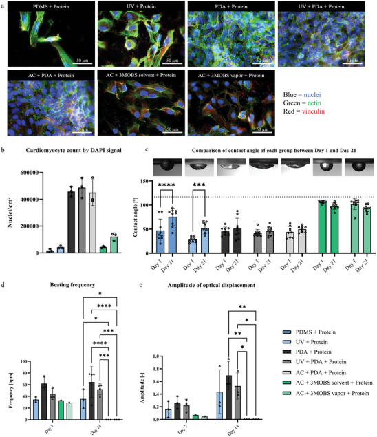
- Polydopamine plus laminin E8 511 outperformed 3MOBS for cardiomyocyte attachment: the paper reports cell densities about 5–10 times higher than after 3MOBS functionalization, with functional beating up to 14 days post-seeding in PDA-treated groups.
- The monoculture ventricle showed stable function through day 28; mean beating frequency ranged from 69.6 bpm on day 7 to 97.5 bpm on day 21, and theoretical cardiac output ranged from 0.5 µL min−1 on day 28 to 1.2 µL min−1 on day 14.
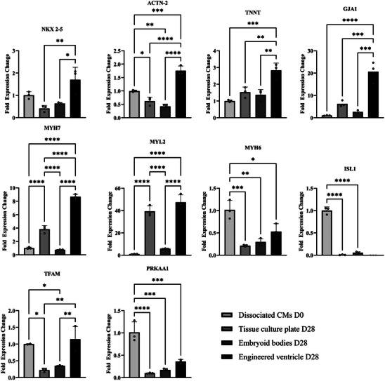
- The model’s cardiomyocytes showed a more ventricular phenotype than controls: MYH7 and MYL2 were upregulated by factors of 8.6 and 47.4, respectively, and GJA1 was upregulated by 20.6 relative to control.
- Drug testing showed expected pharmacology: after 60 min of treatment, isoproterenol (10 µM) increased frequency by 30.6–45.9% and theoretical cardiac output by 98.2–189.1%, while carbachol (10 µM) decreased frequency by 12.6–23.7% and theoretical cardiac output by 44.0–72.7%.
## Methods
- The study engineered a hemispherical PDMS membrane and compared surface functionalization methods, including polydopamine, UV/acid pretreatments, and 3MOBS, then coated the selected surface with laminin E8 511 for cardiomyocyte attachment.
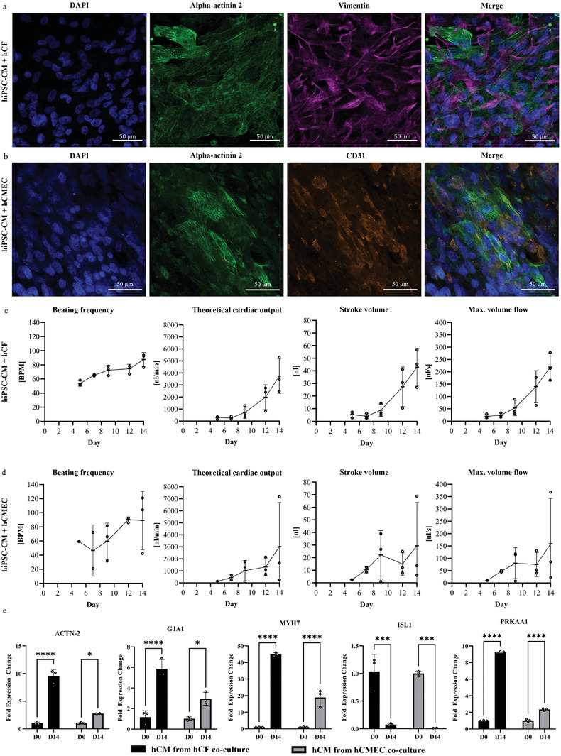
- Human iPSC-derived cardiomyocytes were cultured either alone for 28 days or in co-culture with cardiac fibroblasts or cardiac microvascular endothelial cells for 14 days; the paper also used human primary cardiac fibroblasts and endothelial cells.
- Function was assessed in a custom bioreactor that measured flow from membrane contraction; analyses included brightfield microscopy, immunofluorescence, flow cytometry, qPCR, and drug-response experiments with isoproterenol and carbachol.
## Limitations
- The authors state that co-cultures could only be cultivated until day 14 due to overgrowth, so long-term multi-cell-type maintenance was not achieved.
- The paper says the model is not yet tailored for patient-specific applications because it does not account for the genetic or phenotypic variability of patient-derived cells.
- Evident from the text: some comparisons are limited by different media and culture durations between monoculture and co-culture conditions, and some treatment groups had small sample sizes (for example, n = 5 monoculture ventricles and n = 3 co-culture measurements).
Model-written summary. Check it against the paper before relying on it.
Integrity signals
Registry
Cites
Statistics
Figures



Verification
Claims the paper makes
23 Valid · 5 Not valid · 5 Not checked · 94 Own results and interpretations
At least one cited source doesn’t support this claim or points to a different paper, and none confirmed it — see the verdicts below.
In the paper
“However, they only cultivated pure cardiomyocytes within 14 days after seeding.”
§ Engineered In Vitro Multi‐Cell Type Ventricle Model Generates Long‐Term Pulsatile Flow and Modulates Cardiac Output in Response to Cardioactive Drugs > Discussion
What the sources say
- Disputedcites [20] L. A. MacQueen, S. P. Sheehy, C. O. Chantre, J. F. Zimmerman, F. S. Pasqualini, X. Liu, J. A. Goss, P. H. Campbell, G. M. Gonzalez, S.‐J. Park, A. K. Capulli, J. P. Ferrier, T. F. Kosar, L. Mahadevan, W. T. Pu, K. K. Parker, Nat. Biomed. Eng. 2018, 12, 930. PMC6774355 31015723 sourceExperimental — accuracy not yet measured
The Tissue-engineered ventricle chamber section says experiments were conducted 14 days after seeding, not that culture was limited to 14 days. The Discussion explicitly reports culturing a hiPSC-derived cardiomyocyte line in ventricle scaffolds for six months, contrary to the claim’s ‘only’ 14-day limit. (LLM, conf=0.89)
Checked by a reasoning model — may be wrong.
plain- Asserted: passes — The authors put it forward as true.
- Checkable: passes — It can be checked against evidence.
- Load-bearing: unclear — It is unclear whether the paper’s conclusions depend on it.
- Based: passes — The paper gives a reason why it holds.
- Disputed
The cited sources support only part of this claim — read each source’s verdict below.
In the paper
“hCF contributes to structural integrity and contractility by producing ECM proteins, such as collagen and fibronectin, which support cellular organization and mechanical stability.[ 52 , 53 , 54 ]”
§ Engineered In Vitro Multi‐Cell Type Ventricle Model Generates Long‐Term Pulsatile Flow and Modulates Cardiac Output in Response to Cardioactive Drugs > Discussion
What the sources say
- Partly supportedcites [52] Y. Xuan, C. Chen, Z. Wen, D. W. Wang, Front Cardiovasc. Med. 2022, 9, 882027. 35463742 PMC9022788 sourceExperimental — accuracy not yet measured
The CFs section (page unavailable) says cardiac fibroblasts synthesize a collagen-rich ECM network that provides structural integrity and biomechanical signals. The ECM section (page unavailable) also describes that network as supporting cardiac cells. The passages do not specifically establish this for human cardiac fibroblasts (hCF), name fibronectin, or say that ECM production contributes to contractility. (LLM, conf=0.86)
- Supportedcites [53] D. Fan, A. Takawale, J. Lee, Z. Kassiri, Fibrogenesis Tissue Repair 2012, 1, 15. PMC3464725 22943504 source
The review’s “Cardiac fibroblasts are critical in extracellular matrix homeostasis” section says cardiac fibroblasts are the primary source of ECM proteins, including collagen and fibronectin, and that the ECM provides structural and functional integrity and mechanical connections in the heart. Its “Functions of cardiac fibroblasts” section also links fibroblasts to normal cardiac structure and proper contraction. The quoted passages do not provide page numbers. (LLM, conf=0.88)
- Partly supportedcites [54] P. Beauchamp, C. B. Jackson, L. C. Ozhathil, I. Agarkova, C. L. Galindo, D. B. Sawyer, T. M. Suter, C. Zuppinger, Front. Mol. Biosci. 2020, 7, 14. 32118040 PMC7033479 sourceExperimental — accuracy not yet measured
The Introduction (page not specified) says cardiac fibroblasts produce native ECM proteins that contribute to a tissue-like environment, and the Discussion, “Electrical and Contractile Function” (page not specified), reports an improved contraction rate when fibroblasts were added to spheroids. The paper does not identify collagen and fibronectin as proteins produced by these fibroblasts or show that ECM production caused the contractile improvement or mechanical stability; its fibronectin mentions describe culture coatings. (LLM, conf=0.86)
plainstates a cause- Asserted: passes — The authors put it forward as true.
- Checkable: passes — It can be checked against evidence.
- Load-bearing: unclear — It is unclear whether the paper’s conclusions depend on it.
- Based: passes — The paper gives a reason why it holds.
- Partly supported
The cited sources support only part of this claim — read each source’s verdict below.
In the paper
“Additionally, Lee's ventricle model was not integrated into a bioreactor that supported measurements of the generated flow.”
§ Engineered In Vitro Multi‐Cell Type Ventricle Model Generates Long‐Term Pulsatile Flow and Modulates Cardiac Output in Response to Cardioactive Drugs > Discussion
What the sources say
- Partly supportedcites [19] A. Lee, A. R. Hudson, D. J. Shiwarski, J. W. Tashman, T. J. Hinton, S. Yerneni, J. M. Bliley, P. G. Campbell, A. W. Feinberg, Science 2019, 6452, 482. 31371612 sourceExperimental — accuracy not yet measured
The ventricle-model passage (section labeled Abstract, page unavailable) reports measurements of calcium activity, wall motion, and chamber-area change; it does not report generated-flow measurements. A separate passage in that section describes flow measurements for the printed valve, not the ventricle. Our copy does not explicitly establish that the ventricle was not integrated into a bioreactor, so that part of the claim remains unsupported. (LLM, conf=0.74)
plain- Asserted: passes — The authors put it forward as true.
- Checkable: passes — It can be checked against evidence.
- Load-bearing: unclear — It is unclear whether the paper’s conclusions depend on it.
- Based: passes — The paper gives a reason why it holds.
- Partly supported
The cited sources support only part of this claim — read each source’s verdict below.
In the paper
“In addition, coculture may improve hCM survival and limited proliferation, increasing the number of functional cardiomyocytes.[ 57 , 58 , 59 ]”
§ Engineered In Vitro Multi‐Cell Type Ventricle Model Generates Long‐Term Pulsatile Flow and Modulates Cardiac Output in Response to Cardioactive Drugs > Discussion
What the sources say
- Not checked — no full text of the sourcecites [57] H. K. Voges, S. R. Foster, L. Reynolds, B. L. Parker, L. Devilée, G. A. Quaife‐Ryan, P. R. J. Fortuna, E. Mathieson, R. Fitzsimmons, M. Lor, C. Batho, J. Reid, M. Pocock, C. E. Friedman, D. Mizikovsky, M. Francois, N. J. Palpant, E. J. Needham, M. Peralta, G. Del Monte‐Nieto, L. K. Jones, I. M. Smyth, N. R. Mehdiabadi, F. Bolk, V. Janbandhu, E. Yao, R. P. Harvey, J. J. H. Chong, D. A. Elliott, E. G. Stanley, et al., Cell Rep. 2023, 5, 112322. 37105170 source
- Partly supportedcites [58] Y. Liu, R. Kamran, X. Han, M. Wang, Q. Li, D. Lai, K. Naruse, K. Takahashi, Sci. Rep. 2024, 1, 18063. PMC11310341 39117679 sourceExperimental — accuracy not yet measured
The Results section reports greater contractility and a higher percentage of cells expressing cardiomyocyte markers in the tri-culture group, supporting improved cardiac function under coculture. It does not show that coculture improved cardiomyocyte survival or proliferation, or establish an increase in their absolute number; searches for survival and proliferation found no such results in our copy. Page numbers were not provided. (LLM, conf=0.84)
- Partly supportedcites [59] E. Giacomelli, V. Meraviglia, G. Campostrini, A. Cochrane, X. Cao, R. W. J. van Helden, A. Krotenberg Garcia, M. Mircea, S. Kostidis, R. P. Davis, B. J. van Meer, C. R. Jost, A. J. Koster, H. Mei, D. G. Míguez, A. A. Mulder, M. Ledesma‐Terrón, G. Pompilio, L. Sala, D. C. F. Salvatori, R. C. Slieker, E. Sommariva, V. A. A. F. de, M. Giera, S. Semrau, L. G. J. Tertoolen, V. V. Orlova, M. Bellin, C. L. Mummery, Cell Stem Cell 2020, 6, 862. PMC7284308 32459996 sourceExperimental — accuracy not yet measured
The Results section on establishment of the 3D microtissue model (page unavailable) reports that fibroblast-containing tissues retained more cells than tissues without fibroblasts despite the same input, while proliferation remained low in all groups. The Results on electrical and mechanical maturation report improved cardiomyocyte function with coculture. The paper does not establish that improved cardiomyocyte survival or increased cardiomyocyte proliferation caused an increase in the number of functional cardiomyocytes. (LLM, conf=0.86)
hedgedstates a possible cause- Asserted: passes — The authors put it forward as true.
- Checkable: passes — It can be checked against evidence.
- Load-bearing: unclear — It is unclear whether the paper’s conclusions depend on it.
- Based: passes — The paper gives a reason why it holds.
Every source the paper cites for this claim was checked and supports it.
In the paper
“Notably, to achieve a physiologically consistent model, cells were used that were only from donors of the same gender due to differences in cellular phenotype, behavior, and composition between male and female hearts.[ 41 ]”
§ Engineered In Vitro Multi‐Cell Type Ventricle Model Generates Long‐Term Pulsatile Flow and Modulates Cardiac Output in Response to Cardioactive Drugs > Discussion
What the sources say
- Supportedcites [41] C. J. Walker, M. E. Schroeder, B. A. Aguado, K. S. Anseth, L. A. Leinwand, J. Mol. Cell. Cardiol. 2021, 160, 42. 34166708 PMC8571046 source
The Introduction states that cells in male and female hearts differ in function, interactions, and cellular composition. The Discussion and Forward Thinking says these disparities must be addressed when developing in vitro cardiac models. Using donors of the same sex is the citing authors’ methodological choice, not a procedure attributed to this review. (LLM, conf=0.88)
plainstates a cause- Asserted: passes — The authors put it forward as true.
- Checkable: passes — It can be checked against evidence.
- Load-bearing: unclear — It is unclear whether the paper’s conclusions depend on it.
- Based: passes — The paper gives a reason why it holds.
- Supported
64 more claims · 2 with their cited sources checked
Create a free account to see all 127 claims and to verify the 1 reference still open.
- It may state two separate claims as one. 46
- Probably not a substantial claim. 17
- The quote only partly states this claim. 3
Claims other papers make about this one
Each is checked against this paper’s own text: does it say what the citing paper says it does?
The source backs part of the claim, not all of it — or backs it for a different population, condition or comparison.
In the citing paper
“Functional readouts are a priority in several models; notably, Kuckelhorn et al. uniquely integrated a flow sensor, and Ribeiro et al. visualized fluid pumping through a connected glass capillary, thus enabling non-invasive PV analysis (Kuckelkorn et al., 2025; Ribeiro et al., 2025) (Table 1).”
From Mini-hearts for disease modeling and drug testing – process optimization versus biological functionality (2026) · cites this paper as Kuckelkorn et al., 2025
Experimental — accuracy not yet measuredThe source backs part of the claim, not all of it — or backs it for a different population, condition or comparison.
In the citing paper
“Several ECCs are derived from human induced Pluripotent Stem Cells (hiPSCs), commonly differentiated into CMs and FBs (Li et al., 2018; Michas et al., 2022; Kuckelkorn et al., 2025; Ribeiro et al., 2025).”
From Mini-hearts for disease modeling and drug testing – process optimization versus biological functionality (2026) · cites this paper as Kuckelkorn et al., 2025
Experimental — accuracy not yet measured
Citations
Cites
Heart Disease and Stroke Statistics—2020 Update: A Report From the American Heart Association
Virani, S. S., Alonso, A., Benjamin, E. J., Bittencourt, M. S., Callaway, C. W., Carson, A. P., Chamberlain, A. M., Chang, A. R., Cheng, S., Delling, F. N., et al. (2020) · Circulation · doi:10.1161/cir.0000000000000757 · Cited by 9,175
Primer-BLAST: A tool to design target-specific primers for polymerase chain reaction
Ye, J., Coulouris, G., Zaretskaya, I., Cutcutache, I., Rozen, S., & Madden, T. L. (2012) · BMC Bioinformatics · doi:10.1186/1471-2105-13-134 · Cited by 6,111
3D bioprinting of collagen to rebuild components of the human heart
Lee, A., Hudson, A. R., Shiwarski, D. J., Tashman, J. W., Hinton, T. J., Yerneni, S., Bliley, J. M., Campbell, P. G., & Feinberg, A. W. (2019) · Science · doi:10.1126/science.aav9051 · Cited by 1,782
3D Printing of Personalized Thick and Perfusable Cardiac Patches and Hearts
Noor, N., Shapira, A., Edri, R., Gal, I., Wertheim, L., & Dvir, T. (2019) · Advanced Science · doi:10.1002/advs.201900344 · Cited by 1,042
Cardiac fibroblasts, fibrosis and extracellular matrix remodeling in heart disease
Fan, D., Takawale, A., Lee, J., & Kassiri, Z. (2012) · Fibrogenesis & Tissue Repair · doi:10.1186/1755-1536-5-15 · Cited by 814
Efimenko, K., Wallace, W. E., & Genzer, J. (2002) · Journal of Colloid and Interface Science · doi:10.1006/jcis.2002.8594 · Cited by 757
Brutsaert, D. L. (2003) · Physiological Reviews · doi:10.1152/physrev.00017.2002 · Cited by 697
Giacomelli, E., Meraviglia, V., Campostrini, G., Cochrane, A., Cao, X., van Helden, R. W. J., Krotenberg Garcia, A., Mircea, M., Kostidis, S., Davis, R. P., et al. (2020) · Cell Stem Cell · doi:10.1016/j.stem.2020.05.004 · Cited by 622
Redefining the identity of cardiac fibroblasts
Tallquist, M. D., & Molkentin, J. D. (2017) · Nature Reviews Cardiology · doi:10.1038/nrcardio.2017.57 · Cited by 581
Lewis-Israeli, Y. R., Wasserman, A. H., Gabalski, M. A., Volmert, B. D., Ming, Y., Ball, K. A., Yang, W., Zou, J., Ni, G., Pajares, N., et al. (2021) · Nature Communications · doi:10.1038/s41467-021-25329-5 · Cited by 536
Role of animal models in biomedical research: a review
Mukherjee, P., Roy, S., Ghosh, D., & Nandi, S. K. (2022) · Laboratory Animal Research · doi:10.1186/s42826-022-00128-1 · Cited by 502
Small and large animal models in cardiac contraction research: advantages and disadvantages
Milani-Nejad, N., & Janssen, P. M. L. (2014) · Pharmacology & Therapeutics · doi:10.1016/j.pharmthera.2013.10.007 · Cited by 484
Human cardiac organoids for the modelling of myocardial infarction and drug cardiotoxicity
Richards, D. J., Li, Y., Kerr, C. M., Yao, J., Beeson, G. C., Coyle, R. C., Chen, X., Jia, J., Damon, B., Wilson, R., et al. (2020) · Nature Biomedical Engineering · doi:10.1038/s41551-020-0539-4 · Cited by 439
Cardiac fibroblasts: friend or foe?
Baudino, T. A., Carver, W., Giles, W., & Borg, T. K. (2006) · American Journal of Physiology-Heart and Circulatory Physiology · doi:10.1152/ajpheart.00023.2006 · Cited by 427
Miyazaki, T., Futaki, S., Suemori, H., Taniguchi, Y., Yamada, M., Kawasaki, M., Hayashi, M., Kumagai, H., Nakatsuji, N., Sekiguchi, K., et al. (2012) · Nature Communications · doi:10.1038/ncomms2231 · Cited by 368
Kupfer, M. E., Lin, W.-H., Ravikumar, V., Qiu, K., Wang, L., Gao, L., Bhuiyan, D. B., Lenz, M., Ai, J., Mahutga, R. R., et al. (2020) · Circulation Research · doi:10.1161/circresaha.119.316155 · Cited by 329
Mills, R. J., Parker, B. L., Quaife-Ryan, G. A., Voges, H. K., Needham, E. J., Bornot, A., Ding, M., Andersson, H., Polla, M., Elliott, D. A., et al. (2019) · Cell Stem Cell · doi:10.1016/j.stem.2019.03.009 · Cited by 312
Biomechanical properties and microstructure of human ventricular myocardium
Sommer, G., Schriefl, A. J., Andrä, M., Sacherer, M., Viertler, C., Wolinski, H., & Holzapfel, G. A. (2015) · Acta Biomaterialia · doi:10.1016/j.actbio.2015.06.031 · Cited by 290
Davis, R. P., Casini, S., van den Berg, C. W., Hoekstra, M., Remme, C. A., Dambrot, C., Salvatori, D., Oostwaard, D. W.-V., Wilde, A. A. M., Bezzina, C. R., et al. (2012) · Circulation · doi:10.1161/circulationaha.111.066092 · Cited by 264
Chuah, Y. J., Koh, Y. T., Lim, K., Menon, N. V., Wu, Y., & Kang, Y. (2015) · Scientific Reports · doi:10.1038/srep18162 · Cited by 262
Embryonic Heart Progenitors and Cardiogenesis
Brade, T., Pane, L. S., Moretti, A., Chien, K. R., & Laugwitz, K.-L. (2013) · Cold Spring Harbor Perspectives in Medicine · doi:10.1101/cshperspect.a013847 · Cited by 254
A tissue-engineered scale model of the heart ventricle
MacQueen, L. A., Sheehy, S. P., Chantre, C. O., Zimmerman, J. F., Pasqualini, F. S., Liu, X., Goss, J. A., Campbell, P. H., Gonzalez, G. M., Park, S.-J., et al. (2018) · Nature Biomedical Engineering · doi:10.1038/s41551-018-0271-5 · Cited by 227
An actuated pump on-chip powered by cultured cardiomyocytes
Tanaka, Y., Morishima, K., Shimizu, T., Kikuchi, A., Yamato, M., Okano, T., & Kitamori, T. (2006) · Lab on a Chip · doi:10.1039/b515149j · Cited by 211
Beauchamp, P., Jackson, C. B., Ozhathil, L. C., Agarkova, I., Galindo, C. L., Sawyer, D. B., Suter, T. M., & Zuppinger, C. (2020) · Frontiers in Molecular Biosciences · doi:10.3389/fmolb.2020.00014 · Cited by 202
Zhang, J., Tao, R., Campbell, K. F., Carvalho, J. L., Ruiz, E. C., Kim, G. C., Schmuck, E. G., Raval, A. N., da Rocha, A. M., Herron, T. J., et al. (2019) · Nature Communications · doi:10.1038/s41467-019-09831-5 · Cited by 201
Zhang, H., Tian, L., Shen, M., Tu, C., Wu, H., Gu, M., Paik, D. T., & Wu, J. C. (2019) · Circulation Research · doi:10.1161/circresaha.119.315491 · Cited by 187
Surface Modification Techniques for Endothelial Cell Seeding in PDMS Microfluidic Devices
Akther, F., Yakob, S. B., Nguyen, N.-T., & Ta, H. T. (2020) · Biosensors · doi:10.3390/bios10110182 · Cited by 182
A micro-spherical heart pump powered by cultured cardiomyocytes
Tanaka, Y., Sato, K., Shimizu, T., Yamato, M., Okano, T., & Kitamori, T. (2007) · Lab Chip · doi:10.1039/b612082b · Cited by 182
Challenges and opportunities for the next generation of cardiovascular tissue engineering
Cho, S., Discher, D. E., Leong, K. W., Vunjak-Novakovic, G., & Wu, J. C. (2022) · Nature Methods · doi:10.1038/s41592-022-01591-3 · Cited by 176
Silva, A. C., Matthys, O. B., Joy, D. A., Kauss, M. A., Natarajan, V., Lai, M. H., Turaga, D., Blair, A. P., Alexanian, M., Bruneau, B. G., et al. (2021) · Cell Stem Cell · doi:10.1016/j.stem.2021.11.007 · Cited by 167
Paracrine modulation of heart cell function by endothelial cells
Shah, A. M. (1996) · Cardiovascular Research · doi:10.1016/s0008-6363(96)00025-9 · Cited by 165
(2011) · PubMed · Cited by 159
Vascular cells improve functionality of human cardiac organoids
Voges, H. K., Foster, S. R., Reynolds, L., Parker, B. L., Devilée, L., Quaife-Ryan, G. A., Fortuna, P. R. J., Mathieson, E., Fitzsimmons, R., Lor, M., et al. (2023) · Cell Reports · doi:10.1016/j.celrep.2023.112322 · Cited by 139
Ergir, E., Oliver-De La Cruz, J., Fernandes, S., Cassani, M., Niro, F., Pereira-Sousa, D., Vrbský, J., Vinarský, V., Perestrelo, A. R., Debellis, D., et al. (2022) · Scientific Reports · doi:10.1038/s41598-022-22225-w · Cited by 99
Hamad, S., Derichsweiler, D., Papadopoulos, S., Nguemo, F., Šarić, T., Sachinidis, A., Brockmeier, K., Hescheler, J., Boukens, B. J., & Pfannkuche, K. (2019) · Theranostics · doi:10.7150/thno.32058 · Cited by 83
Self-citationEngineering a living cardiac pump on a chip using high-precision fabrication
Michas, C., Karakan, M. Ç., Nautiyal, P., Seidman, J. G., Seidman, C. E., Agarwal, A., Ekinci, K., Eyckmans, J., White, A. E., & Chen, C. S. (2022) · Science Advances · doi:10.1126/sciadv.abm3791 · Cited by 82
Matters of the heart: Cellular sex differences
Walker, C. J., Schroeder, M. E., Aguado, B. A., Anseth, K. S., & Leinwand, L. A. (2021) · Journal of Molecular and Cellular Cardiology · doi:10.1016/j.yjmcc.2021.04.010 · Cited by 74
Development of a porous 3D graphene-PDMS scaffold for improved osseointegration
Li, J., Liu, X., Crook, J. M., & Wallace, G. G. (2017) · Colloids and Surfaces B: Biointerfaces · doi:10.1016/j.colsurfb.2017.07.087 · Cited by 69
Differentiation of pluripotent stem cells into endothelial cells
Yoder, M. C. (2015) · Current Opinion in Hematology · doi:10.1097/moh.0000000000000140 · Cited by 67
Grancharova, T., Gerbin, K. A., Rosenberg, A. B., Roco, C. M., Arakaki, J. E., DeLizo, C. M., Dinh, S. Q., Donovan-Maiye, R. M., Hirano, M., Nelson, A. M., et al. (2021) · Scientific Reports · doi:10.1038/s41598-021-94732-1 · Cited by 61
Piccini, I., Rao, J., Seebohm, G., & Greber, B. (2015) · Genomics Data · doi:10.1016/j.gdata.2015.03.008 · Cited by 60
Gezer, P. G., Brodsky, S., Hsiao, A., Liu, G. L., & Kokini, J. L. (2015) · Colloids and Surfaces B Biointerfaces · doi:10.1016/j.colsurfb.2015.07.006 · Cited by 57
Liu, Y., Kamran, R., Han, X., Wang, M., Li, Q., Lai, D., Naruse, K., & Takahashi, K. (2024) · Scientific Reports · doi:10.1038/s41598-024-68275-0 · Cited by 47
Tsuzuki, T., Baassiri, K., Mahmoudi, Z., Perumal, A. S., Rajendran, K., Rubies, G. M., & Nicolau, D. V. (2022) · Materials · doi:10.3390/ma15062313 · Cited by 45
Goßmann, M., Frotscher, R., Linder, P. W., Neumann, S., Bayer, R., Epple, M., Staat, M., Artmann, A. T., & Artmann, G. M. (2016) · Cellular Physiology and Biochemistry · doi:10.1159/000443124 · Cited by 42
Functional microvascularization of human myocardium in vitro
King, O., Cruz-Moreira, D., Sayed, A., Kermani, F., Kit-Anan, W., Sunyovszki, I., Wang, B. X., Downing, B., Fourre, J., Hachim, D., et al. (2022) · Cell Reports Methods · doi:10.1016/j.crmeth.2022.100280 · Cited by 42
Dunn, K. K., & Palecek, S. P. (2018) · Frontiers in Medicine · doi:10.3389/fmed.2018.00110 · Cited by 41
Micropatterning neuronal networks
Hardelauf, H., Waide, S., Sisnaiske, J., et al. (2014) · The Analyst · doi:10.1039/c4an00608a · Cited by 40
Fang, Y., Sun, W., Zhang, T., & Xiong, Z. (2022) · Biomaterials · doi:10.1016/j.biomaterials.2021.121298 · Cited by 40
Chanthra, N., Abe, T., Miyamoto, M., Sekiguchi, K., Kwon, C., Hanazono, Y., & Uosaki, H. (2020) · Scientific Reports · doi:10.1038/s41598-020-61163-3 · Cited by 36
The Roles of Cardiac Fibroblasts and Endothelial Cells in Myocarditis
Xuan, Y., Chen, C., Wen, Z., & Wang, D. W. (2022) · Frontiers in Cardiovascular Medicine · doi:10.3389/fcvm.2022.882027 · Cited by 35
Etezadi, F., Le, M. N. T., Shahsavarani, H., Alipour, A., Moazzezy, N., Samani, S., Amanzadeh, A., Pahlavan, S., Bonakdar, S., Shokrgozar, M. A., et al. (2022) · ACS Biomaterials Science & Engineering · doi:10.1021/acsbiomaterials.2c00162 · Cited by 33
Chemically defined, ultrasoft PDMS elastomers with selectable elasticity for mechanobiology
Heinrichs, V., Dieluweit, S., Stellbrink, J., Pyckhout-Hintzen, W., Hersch, N., Richter, D., & Merkel, R. (2018) · PLOS ONE · doi:10.1371/journal.pone.0195180 · Cited by 30
Heart in a dish – choosing the rightin vitromodel
Drakhlis, L., & Zweigerdt, R. (2023) · Disease Models & Mechanisms · doi:10.1242/dmm.049961 · Cited by 25
Yue, X., Acun, A., & Zorlutuna, P. (2017) · Acta Biomaterialia · doi:10.1016/j.actbio.2017.06.031 · Cited by 16
Simplified platform for mosaic in vivo analysis of cellular maturation in the developing heart
Goudy, J., Henley, T., Méndez, H. G., & Bressan, M. C. (2019) · Scientific Reports · doi:10.1038/s41598-019-47009-7 · Cited by 12
Fabrication and Characterization of Graphene Oxide-Coated Plate for Efficient Culture of Stem Cells
Park, D., Park, J., Lee, J., Shim, C. J., Kim, M. S., Lee, T. Y., & Lim, J. O. (2021) · Tissue Engineering and Regenerative Medicine · doi:10.1007/s13770-021-00370-z · Cited by 9
Progress of Mitochondrial Function Regulation in Cardiac Regeneration
Chen, Y. G., Zhao, A.-R., Wei, T., Wang, H., & Wang, L.-S. (2024) · Journal of Cardiovascular Translational Research · doi:10.1007/s12265-024-10514-w · Cited by 4
Zhou, Y., Zhou, R., Huang, W., Wang, J., Jiang, C., Li, A., Huang, C., & Zhang, Y. (2024) · Biologics: Targets and Therapy · doi:10.2147/btt.s448054 · Cited by 4
Research works printed but not linked: 6
- Virani S. S., 2020, Circulation, 9
- Drakhlis L., 2023, Dis. Model Mech., 5
- Gupta S., 2018, Tissue Eng. BioNanoSci., 3, 868
- Tsuzuki T., 2022, Materials, 6
- Ostchega Y., 2011, Natl Health Stat Report., 1, 1
- Zhou Y., 2024, Biologics, 18, 115
Cited by
Kim, Y. H., Son, Y. H., Choi, Y., Kim, M. S., Park, S., & Lee, K. Y. (2026) · Microsystems & Nanoengineering · doi:10.1038/s41378-026-01249-6 · Cited by 8
Hecking, J., Cano-Jorge, M., Passier, R., & Rivera-Arbeláez, J. M. (2026) · Frontiers in Bioengineering and Biotechnology · doi:10.3389/fbioe.2025.1719533
Metadata
- Authors
- Kuckelkorn, C., Aksoy, E., Stojanovic, N., Oulahyane, L., Ritter, M., Pfannkuche, K., & Fischer, H.
- Affiliations
- Universitätsklinikum Aachen (DE)
- University of Cologne (DE)
- Corresponding author
- Fischer, Horst
- Venue
- Advanced Healthcare Materials
- Publisher
- Wiley
- Published
- 2025-02-13
- Volume
- 14
- Issue
- 10
- Pages
- e2403897-e2403897
- ISSN
- 2192-2640 (ISSN-L)
- 2192-2659
- PMC
- PMC12004430
- OpenAlex
- W4407426312
- Cited by
- 3
- References
- 55
- Citations by year
- 2026: 3
- FWCI
- 2.80
- Type
- journal article
- Language
- en
- Open access
- hybrid
- doi.org
- Licence
- cc-by
- creativecommons.org/licenses/by/4.0/ (vor)
- doi.wiley.com/10.1002/tdm_license_1.1 (tdm)
- Locations
- Advanced Healthcare Materials (publishedVersion, cc-by)
- PubMed Central (publishedVersion, cc-by)
- PubMed (publishedVersion)
- RWTH Publications (RWTH Aachen) (submittedVersion, other-oa)
- Funders
- RWTH Aachen University
- Topics
- Health Sciences › Medicine › Surgery › Tissue Engineering and Regenerative Medicine
- Physical Sciences › Engineering › Biomedical Engineering › 3D Printing in Biomedical Research
- Life Sciences › Biochemistry, Genetics and Molecular Biology › Molecular Biology › Pluripotent Stem Cells Research
- Keywords
- iPSC-derived cardiomyocytes, pulsatile flow, cardiac output, polydopamine coating, PDMS membrane, bioreactors, cardioactive drugs, carbachol, isoproterenol, heart rate response, cardiomyocyte beating
- MeSH terms
- Cardiac Output, Dimethylpolysiloxanes, Fibroblasts, Heart Ventricles, Humans, Pulsatile Flow, Coculture Techniques, Tissue Engineering, Myocytes, Cardiac, Induced Pluripotent Stem Cells
About these checks
Registry, retraction, Cites and statistics results are computed, not judged, with one exception: a printed reference that no index lists is read once by a model before it counts as a citation error. Claims are extracted by a model and kept only when their quote appears verbatim in the text; each cited source is then checked, with a model’s help, for whether it says what the claim relies on. Those verdicts can be wrong — read the claim’s quote and the cited source before relying on one.
The claim verdicts this report counts are experimental: their accuracy is not yet measured.
A language model drew the claims from the paper on 2026-10-10, under version 3 of the claim definition (model deepseek-v4.1-flash, prompt extract-v3).
A second model labelled each claim on 2026-10-10 (model jev-1.13-20260917, definition revision gold-guideline-v0.2).
Cited sources are checked with a language model’s help (gpt-6-sol).
Statistics and arithmetic are recomputed, not judged.
Confidence bands are shown only for fields calibrated on our annotated test set. None is calibrated yet, so no band is shown.
What counts as a claim ›How we measure confidence ›How we check this ›
Are you an author, or do you see a problem with this page? Request a review
Data sources
Bibliographic data: Semantic Scholar, OpenAlex, Crossref.
Contains information from the Semantic Scholar Academic Graph Datasets, made available under the ODC Attribution License (ODC-BY 1.0).
Semantic Scholar’s data is described in: Kinney, R., et al. (2023). The Semantic Scholar Open Data Platform. arXiv:2301.10140.
