

The DOI matches its Crossref record; 38 of the 38 references it cites were found in a registry or an index.
iPSC ‐derived microglia carrying the TREM2 R47H /+ mutation are proinflammatory and promote synapse loss
Abstract
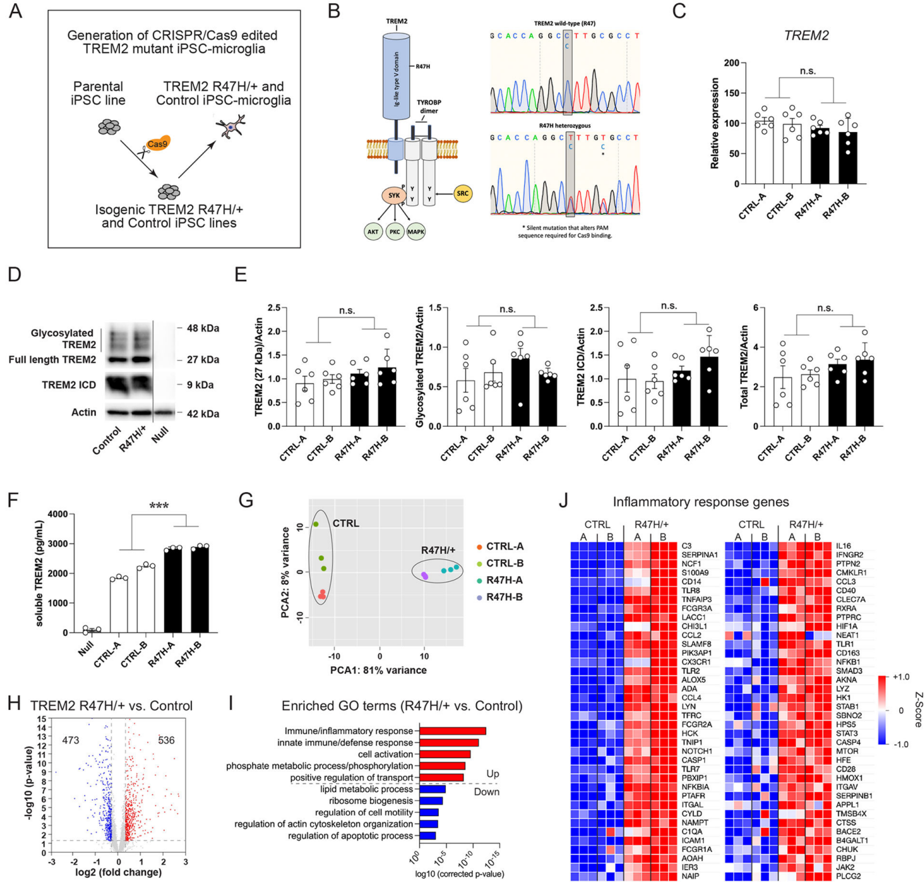
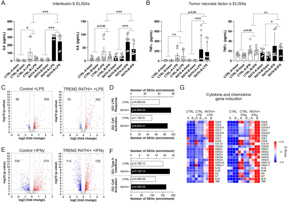
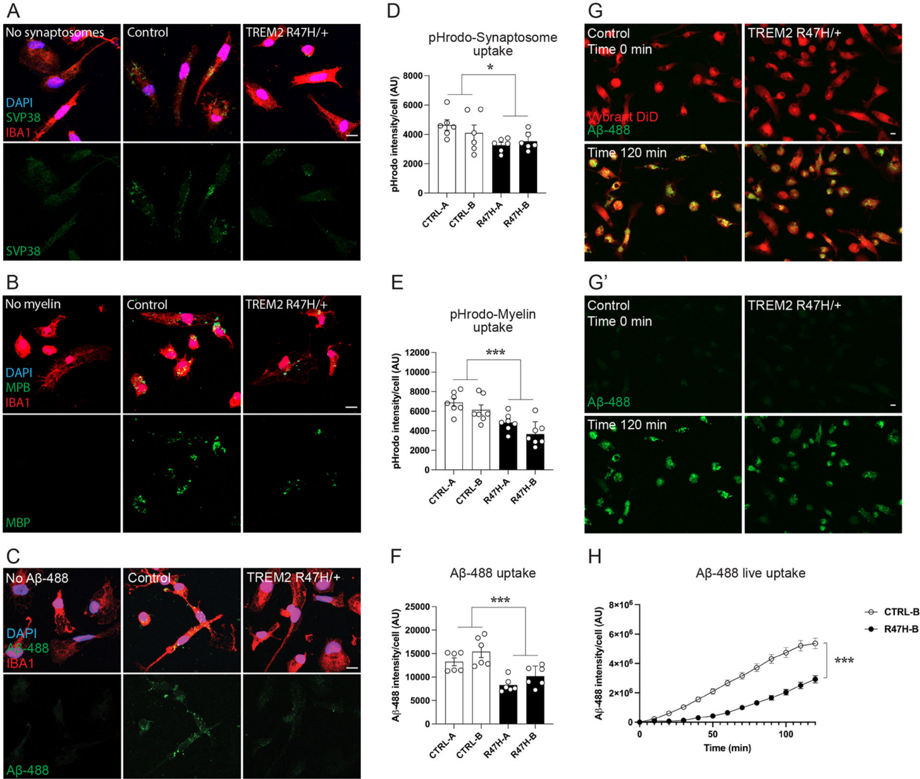
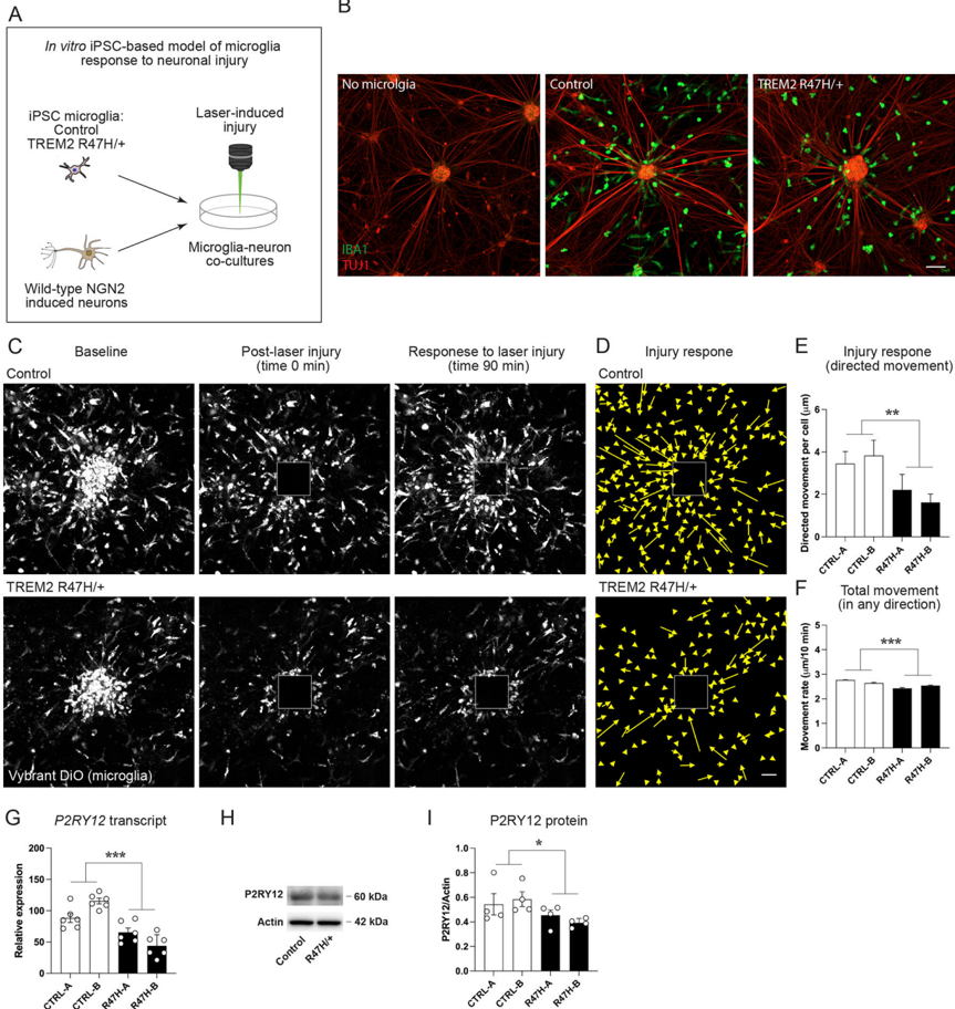
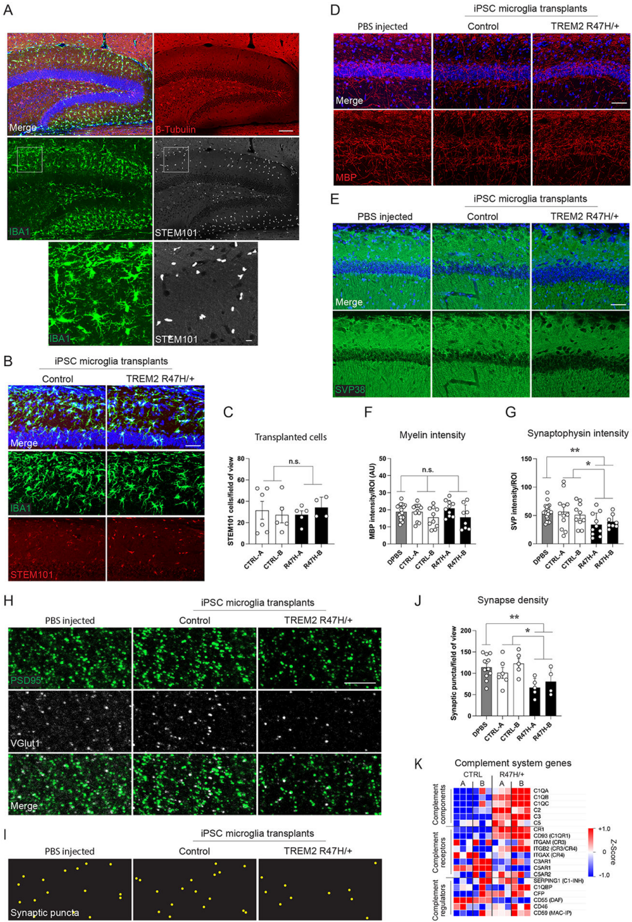
Genetic findings have highlighted key roles for microglia in the pathology of neurodegenerative conditions such as Alzheimer's disease (AD). A number of mutations in the microglial protein triggering receptor expressed on myeloid cells 2 (TREM2) have been associated with increased risk for developing AD, most notably the R47H/+ substitution. We employed gene editing and stem cell models to gain insight into the effects of the TREM2 R47H/+ mutation on human‐induced pluripotent stem cell‐derived microglia. We found transcriptional changes affecting numerous cellular processes, with R47H/+ cells exhibiting a proinflammatory gene expression signature. TREM2 R47H/+ also caused impairments in microglial movement and the uptake of multiple substrates, as well as rendering microglia hyperresponsive to inflammatory stimuli. We developed an in vitro laser‐induced injury model in neuron–microglia cocultures, finding an impaired injury response by TREM2 R47H/+ microglia. Furthermore, mouse brains transplanted with TREM2 R47H/+ microglia exhibited reduced synaptic density, with upregulation of multiple complement cascade components in TREM2 R47H/+ microglia suggesting inappropriate synaptic pruning as one potential mechanism. These findings identify a number of potentially detrimental effects of the TREM2 R47H/+ mutation on microglial gene expression and function likely to underlie its association with AD.
Overview
The claims and statistics of this paper haven’t been checked yet.
Run the check — free, no account needed. It reads the open-access text and checks the sources it cites.
Integrity signals
Registry
Cites
Statistics
Figures
Verification
Claims the paper makes
The claims and statistics of this paper haven’t been checked yet.
Run the check — free, no account needed. It reads the open-access text and checks the sources it cites.
Claims other papers make about this one
Each is checked against this paper’s own text: does it say what the citing paper says it does?
The source backs part of the claim, not all of it — or backs it for a different population, condition or comparison.
In the citing paper
“Notably, iPSC-derived microglia with altered expression of the sAD risk genes APOE4 and TREM2 display pronounced phenotypic differences from control microglia, such as impaired phagocytosis, whereas mutations involved in familial AD have a more limited impact on microglial function [51, 60–63, 119–122].”
From microRNA-132 attenuates inflammation in induced pluripotent stem cell-derived microglia from Alzheimer’s disease patients (2026) · cites this paper as [122]
Experimental — accuracy not yet measured
Self-check
Percentages, sums and reported differences re-computed from the paper’s own numbers.
The self-check needs the paper’s full text.
Citations
Cites
Complement and microglia mediate early synapse loss in Alzheimer mouse models
Hong, S., Beja-Glasser, V. F., Nfonoyim, B. M., Frouin, A., Li, S., Ramakrishnan, S., Merry, K. M., Shi, Q., Rosenthal, A., Barres, B. A., et al. (2016) · Science · doi:10.1126/science.aad8373 · Cited by 3,343 · cited 3×
“TREM2 has been shown to play a role in developmental synaptic pruning in the mouse brain, and microglia have been reported to engage in inappropriate synaptic pruning in adult AD model mice (Filipello et al., 2018; Hong et al., 2016; Salter & Stevens, 2017).”
BackgroundHarold, D., Abraham, R., Hollingworth, P., Sims, R., Gerrish, A., Hamshere, M. L., Pahwa, J. S., Moskvina, V., Dowzell, K., Williams, A., et al. (2009) · Nature Genetics · doi:10.1038/ng.440 · Cited by 3,326
Variant of TREM2 Associated with the Risk of Alzheimer's Disease
Jonsson, T., Stefansson, H., Steinberg, S., Jonsdottir, I., Jonsson, P. V., Snaedal, J., Bjornsson, S., Huttenlocher, J., Levey, A. I., Lah, J. J., et al. (2013) · New England Journal of Medicine · doi:10.1056/nejmoa1211103 · Cited by 2,558
The Cellular Phase of Alzheimer’s Disease
De Strooper, B., & Karran, E. (2016) · Cell · doi:10.1016/j.cell.2015.12.056 · Cited by 1,990
The P2Y12 receptor regulates microglial activation by extracellular nucleotides
Haynes, S. E., Hollopeter, G., Yang, G., Kurpius, D., Dailey, M. E., Gan, W.-B., & Julius, D. (2006) · Nature Neuroscience · doi:10.1038/nn1805 · Cited by 1,542
Lin, Y.-T., Seo, J., Gao, F., Feldman, H. M., Wen, H.-L., Penney, J., Cam, H. P., Gjoneska, E., Raja, W. K., Cheng, J., et al. (2018) · Neuron · doi:10.1016/j.neuron.2018.05.008 · Cited by 1,125
Self-citationYeh, F. L., Wang, Y., Tom, I., Gonzalez, L. C., & Sheng, M. (2016) · Neuron · doi:10.1016/j.neuron.2016.06.015 · Cited by 943
TREM2-mediated early microglial response limits diffusion and toxicity of amyloid plaques
Wang, Y., Ulland, T. K., Ulrich, J. D., Song, W., Tzaferis, J. A., Hole, J. T., Yuan, P., Mahan, T. E., Shi, Y., Gilfillan, S., et al. (2016) · Journal of Experimental Medicine · doi:10.1084/jem.20151948 · Cited by 840
TREM2 mutations implicated in neurodegeneration impair cell surface transport and phagocytosis
Kleinberger, G., Yamanishi, Y., Suárez-Calvet, M., Czirr, E., Lohmann, E., Cuyvers, E., Struyfs, H., Pettkus, N., Wenninger-Weinzierl, A., Mazaheri, F., et al. (2014) · Science Translational Medicine · doi:10.1126/scitranslmed.3009093 · Cited by 798
TREM2 regulates microglial cholesterol metabolism upon chronic phagocytic challenge
Nugent, A. A., Lin, K., van Lengerich, B., Lianoglou, S., Przybyla, L., Davis, S. S., Llapashtica, C., Wang, J., Kim, D. J., Xia, D., et al. (2020) · Neuron · doi:10.1016/j.neuron.2019.12.007 · Cited by 745
Filipello, F., Morini, R., Corradini, I., Zerbi, V., Canzi, A., Michalski, B., Erreni, M., Markicevic, M., Starvaggi-Cucuzza, C., Otero, K., et al. (2018) · Immunity · doi:10.1016/j.immuni.2018.04.016 · Cited by 695
The road to restoring neural circuits for the treatment of Alzheimer's disease
Canter, R. G., Penney, J., & Tsai, L.-H. (2016) · Nature · doi:10.1038/nature20412 · Cited by 565
Self-citationMicroglial Cx3cr1 knockout prevents neuron loss in a mouse model of Alzheimer's disease
Fuhrmann, M., Bittner, T., Jung, C. K. E., Burgold, S., Page, R. M., Mitteregger, G., Haass, C., LaFerla, F. M., Kretzschmar, H., & Herms, J. (2010) · Nature Neuroscience · doi:10.1038/nn.2511 · Cited by 562
TREM2 sustains microglial expansion during aging and response to demyelination
Poliani, P. L., Wang, Y., Fontana, E., Robinette, M. L., Yamanishi, Y., Gilfillan, S., & Colonna, M. (2015) · Journal of Clinical Investigation · doi:10.1172/jci77983 · Cited by 525
Leyns, C. E. G., Ulrich, J. D., Finn, M. B., Stewart, F. R., Koscal, L. J., Remolina Serrano, J., Robinson, G. O., Anderson, E., Colonna, M., & Holtzman, D. M. (2017) · Proceedings of the National Academy of Sciences · doi:10.1073/pnas.1710311114 · Cited by 517 · cited 2×
“Inflammatory responses by microglia in amyloid and tau models are generally dampened by the loss of TREM2 (Griciuc et al., 2019; Jay et al., 2017; Leyns et al., 2017).”
BackgroundTranscriptional and Translational Differences of Microglia from Male and Female Brains
Guneykaya, D., Ivanov, A., Hernandez, D. P., Haage, V., Wojtas, B., Meyer, N., Maricos, M., Jordan, P., Buonfiglioli, A., Gielniewski, B., et al. (2018) · Cell Reports · doi:10.1016/j.celrep.2018.08.001 · Cited by 477
Modeling Alzheimer’s disease with iPSC-derived brain cells
Penney, J., Ralvenius, W. T., & Tsai, L.-H. (2020) · Molecular Psychiatry · doi:10.1038/s41380-019-0468-3 · Cited by 471
Self-citationMadry, C., Kyrargyri, V., Arancibia-Cárcamo, I. L., Jolivet, R., Kohsaka, S., Bryan, R. M., & Attwell, D. (2018) · Neuron · doi:10.1016/j.neuron.2017.12.002 · Cited by 464
McQuade, A., Coburn, M., Tu, C. H., Hasselmann, J., Davtyan, H., & Blurton-Jones, M. (2018) · Molecular Neurodegeneration · doi:10.1186/s13024-018-0297-x · Cited by 449
Sosna, J., Philipp, S., Albay, R., Reyes-Ruiz, J. M., Baglietto-Vargas, D., LaFerla, F. M., & Glabe, C. G. (2018) · Molecular Neurodegeneration · doi:10.1186/s13024-018-0244-x · Cited by 406
TREM2 Acts Downstream of CD33 in Modulating Microglial Pathology in Alzheimer's Disease
Griciuc, A., Patel, S., Federico, A. N., Choi, S. H., Innes, B. J., Oram, M. K., Cereghetti, G., McGinty, D., Anselmo, A., Sadreyev, R. I., et al. (2019) · Neuron · doi:10.1016/j.neuron.2019.06.010 · Cited by 378
McQuade, A., Kang, Y. J., Hasselmann, J., Jairaman, A., Sotelo, A., Coburn, M., Shabestari, S. K., Chadarevian, J. P., Fote, G., Tu, C. H., et al. (2020) · Nature Communications · doi:10.1038/s41467-020-19227-5 · Cited by 361
TREM2 deficiency impairs chemotaxis and microglial responses to neuronal injury
Mazaheri, F., Snaidero, N., Kleinberger, G., Madore, C., Daria, A., Werner, G., Krasemann, S., Capell, A., Trümbach, D., Wurst, W., et al. (2017) · The EMBO Reports · doi:10.15252/embr.201743922 · Cited by 351
Immune Signaling in Neurodegeneration
Hammond, T. R., Marsh, S. E., & Stevens, B. (2019) · Immunity · doi:10.1016/j.immuni.2019.03.016 · Cited by 336
TREM2 function impedes tau seeding in neuritic plaques
Leyns, C. E. G., Gratuze, M., Narasimhan, S., Jain, N., Koscal, L. J., Jiang, H., Manis, M., Colonna, M., Lee, V. M. Y., Ulrich, J. D., et al. (2019) · Nature Neuroscience · doi:10.1038/s41593-019-0433-0 · Cited by 332
Humanized TREM2 mice reveal microglia-intrinsic and -extrinsic effects of R47H polymorphism
Song, W. M., Joshita, S., Zhou, Y., Ulland, T. K., Gilfillan, S., & Colonna, M. (2018) · Journal of Experimental Medicine · doi:10.1084/jem.20171529 · Cited by 305
Trem2 restrains the enhancement of tau accumulation and neurodegeneration by β-amyloid pathology
Lee, S.-H., Meilandt, W. J., Xie, L., Gandham, V. D., Ngu, H., Barck, K. H., Rezzonico, M. G., Imperio, J., Lalehzadeh, G., Huntley, M. A., et al. (2021) · Neuron · doi:10.1016/j.neuron.2021.02.010 · Cited by 300
Song, W., Hooli, B., Mullin, K., Jin, S. C., Cella, M., Ulland, T. K., Wang, Y., Tanzi, R. E., & Colonna, M. (2017) · Alzheimer's & Dementia · doi:10.1016/j.jalz.2016.07.004 · Cited by 297
The Trem2 R47H variant confers loss-of-function-like phenotypes in Alzheimer's disease
Cheng-Hathaway, P. J., Reed-Geaghan, E. G., Jay, T. R., Casali, B. T., Bemiller, S. M., Puntambekar, S. S., von Saucken, V. E., Williams, R. Y., Karlo, J. C., Moutinho, M., et al. (2018) · Molecular Neurodegeneration · doi:10.1186/s13024-018-0262-8 · Cited by 252
Impact of TREM2R47H variant on tau pathology–induced gliosis and neurodegeneration
Gratuze, M., Leyns, C. E. G., Sauerbeck, A. D., St-Pierre, M.-K., Xiong, M., Kim, N., Serrano, J. R., Tremblay, M.-È., Kummer, T. T., Colonna, M., et al. (2020) · Journal of Clinical Investigation · doi:10.1172/jci138179 · Cited by 252
Andreone, B. J., Przybyla, L., Llapashtica, C., Rana, A., Davis, S. S., van Lengerich, B., Lin, K., Shi, J., Mei, Y., Astarita, G., et al. (2020) · Nature Neuroscience · doi:10.1038/s41593-020-0650-6 · Cited by 233
Sayed, F. A., Telpoukhovskaia, M., Kodama, L., Li, Y., Zhou, Y., Le, D., Hauduc, A., Ludwig, C., Gao, F., Clelland, C., et al. (2018) · Proceedings of the National Academy of Sciences · doi:10.1073/pnas.1811411115 · Cited by 229 · cited 2×
“Impaired microglial responses to laser-induced injury have been reported in the brains of TREM2 heterozygous and homozygous KO mice (Mazaheri et al., 2017; Sayed et al., 2018).”
BackgroundThornton, P., Sevalle, J., Deery, M. J., Fraser, G., Zhou, Y., Ståhl, S., Franssen, E. H., Dodd, R. B., Qamar, S., Gomez Perez‐Nievas, B., et al. (2017) · EMBO Molecular Medicine · doi:10.15252/emmm.201707673 · Cited by 200
Garcia-Reitboeck, P., Phillips, A., Piers, T. M., Villegas-Llerena, C., Butler, M., Mallach, A., Rodrigues, C., Arber, C. E., Heslegrave, A., Zetterberg, H., et al. (2018) · Cell Reports · doi:10.1016/j.celrep.2018.07.094 · Cited by 162
AD-linked R47H- TREM2 mutation induces disease-enhancing microglial states via AKT hyperactivation
Sayed, F. A., Kodama, L., Fan, L., Carling, G. K., Udeochu, J. C., Le, D., Li, Q., Zhou, L., Wong, M. Y., Horowitz, R., et al. (2021) · Science Translational Medicine · doi:10.1126/scitranslmed.abe3947 · Cited by 155 · cited 6×
“The apparent effects of the R47H mutation in tau mouse models have varied, with reports of increased or decreased tau seeding, microglial inflammation, and synaptic/ cognitive effects (Gratuze et al., 2020; Leyns et al., 2019; Sayed et al., 2021; Tran et al., 2023).”
BackgroundResultInfluentialPiers, T. M., Cosker, K., Mallach, A., Johnson, G. T., Guerreiro, R., Hardy, J., & Pocock, J. M. (2020) · The FASEB Journal · doi:10.1096/fj.201902447r · Cited by 135
Genome-wide association studies for Alzheimer’s disease: bigger is not always better
Escott-Price, V., & Hardy, J. (2022) · Brain Communications · doi:10.1093/braincomms/fcac125 · Cited by 119
Hall-Roberts, H., Agarwal, D., Obst, J., Smith, T. B., Monzón-Sandoval, J., Di Daniel, E., Webber, C., James, W. S., Mead, E., Davis, J. B., et al. (2020) · Alzheimer's Research & Therapy · doi:10.1186/s13195-020-00709-z · Cited by 62
Research works printed but not linked: 49
- 10.1186/s13229-020-0320-2 · DOI
- 10.1038/s41588-022-01024-z · DOI · Paper page
- 10.1016/j.stemcr.2018.03.003 · DOI · Paper page
- 10.1093/nar/gkp427 · DOI · Paper page
- 10.1126/science.1231143 · DOI · Paper page
- 10.1038/s41598-021-91207-1 · DOI
- 10.1016/j.neurobiolaging.2017.01.015 · DOI
- 10.1111/acel.13761 · DOI
- 10.1111/imr.12896 · DOI
- 10.3389/fimmu.2019.01685 · DOI
- 10.1056/nejmoa1211851 · DOI · Paper page
- 10.1126/science.1566067 · DOI · Paper page
- 10.1016/j.neuron.2019.07.002 · DOI · Paper page
- 10.7554/ELIFE.73021 · DOI
- 10.1523/jneurosci.2110-16.2017 · DOI · Paper page
- 10.1016/j.cell.2017.05.018 · DOI · Paper page
- 10.7554/eLife.20391 · DOI · Paper page
- 10.3389/fimmu.2020.559342 · DOI
- 10.1038/s41588-019-0358-2 · DOI · Paper page
- 10.1038/ng.439 · DOI · Paper page
- 10.1038/ng.2802 · DOI · Paper page
- 10.1002/0471143030.cb0325s33 · DOI
- 10.1016/j.smim.2019.101340 · DOI
- 10.1016/j.neuron.2020.09.029 · DOI · Paper page
- 10.1038/nri.2017.125 · DOI · Paper page
- 10.1093/nar/gkz114 · DOI
- 10.1084/jem.20200474 · DOI · Paper page
- 10.1186/s13059-014-0550-8 · DOI · Paper page
- 10.1038/d41586-018-05719-4 · DOI · Paper page
- 10.1086/342259 · DOI · Paper page
- 10.1523/JNEUROSCI.0591-17.2017 · DOI
- 10.1007/s00401-016-1533-5 · DOI · Paper page
- 10.1002/glia.24318 · DOI
- 10.1146/annurev-pathol-031620-113409 · DOI
- 10.1016/j.celrep.2021.110148 · DOI
- 10.1007/s11064-012-0708-2 · DOI
- 10.3389/fimmu.2020.617860 · DOI · Paper page
- 10.1038/nm.4397 · DOI · Paper page
- 10.1038/nature16549 · DOI · Paper page
- 10.1016/j.ymeth.2016.09.016 · DOI
- 10.1186/s13024-023-00598-4 · DOI
- 10.1016/j.neurobiolaging.2007.11.014 · DOI · Paper page
- 10.1016/j.stemcr.2022.10.017 · DOI
- 10.1016/j.cell.2022.09.033 · DOI · Paper page
- 10.1016/j.cell.2015.01.049 · DOI · Paper page
- 10.1016/j.neuron.2015.11.013 · DOI · Paper page
- 10.1016/j.neuron.2018.01.031 · DOI · Paper page
- 10.3233/JAD-181038 · DOI
- 10.1038/s41591-019-0695-9 · DOI · Paper page
Cited by
Sorted by citations
Yang, G., Xu, X., Gao, W., Wang, X., Zhao, Y., & Xu, Y. (2025) · Frontiers in Cellular Neuroscience · doi:10.3389/fncel.2025.1700692 · Cited by 85
Microglia, Trem2, and Neurodegeneration
Shi, Q., Gutierrez, R. A., & Bhat, M. A. (2025) · The Neuroscientist · doi:10.1177/10738584241254118 · Cited by 69
Pocock, J., Vasilopoulou, F., Svensson, E., & Cosker, K. (2024) · Neuropharmacology · doi:10.1016/j.neuropharm.2024.110020 · Cited by 54
Novel human iPSC models of neuroinflammation in neurodegenerative disease and regenerative medicine
Summers, R. A., Fagiani, F., Rowitch, D. H., Absinta, M., & Reich, D. S. (2024) · Trends in Immunology · doi:10.1016/j.it.2024.08.004 · Cited by 39
Microglial phagocytosis in Alzheimer disease
Brown, G. C., St George-Hyslop, P., Paolicelli, R. C., & Lemke, G. (2026) · Nature Reviews Neurology · doi:10.1038/s41582-025-01162-y · Cited by 36
Metadata
- Authors
- Penney, J., Ralvenius, W. T., Loon, A., Cerit, O., Dileep, V., Milo, B., Pao, P., Woolf, H., & Tsai, L.
- Affiliations
- Picower Institute for Learning and Memory, Massachusetts Institute of Technology Cambridge Massachusetts USA
- ORCID
- Penney, Jay
- Venue
- Glia
- Publisher
- Wiley
- Published
- 2023-11-15
- Volume
- 72
- Issue
- 2
- Pages
- 452-469
- ISSN
- 0894-1491
- 1098-1136
- Cited by
- 52
- References
- 38
- Language
- en
About these checks
Registry, retraction, Cites and statistics results are computed, not judged, with one exception: a printed reference that no index lists is read once by a model before it counts as a citation error. Claims are extracted by a model and kept only when their quote appears verbatim in the text; each cited source is then checked, with a model’s help, for whether it says what the claim relies on. Those verdicts can be wrong — read the claim’s quote and the cited source before relying on one.
Statistics and arithmetic are recomputed, not judged.
Confidence bands are shown only for fields calibrated on our annotated test set. None is calibrated yet, so no band is shown.
What counts as a claim ›How we measure confidence ›How we check this ›
Are you an author, or do you see a problem with this page? Request a review
