

The DOI matches its Crossref record; 79 of the 79 references it cites were found in a registry or an index.
Fox-1 family of RNA-binding proteins
Open accessAbstract
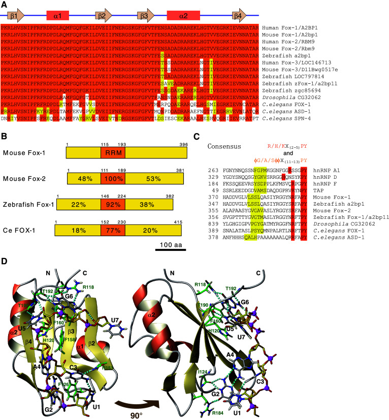
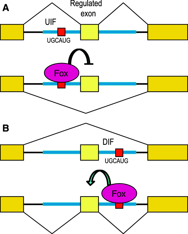
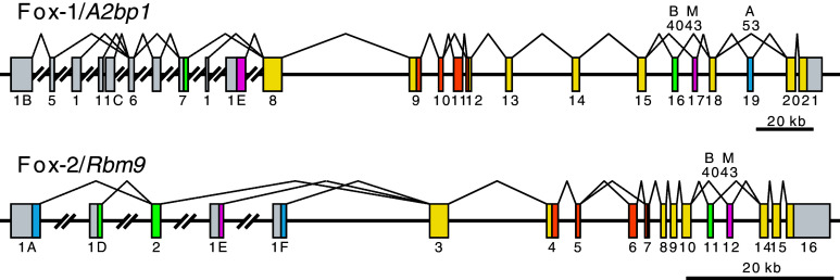
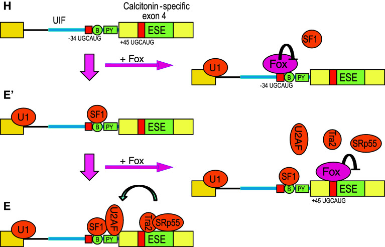
The Fox-1 family of RNA-binding proteins are evolutionarily conserved regulators of tissue-specific alternative splicing in metazoans. The Fox-1 family specifically recognizes the (U)GCAUG stretch in regulated exons or in flanking introns, and either promotes or represses target exons. Recent unbiased bioinformatics analyses of alternatively spliced exons and comparison of various vertebrate genomes identified the (U)GCAUG stretch as a highly conserved and widely distributed element enriched in intronic regions surrounding exons with altered inclusion in muscle, heart, and brain, consistent with specific expression of Fox-1 and Fox-2 in these tissues. Global identification of Fox-2 target RNAs in living cells revealed that many of the Fox-2 target genes themselves encode splicing regulators. Further systematic elucidation of target genes of the Fox-1 family and other splicing regulators in various tissues will lead to a comprehensive understanding of splicing regulatory networks.
Overview
The claims and statistics of this paper haven’t been checked yet.
Run the check — free, no account needed. It reads the open-access text and checks the sources it cites.
Integrity signals
Registry
Cites
Statistics
Figures
Verification
Claims the paper makes
The claims and statistics of this paper haven’t been checked yet.
Run the check — free, no account needed. It reads the open-access text and checks the sources it cites.
Claims other papers make about this one
Each is checked against this paper’s own text: does it say what the citing paper says it does?
In the citing paper
“RBFOX binding upstream of, or within, an alternative exon typically inhibits exon inclusion, whereas downstream binding usually promotes splicing48.”
From The neurogenetics of alternative splicing (2016) · cites this paper as [48]
Self-check
Percentages, sums and reported differences re-computed from the paper’s own numbers.
The self-check needs the paper’s full text.
Citations
Cites
MOLMOL: A program for display and analysis of macromolecular structures
Koradi, R., Billeter, M., & Wüthrich, K. (1996) · Journal of Molecular Graphics · doi:10.1016/0263-7855(96)00009-4 · Cited by 7,156
Alternative isoform regulation in human tissue transcriptomes
Wang, E. T., Sandberg, R., Luo, S., Khrebtukova, I., Zhang, L., Mayr, C., Kingsmore, S. F., Schroth, G. P., & Burge, C. B. (2008) · Nature · doi:10.1038/nature07509 · Cited by 5,181
Pan, Q., Shai, O., Lee, L. J., Frey, B. J., & Blencowe, B. J. (2008) · Nature Genetics · doi:10.1038/ng.259 · Cited by 3,951
Mechanisms of Alternative Pre-Messenger RNA Splicing
Black, D. L. (2003) · Annual Review of Biochemistry · doi:10.1146/annurev.biochem.72.121801.161720 · Cited by 2,704
HITS-CLIP yields genome-wide insights into brain alternative RNA processing
Licatalosi, D. D., Mele, A., Fak, J. J., Ule, J., Kayikci, M., Chi, S. W., Clark, T. A., Schweitzer, A. C., Blume, J. E., Wang, X., et al. (2008) · Nature · doi:10.1038/nature07488 · Cited by 1,352 · cited 2×
“These stories are reminiscent of the CLIP-seq analysis of NOVA that led to the discovery that NOVA interacts with 30 UTRs of target mRNAs and regulates alternative polyadenylation in the brain [43].”
MethodUnderstanding alternative splicing: towards a cellular code
Matlin, A. J., Clark, F., & Smith, C. W. J. (2005) · Nature Reviews Molecular Cell Biology · doi:10.1038/nrm1645 · Cited by 1,299
Alternative splicing: increasing diversity in the proteomic world
Graveley, B. R. (2001) · Trends in Genetics · doi:10.1016/s0168-9525(00)02176-4 · Cited by 1,228
Maris, C., Dominguez, C., & Allain, F. H. ‐. (2005) · FEBS Journal · doi:10.1111/j.1742-4658.2005.04653.x · Cited by 1,135
Lim, J., Hao, T., Shaw, C., Patel, A. J., Szabó, G., Rual, J.-F., Fisk, C. J., Li, N., Smolyar, A., Hill, D. E., et al. (2006) · Cell · doi:10.1016/j.cell.2006.03.032 · Cited by 789
ESRP1 and ESRP2 Are Epithelial Cell-Type-Specific Regulators of FGFR2 Splicing
Warzecha, C. C., Sato, T. K., Nabet, B., Hogenesch, J. B., & Carstens, R. P. (2009) · Molecular Cell · doi:10.1016/j.molcel.2009.01.025 · Cited by 580
Cited by
Sorted by citations
Mechanisms and Consequences of Alternative Polyadenylation
Di Giammartino, D. C., Nishida, K., & Manley, J. L. (2011) · Molecular Cell · doi:10.1016/j.molcel.2011.08.017 · Cited by 737
Evolution of SR protein and hnRNP splicing regulatory factors
Busch, A., & Hertel, K. J. (2011) · Wiley Interdisciplinary Reviews - RNA · doi:10.1002/wrna.100 · Cited by 418
Cell-Type-Specific Alternative Splicing Governs Cell Fate in the Developing Cerebral Cortex
Zhang, X., Chen, M. H., Wu, X., Kodani, A., Fan, J., Doan, R. N., Ozawa, M., Ma, J., Yoshida, N., Reiter, J. F., et al. (2016) · Cell · doi:10.1016/j.cell.2016.07.025 · Cited by 363
The neurogenetics of alternative splicing
Vuong, C. K., Black, D. L., & Zheng, S. (2016) · Nature Reviews Neuroscience · doi:10.1038/nrn.2016.27 · Cited by 340
The splicing regulator Rbfox1 (A2BP1) controls neuronal excitation in the mammalian brain
Gehman, L. T., Stoilov, P., Maguire, J., Damianov, A., Lin, C.-H., Shiue, L., Ares, M., Mody, I., & Black, D. L. (2011) · Nature Genetics · doi:10.1038/ng.841 · Cited by 331
Splicing-factor alterations in cancers
Anczuków, O., & Krainer, A. R. (2016) · RNA · doi:10.1261/rna.057919.116 · Cited by 304
Novel Insights into NeuN: from Neuronal Marker to Splicing Regulator
Duan, W., Zhang, Y., Hou, Z., Huang, C. L., Zhu, H., Zhang, C., & Yin, Q. (2015) · Molecular Neurobiology · doi:10.1007/s12035-015-9122-5 · Cited by 312
Cytoplasmic Rbfox1 Regulates the Expression of Synaptic and Autism-Related Genes
Lee, J.-A., Damianov, A., Lin, C.-H., Fontes, M., Parikshak, N. N., Anderson, E. S., Geschwind, D. H., Black, D. L., & Martin, K. C. (2016) · Neuron · doi:10.1016/j.neuron.2015.11.025 · Cited by 232
The splicing regulator Rbfox2 is required for both cerebellar development and mature motor function
Gehman, L. T., Meera, P., Stoilov, P., Shiue, L., O'Brien, J. E., Meisler, M. H., Ares, M., Otis, T. S., & Black, D. L. (2012) · Genes & Development · doi:10.1101/gad.182477.111 · Cited by 213
RBFOX1 regulates both splicing and transcriptional networks in human neuronal development
Fogel, B. L., Wexler, E., Wahnich, A., Friedrich, T., Vijayendran, C., Gao, F., Parikshak, N., Konopka, G., & Geschwind, D. H. (2012) · Human Molecular Genetics · doi:10.1093/hmg/dds240 · Cited by 216
Rbfox Proteins Regulate Splicing as Part of a Large Multiprotein Complex LASR
Damianov, A., Yi, Y., Lin, C.-H., Lee, J.-A., Tran, D., Vashisht, A. A., Bahrami‐Samani, E., Xing, Y., Martin, K. C., Wohlschlegel, J. A., et al. (2016) · Cell · doi:10.1016/j.cell.2016.03.040 · Cited by 210
Chronic cocaine-regulated epigenomic changes in mouse nucleus accumbens
Feng, J., Wilkinson, M., Liu, X., Purushothaman, I., Ferguson, D., Vialou, V., Maze, I., Shao, N., Kennedy, P., Koo, J., et al. (2014) · Genome Biology · doi:10.1186/gb-2014-15-4-r65 · Cited by 198
Genes and Athletic Performance: An Update
Ahmetov, I. I., Egorova, E. S., Gabdrakhmanova, L. J., & Fedotovskaya, O. N. (2016) · Medicine and sport science/Medicine and sport · doi:10.1159/000445240 · Cited by 183
RNA Protein Interaction in Neurons
Darnell, R. B. (2013) · Annual Review of Neuroscience · doi:10.1146/annurev-neuro-062912-114322 · Cited by 181
Vascular smooth muscle phenotypic diversity and function
Fisher, S. A. (2010) · Physiological Genomics · doi:10.1152/physiolgenomics.00111.2010 · Cited by 180
Bräutigam, C., Rago, L., Rolke, A., Waldmeier, L., Christofori, G., & Winter, J. (2013) · Oncogene · doi:10.1038/onc.2013.50 · Cited by 175
Developmental regulation of RNA processing by Rbfox proteins
Conboy, J. G. (2016) · Wiley Interdisciplinary Reviews - RNA · doi:10.1002/wrna.1398 · Cited by 133
Rbfox2 controls autoregulation in RNA-binding protein networksPreprint
Jangi, M., Boutz, P. L., Paul, P., & Sharp, P. A. (2014) · Genes & Development · doi:10.1101/gad.235770.113 · Cited by 136
RNA-binding proteins in neurological development and disease
Prashad, S., & Gopal, P. P. (2020) · RNA Biology · doi:10.1080/15476286.2020.1809186 · Cited by 124
Rbfox Splicing Factors Promote Neuronal Maturation and Axon Initial Segment Assembly
Jacko, M., Weyn‐Vanhentenryck, S. M., Smerdon, J. W., Yan, R., Feng, H., Williams, D. J., Pai, J. A., Xu, K., Wichterle, H., & Zhang, C. (2018) · Neuron · doi:10.1016/j.neuron.2018.01.020 · Cited by 131
Dysregulation of mRNA Localization and Translation in Genetic Disease
Wang, E. T., Taliaferro, J. M., Lee, J.-A., Sudhakaran, I. P., Rossoll, W., Groß, C., Moss, K. R., & Bassell, G. J. (2016) · Journal of Neuroscience · doi:10.1523/jneurosci.2352-16.2016 · Cited by 114
Rbfox1 Regulates Synaptic Transmission through the Inhibitory Neuron-Specific vSNARE Vamp1
Vuong, C. K., Wei, W., Lee, J.-A., Lin, C.-H., Damianov, A., de la Torre-Ubieta, L., Halabi, R., Otis, K. O., Martin, K. C., O’Dell, T. J., et al. (2018) · Neuron · doi:10.1016/j.neuron.2018.03.008 · Cited by 102
Mechanisms of activation and repression by the alternative splicing factors RBFOX1/2
Sun, S., Zhang, Z., Fregoso, O. I., & Krainer, A. R. (2011) · RNA · doi:10.1261/rna.030486.111 · Cited by 105
Alternative splicing of the cell fate determinant Numb in hepatocellular carcinoma
Lu, Y., Xu, W., Ji, J., Feng, D., Sourbier, C., Yang, Y., Qu, J., Zeng, Z., Wang, C., Chang, X., et al. (2015) · Hepatology · doi:10.1002/hep.27923 · Cited by 101
Dredge, B. K., & Jensen, K. B. (2011) · PLoS ONE · doi:10.1371/journal.pone.0021585 · Cited by 101
Microexons: discovery, regulation, and function
Ustianenko, D., Weyn‐Vanhentenryck, S. M., & Zhang, C. (2017) · Wiley Interdisciplinary Reviews - RNA · doi:10.1002/wrna.1418 · Cited by 104
Inference of transcriptional regulation in cancers
Jiang, P., Freedman, M. L., Liu, J. S., & Liu, X. S. (2015) · Proceedings of the National Academy of Sciences · doi:10.1073/pnas.1424272112 · Cited by 93
Orchestration of Neurodevelopmental Programs by RBFOX1
Bill, B. R., Lowe, J. K., DyBuncio, C. T., & Fogel, B. L. (2013) · International Review of Neurobiology · doi:10.1016/b978-0-12-418700-9.00008-3 · Cited by 94
Wei, C., Qiu, J., Zhou, Y., Xue, Y., Hu, J., Ouyang, K., Banerjee, I., Zhang, C., Chen, B., Li, H., et al. (2015) · Cell Reports · doi:10.1016/j.celrep.2015.02.013 · Cited by 95
Dou, X., Xiao, Y., Shen, C., Wang, K., Wu, T., Liu, C., Li, Y., Yu, X., Liu, J., Dai, Q., et al. (2023) · Nature Cell Biology · doi:10.1038/s41556-023-01213-w · Cited by 100
Diversification of the muscle proteome through alternative splicing
Nakka, K. K., Ghigna, C., Gabellini, D., & Dilworth, F. J. (2018) · Skeletal Muscle · doi:10.1186/s13395-018-0152-3 · Cited by 97
RBFox2 Binds Nascent RNA to Globally Regulate Polycomb Complex 2 Targeting in Mammalian Genomes
Wei, C., Xiao, R., Chen, L., Cui, H., Zhou, Y., Xue, Y., Hu, J., Zhou, B., Tsutsui, T., Qiu, J., et al. (2016) · Molecular Cell · doi:10.1016/j.molcel.2016.04.013 · Cited by 91
Repression of Pumilio Protein Expression by Rbfox1 Promotes Germ Cell Differentiation
Carreira-Rosario, A., Bhargava, V., Hillebrand, J., Kollipara, R. K., Ramaswami, M., & Buszczak, M. (2016) · Developmental Cell · doi:10.1016/j.devcel.2016.02.010 · Cited by 89
Satellite-like cells contribute to pax7-dependent skeletal muscle repair in adult zebrafish
Berberoglu, M. A., Gallagher, T. L., Morrow, Z. T., Talbot, J. C., Hromowyk, K. J., Tenente, I. M., Langenau, D. M., & Amacher, S. L. (2017) · Developmental Biology · doi:10.1016/j.ydbio.2017.03.004 · Cited by 85
Ahmetov, I. I., Kulemin, N. A., Викторович, П. Д., Naumov, V., Akimov, E. B., Bravyy, Y. R., Egorova, E. S., Galeeva, A. R., Generozov, E. V., Kostryukova, E., et al. (2014) · Biology of Sport · doi:10.5604/20831862.1124568 · Cited by 82
RNA ‐binding proteins, neural development and the addictions
Bryant, C. D., & Yazdani, N. (2015) · Genes Brain & Behavior · doi:10.1111/gbb.12273 · Cited by 82
RBFox2 Binds Nascent RNA to Globally Regulate Polycomb Complex 2 Targeting in Mammalian Genomes
Wei, C., Xiao, R., Chen, L., Cui, H., Zhou, Y., Xue, Y., Hu, J., Zhou, B., Tsutsui, T., Qiu, J., et al. (2016) · Molecular Cell · doi:10.1016/j.molcel.2016.06.003 · Cited by 81
Park, J. Y., Li, W., Zheng, D., Zhai, P., Zhao, Y., Matsuda, T., Vatner, S. F., Sadoshima, J., & Tian, B. (2011) · PLoS ONE · doi:10.1371/journal.pone.0022391 · Cited by 79
Shumay, E., Fowler, J. S., & Volkow, N. D. (2010) · PLoS ONE · doi:10.1371/journal.pone.0011067 · Cited by 80
Janakiraman, H., House, R. P., Gangaraju, V. K., Diehl, J. A., Howe, P. H., & Palanisamy, V. (2018) · Molecular Cancer Research · doi:10.1158/1541-7786.mcr-17-0547 · Cited by 74
Kuroyanagi, H., Ohno, G., Sakane, H., Maruoka, H., & Hagiwara, M. (2010) · Nature Protocols · doi:10.1038/nprot.2010.107 · Cited by 75
Self-citationRNA binding proteins in the regulation of heart development
Blech-Hermoni, Y. N., & Ladd, A. N. (2013) · The International Journal of Biochemistry & Cell Biology · doi:10.1016/j.biocel.2013.08.008 · Cited by 72
Chen, Y., Zubović, L., Yang, F., Godin, K. S., Pavelitz, T., Castellanos, J., Macchi, P., & Varani, G. (2016) · Nucleic Acids Research · doi:10.1093/nar/gkw177 · Cited by 66
RNA‐binding proteins in neurodegenerative disease: TDP‐43 and beyond
Hanson, K. A., Kim, S. H., & Tibbetts, R. S. (2011) · Wiley Interdisciplinary Reviews - RNA · doi:10.1002/wrna.111 · Cited by 68
Huntington’s disease-specific mis-splicing unveils key effector genes and altered splicing factors
Elorza, A., Márquez, Y., Cabrera, J. R., Sanchez‐Trincado, J. L., Santos‐Galindo, M., Hernández, I. H., Picó, S., Díaz‐Hernandéz, J. I., García‐Escudero, R., Irimia, M., et al. (2021) · Brain · doi:10.1093/brain/awab087 · Cited by 64
Decoding muscle alternative splicing
Llorian, M., & Smith, C. W. J. (2011) · Current Opinion in Genetics & Development · doi:10.1016/j.gde.2011.03.006 · Cited by 59
Concentration-dependent splicing is enabled by Rbfox motifs of intermediate affinity
Begg, B. E., Jens, M., Wang, P. Y., Minor, C. M., & Burge, C. B. (2020) · Nature Structural & Molecular Biology · doi:10.1038/s41594-020-0475-8 · Cited by 60
Bozack, A. K., Rifas‐Shiman, S. L., Coull, B. A., Baccarelli, A., Wright, R. O., Amarasiriwardena, C. J., Gold, D. R., Oken, E., Hivert, M., & Cárdenas, A. (2021) · Clinical Epigenetics · doi:10.1186/s13148-021-01198-z · Cited by 55
Runfola, V., Sebastian, S., Dilworth, F. J., & Gabellini, D. (2015) · Journal of Cell Science · doi:10.1242/jcs.161059 · Cited by 54
Maurin, M. M., Ranjouri, M., Megino‐Luque, C., Newberg, J. Y., Du, D., Martin, K., Miner, R. E., Prater, M. S., Wee, D. K. B., Centeno, B. A., et al. (2023) · Nature Communications · doi:10.1038/s41467-023-44126-w · Cited by 53
RBFox2-miR-34a-Jph2 axis contributes to cardiac decompensation during heart failure
Hu, J., Gao, C., Wei, C., Xue, Y., Shao, C., Hao, Y., Gou, L., Zhou, Y., Zhang, J., Ren, S. V., et al. (2019) · Proceedings of the National Academy of Sciences · doi:10.1073/pnas.1822176116 · Cited by 50
Rbfox proteins regulate alternative splicing of neuronal sodium channel SCN8A
O'Brien, J. E., Drews, V. L., Jones, J. M., Dugas, J. C., Barres, B. A., & Meisler, M. H. (2012) · Molecular and Cellular Neuroscience · doi:10.1016/j.mcn.2011.10.005 · Cited by 49
Stress Granules Contain Rbfox2 with Cell Cycle-related mRNAs
Park, C., Choi, S., Kim, Y.-E., Lee, S., Park, S.-H., Adelstein, R. S., Kawamoto, S., & Kim, K. K. (2017) · Scientific Reports · doi:10.1038/s41598-017-11651-w · Cited by 49
Kuroyanagi, H., Watanabe, Y., & Hagiwara, M. (2013) · PLoS Genetics · doi:10.1371/journal.pgen.1003337 · Cited by 46
Self-citationOhno, G., Ono, K., Togo, M., Watanabe, Y., Ono, S., Hagiwara, M., & Kuroyanagi, H. (2012) · PLoS Genetics · doi:10.1371/journal.pgen.1002991 · Cited by 45
Self-citationThe transcription factor ERG recruits CCR4–NOT to control mRNA decay and mitotic progression
Rambout, X., Detiffe, C., Bruyr, J., Mariavelle, E., Cherkaoui, M., Brohée, S., Demoitié, P., Lebrun, M., Soin, R., Lesage, B., et al. (2016) · Nature Structural & Molecular Biology · doi:10.1038/nsmb.3243 · Cited by 48
The function of spliceosome components in open mitosis
Hofmann, J. C., Husedzinovic, A., & Gruß, O. J. (2010) · Nucleus · doi:10.4161/nucl.1.6.13328 · Cited by 44
Maxeiner, S., Glassmann, A., Kao, H., & Schilling, K. (2013) · Histochemistry and Cell Biology · doi:10.1007/s00418-013-1159-9 · Cited by 45
Alternative splicing interference by xenobiotics
Zaharieva, E. E., Chipman, J. K., & Soller, M. (2012) · Toxicology · doi:10.1016/j.tox.2012.01.014 · Cited by 46
RNA splicing regulators play critical roles in neurogenesis
Fisher, E., & Feng, J. (2022) · Wiley Interdisciplinary Reviews - RNA · doi:10.1002/wrna.1728 · Cited by 45
Targeted inhibition of oncogenic miR-21 maturation with designed RNA-binding proteins
Chen, Y., Yang, F., Zubović, L., Pavelitz, T., Yang, W. J., Godin, K. S., Walker, M. J. P., Zheng, S., Macchi, P., & Varani, G. (2016) · Nature Chemical Biology · doi:10.1038/nchembio.2128 · Cited by 42
Zhou, Y., Fan, J., Zhu, H., Li, J., Fan, W., Kapoor, I., Wang, Y., Wang, Y., Zhu, G., & Wang, J. (2017) · Hypertension · doi:10.1161/hypertensionaha.117.09301 · Cited by 41
Cibi, D. M., Mia, M. M., Shekeran, S. G., Yun, L. S., Sandireddy, R., Gupta, P. S., Hota, M., Sun, L., Ghosh, S., & Singh, M. K. (2019) · eLife · doi:10.7554/elife.45418 · Cited by 46
Pistoni, M., Shiue, L., Cline, M., Bortolanza, S., Neguembor, M. V., Xynos, A., Ares, M., & Gabellini, D. (2013) · PLoS Genetics · doi:10.1371/journal.pgen.1003186 · Cited by 40
Liver RBFOX2 regulates cholesterol homeostasis via Scarb1 alternative splicing in mice
Paterson, H. A. B., Yu, S., Artigas, N., Prado, M. A., Haberman, N., Wang, Y.-F., Jobbins, A. M., Pahita, E., Mokochinski, J. B., Hall, Z., et al. (2022) · Nature Metabolism · doi:10.1038/s42255-022-00681-y · Cited by 45
Two Distinct Categories of Focal Deletions in Cancer Genomes
Rajaram, M., Zhang, J., Wang, T., Li, J., Kuscu, C., Qi, H., Kato, M., Grubor, V. D., Weil, R. J., Helland, Å., et al. (2013) · PLoS ONE · doi:10.1371/journal.pone.0066264 · Cited by 45
Evaluation of A2BP1 as an Obesity Gene
Ma, L., Hanson, R. L., Traurig, M. T., Muller, Y. L., Kaur, B. P., Perez, J. M., Meyre, D., Fu, M., Körner, A., Franks, P. W., et al. (2010) · Diabetes · doi:10.2337/db09-1604 · Cited by 41
Parallels between neuron and lens fiber cell structure and molecular regulatory networks
Frederikse, P. H., Kasinathan, C., & Kleiman, N. J. (2012) · Developmental Biology · doi:10.1016/j.ydbio.2012.05.022 · Cited by 38
Jiang, P., Singh, M., & Coller, H. A. (2013) · PLoS Computational Biology · doi:10.1371/journal.pcbi.1003075 · Cited by 38
Li, P., Yu, Z., He, L., Zhou, D., Xie, S., Hou, H., & Geng, X. (2017) · Biomedicine & Pharmacotherapy · doi:10.1016/j.biopha.2017.05.087 · Cited by 36
Alternative mRNA Splicing in the Pathogenesis of Obesity
Wong, C., Xu, L., & Yau, M. Y. C. (2018) · International Journal of Molecular Sciences · doi:10.3390/ijms19020632 · Cited by 34
RBFOX and SUP-12 sandwich a G base to cooperatively regulate tissue-specific splicing
Kuwasako, K., Takahashi, M., Unzai, S., Tsuda, K., Yoshikawa, S., He, F., Kobayashi, N., Güntert, P., Shirouzu, M., Ito, T., et al. (2014) · Nature Structural & Molecular Biology · doi:10.1038/nsmb.2870 · Cited by 33
Self-citationLucas, C. G., Calvez, M., Babu, R., & Brown, A. M. (2013) · Neuroscience Letters · doi:10.1016/j.neulet.2013.10.037 · Cited by 38
Shen, F., Xu, X. M., Yu, Z., Li, H., Shen, H., Li, X., Shen, M., & Chen, G. (2020) · Journal of Cerebral Blood Flow & Metabolism · doi:10.1177/0271678x20916860 · Cited by 33
Zubović, L., Baralle, M., & Baralle, F. E. (2012) · Nucleic Acids Research · doi:10.1093/nar/gks249 · Cited by 30
RetractedNlend, R. N., Meyer, K., & Schümperli, D. (2010) · RNA Biology · doi:10.4161/rna.7.4.12206 · Cited by 30
A widespread length-dependent splicing dysregulation in cancer
Zhang, S., Mao, M., Lv, Y., Yang, Y., He, W., Song, Y., Wang, Y., Yang, Y., Abo, M. A., Freedman, J. A., et al. (2022) · Science Advances · doi:10.1126/sciadv.abn9232 · Cited by 26
RNA splicing regulated by RBFOX1 is essential for cardiac function in zebrafish
Frese, K., Meder, B., Keller, A., Just, S., Haas, J., Vogel, B., Fischer, S., Backes, C., Matzas, M., Köhler, D., et al. (2015) · Journal of Cell Science · doi:10.1242/jcs.166850 · Cited by 27
RNA and neuronal function: the importance of post-transcriptional regulation
Bhat, V. D., Jayaraj, J., & Babu, K. (2022) · Oxford Open Neuroscience · doi:10.1093/oons/kvac011 · Cited by 26
RBFOX2 protein domains and cellular activities
Arya, A. D., Wilson, D. I., Baralle, D., & Raponi, M. (2014) · Biochemical Society Transactions · doi:10.1042/bst20140050 · Cited by 26
Conserved functions of RNA-binding proteins in muscle
Nikonova, E. V., Kao, S.-Y., Ravichandran, K., Wittner, A., & Spletter, M. L. (2019) · The International Journal of Biochemistry & Cell Biology · doi:10.1016/j.biocel.2019.02.008 · Cited by 24
Parra, M., Gee, S. L., Mohandas, N., & Conboy, J. G. (2010) · Journal of Biological Chemistry · doi:10.1074/jbc.m110.158154 · Cited by 25
Protein kinase WNK3 regulates the neuronal splicing factor Fox-1
Lee, A.-Y., Chen, W., Stippec, S., Self, J., Yang, F., Ding, X., Chen, S., Juang, Y.-C. A., & Cobb, M. H. (2012) · Proceedings of the National Academy of Sciences · doi:10.1073/pnas.1215406109 · Cited by 28
Analysis of PI3K pathway components in human cancers
DARAGMEH, J., Barriah, W., Saad, B., & Zaid, H. (2016) · Oncology Letters · doi:10.3892/ol.2016.4309 · Cited by 25
Tissue-Specific Alternative Splicing of Tak1 Is Conserved in Deuterostomes
Venables, J. P., Vignal, E., Baghdiguian, S., Fort, P., & Tazi, J. (2011) · Molecular Biology and Evolution · doi:10.1093/molbev/msr193 · Cited by 25
Amrane, S., Rébora, K., Zniber, I., Dupuy, D., & Mackereth, C. D. (2014) · Nature Communications · doi:10.1038/ncomms5595 · Cited by 23
Su, W., Lv, M., Wang, D., He, Y., Han, H., Zhang, Y., Zhang, X., Lv, S., & Yao, L. (2024) · Mediators of Inflammation · doi:10.1155/2024/7459054 · Cited by 22
RNA-Binding Proteins in Heart Development
Giudice, J., & Cooper, T. A. (2014) · Advances in experimental medicine and biology · doi:10.1007/978-1-4939-1221-6_11 · Cited by 23
RNA-binding proteins regulating the CD44 alternative splicing
Maltseva, D. V., & Tonevitsky, A. (2023) · Frontiers in Molecular Biosciences · doi:10.3389/fmolb.2023.1326148 · Cited by 23
Hagen, E. M., Sicko, R. J., Kay, D. M., Rigler, S. L., Dimopoulos, A., Ahmad, S., Doleman, M. H., Fan, R., Romitti, P. A., Browne, M. L., et al. (2016) · Human Genetics · doi:10.1007/s00439-016-1727-x · Cited by 21
Chen, P.-F., Hsiao, J. S., Sirois, C. L., & Chamberlain, S. J. (2016) · Scientific Reports · doi:10.1038/srep25368 · Cited by 21
The Role of Genetic Polymorphisms in Diabetic Retinopathy: Narrative Review
Sienkiewicz‐Szłapka, E., Fiedorowicz, E., Król-Grzymała, A., Kordulewska, N. K., Rozmus, D., Cieślińska, A., & Grzybowski, A. E. (2023) · International Journal of Molecular Sciences · doi:10.3390/ijms242115865 · Cited by 20
Improved discovery of RNA-binding protein binding sites in eCLIP data using DEWSeq
Schwarzl, T., Sahadevan, S., Lang, B., Miladi, M., Backofen, R., Huber, W. K., Hentze, M. W., & Tartaglia, G. G. (2023) · Nucleic Acids Research · doi:10.1093/nar/gkad998 · Cited by 21
MEF2C exon α: Role in gene activation and differentiation
Infantino, V., Convertini, P., Menga, A., & Iacobazzi, V. (2013) · Gene · doi:10.1016/j.gene.2013.08.044 · Cited by 19
Wen, J., Chiba, A., & Cai, X. (2010) · Nucleic Acids Research · doi:10.1093/nar/gkq679 · Cited by 19
Kim, Y., Kim, J. O., Park, K.-S., Won, M., Kim, K. E., & Kim, K. K. (2016) · Molecules and Cells · doi:10.14348/molcells.2016.0150 · Cited by 20
Wang, J., Li, G., Yu, D., Wong, Y. P., Yong, T. F., Liang, M. C., Liao, P. H., Foo, R., Hoppe, U. C., & Soong, T. W. (2017) · Channels · doi:10.1080/19336950.2017.1381805 · Cited by 19
The combinatorial control of alternative splicing in C. elegans
Tan, J. H., & Fraser, A. G. (2017) · PLoS Genetics · doi:10.1371/journal.pgen.1007033 · Cited by 18
Bitel, C. L., Nathan, R. S., Wong, P., Kuppasani, S., Matsushita, M., Kanazawa, H., & Frederikse, P. H. (2010) · Current Eye Research · doi:10.3109/02713683.2010.500114 · Cited by 18
Hakim, N. H. A., Majlis, B. Y., Suzuki, H., & Tsukahara, T. (2016) · BioScience Trends · doi:10.5582/bst.2016.01169 · Cited by 18
Metadata
- Authors
- Kuroyanagi, H.
- Affiliations
- Tokyo Medical and Dental University (JP)
- Corresponding author
- Kuroyanagi, Hidehito
- ORCID
- Kuroyanagi, Hidehito
- Venue
- Cellular and Molecular Life Sciences
- Published
- 2009-08-18
- Volume
- 66
- Issue
- 24
- Pages
- 3895-3907
- ISSN
- 1420-682X (ISSN-L)
- 1420-9071
- PMC
- PMC2777236
- OpenAlex
- W2057774013
- Cited by
- 187
- References
- 78
- Citations by year
- 2026: 5 · 2025: 6 · 2024: 12 · 2023: 10 · 2022: 13 · 2021: 7 · 2020: 9 · 2019: 9 · 2018: 12 · 2017: 11 · 2016: 23 · 2015: 10 · 2014: 10 · 2013: 17 · 2012: 11
- FWCI
- 0.84
- Type
- journal article
- Language
- en
- Open access
- hybrid
- doi.org
- Licence
- cc-by-nc
- Locations
- Cellular and Molecular Life Sciences (publishedVersion, cc-by-nc)
- PubMed Central (publishedVersion, cc-by-nc)
- PubMed (publishedVersion)
- Funders
- Tokyo Medical and Dental University
- Japan Society for the Promotion of Science
- Topics
- Life Sciences › Biochemistry, Genetics and Molecular Biology › Molecular Biology › RNA Research and Splicing
- Life Sciences › Biochemistry, Genetics and Molecular Biology › Molecular Biology › RNA regulation and disease
- Life Sciences › Biochemistry, Genetics and Molecular Biology › Molecular Biology › RNA and protein synthesis mechanisms
- Keywords
- RNA-binding proteins, alternative splicing, intronic splicing enhancer, muscle, heart, brain, splicing regulators, splicing regulatory network, vertebrate genomes
- MeSH terms
- RNA Splicing Factors, Amino Acid Sequence, Animals, Base Sequence, Binding Sites, Humans, Molecular Sequence Data, RNA-Binding Proteins, Spliceosomes, Sequence Homology, Amino Acid, Alternative Splicing
About these checks
Registry, retraction, Cites and statistics results are computed, not judged, with one exception: a printed reference that no index lists is read once by a model before it counts as a citation error. Claims are extracted by a model and kept only when their quote appears verbatim in the text; each cited source is then checked, with a model’s help, for whether it says what the claim relies on. Those verdicts can be wrong — read the claim’s quote and the cited source before relying on one.
Statistics and arithmetic are recomputed, not judged.
Confidence bands are shown only for fields calibrated on our annotated test set. None is calibrated yet, so no band is shown.
What counts as a claim ›How we measure confidence ›How we check this ›
Are you an author, or do you see a problem with this page? Request a review
