

The DOI matches its Crossref record; 208 of the 208 references it cites were found in a registry or an index.
Cadherins in early neural development
Open accessAbstract
During early neural development, changes in signalling inform the expression of transcription factors that in turn instruct changes in cell identity. At the same time, switches in adhesion molecule expression result in cellular rearrangements that define the morphology of the emerging neural tube. It is becoming increasingly clear that these two processes influence each other; adhesion molecules do not simply operate downstream of or in parallel with changes in cell identity but rather actively feed into cell fate decisions. Why are differentiation and adhesion so tightly linked? It is now over 60 years since Conrad Waddington noted the remarkable "Constancy of the Wild Type” (Waddington in Nature 183: 1654–1655, 1959) yet we still do not fully understand the mechanisms that make development so reproducible. Conversely, we do not understand why directed differentiation of cells in a dish is sometimes unpredictable and difficult to control. It has long been suggested that cells make decisions as 'local cooperatives' rather than as individuals (Gurdon in Nature 336: 772–774, 1988; Lander in Cell 144: 955–969, 2011). Given that the cadherin family of adhesion molecules can simultaneously influence morphogenesis and signalling, it is tempting to speculate that they may help coordinate cell fate decisions between neighbouring cells in the embryo to ensure fidelity of patterning, and that the uncoupling of these processes in a culture dish might underlie some of the problems with controlling cell fate decisions ex-vivo. Here we review the expression and function of cadherins during early neural development and discuss how and why they might modulate signalling and differentiation as neural tissues are formed.
Overview
The claims and statistics of this paper haven’t been checked yet.
Run the check — free, no account needed. It reads the open-access text and checks the sources it cites.
Integrity signals
Registry
Cites
Statistics
Figures
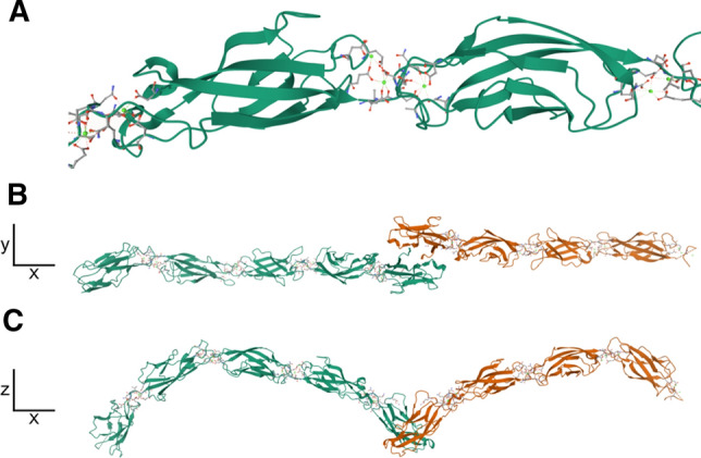
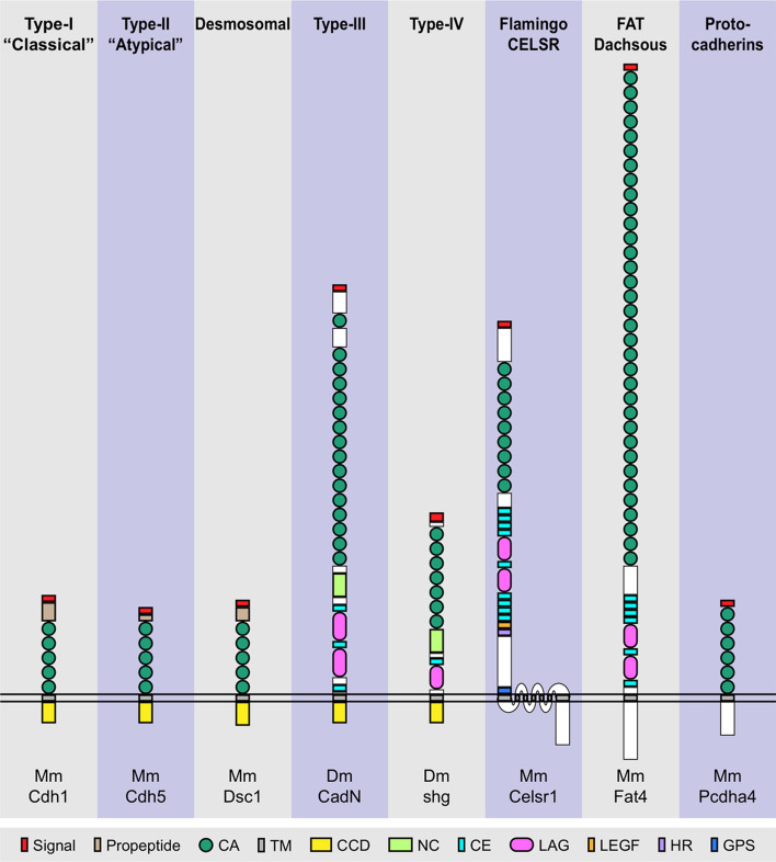
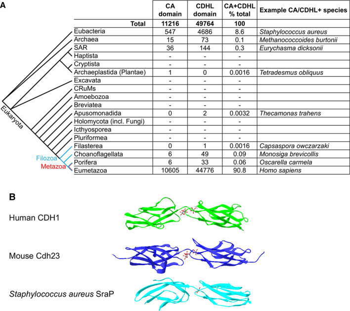

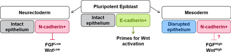
Verification
Claims the paper makes
The claims and statistics of this paper haven’t been checked yet.
Run the check — free, no account needed. It reads the open-access text and checks the sources it cites.
Claims other papers make about this one
Each is checked against this paper’s own text: does it say what the citing paper says it does?
In the citing paper
“Cadherins are a family of calcium-dependent transmembrane adhesion proteins that link β- and α-catenin to the actin cytoskeletal network (Punovuori et al., 2021) and also regulate cellular homeostasis through signaling mediating development, proliferation, apoptosis, and disease pathology (Yulis et al., 2018).”
From APOE3 Christchurch modulates β-catenin/Wnt signaling in iPS cell-derived cerebral organoids from Alzheimer’s cases (2024) · cites this paper as Punovuori et al., 2021
Self-check
Percentages, sums and reported differences re-computed from the paper’s own numbers.
The self-check needs the paper’s full text.
Citations
Cites
SWISS‐MODEL and the Swiss‐Pdb Viewer: An environment for comparative protein modeling
Guex, N., & Peitsch, M. C. (1997) · Electrophoresis · doi:10.1002/elps.1150181505 · Cited by 11,312
Epithelial-Mesenchymal Transitions in Development and Disease
Thiery, J. P., Acloque, H., Huang, R. Y. J., & Nieto, M. A. (2009) · Cell · doi:10.1016/j.cell.2009.11.007 · Cited by 9,931
Highly efficient neural conversion of human ES and iPS cells by dual inhibition of SMAD signaling
Chambers, S. M., Fasano, C. A., Papapetrou, E. P., Tomishima, M., Sadelain, M., & Studer, L. (2009) · Nature Biotechnology · doi:10.1038/nbt.1529 · Cited by 3,620
New cell lines from mouse epiblast share defining features with human embryonic stem cells
Tesar, P. J., Chenoweth, J. G., Brook, F. A., Davies, T. J., Evans, E. P., Mack, D. L., Gardner, R. L., & McKay, R. D. G. (2007) · Nature · doi:10.1038/nature05972 · Cited by 2,187
Ying, Q.-L., Nichols, J., Chambers, I., & Smith, A. G. (2003) · Cell · doi:10.1016/s0092-8674(03)00847-x · Cited by 2,114
Derivation of pluripotent epiblast stem cells from mammalian embryos
Brons, I. G. M., Smithers, L. E., Trotter, M. W. B., Rugg‐Gunn, P. J., Sun, B., Lopes, S. M. C. D. S., Howlett, S., Clarkson, A., Ährlund‐Richter, L., Pedersen, R. A., et al. (2007) · Nature · doi:10.1038/nature05950 · Cited by 2,007
In vitro differentiation of transplantable neural precursors from human embryonic stem cells
Zhang, S., Wernig, M., Duncan, I. D., Brüstle, O., & Thomson, J. A. (2001) · Nature Biotechnology · doi:10.1038/nbt1201-1129 · Cited by 1,935
The New Higher Level Classification of Eukaryotes with Emphasis on the Taxonomy of Protists
Adl, S., Simpson, A., Farmer, M. A., Andersen, R., Anderson, O., Barta, J. R., Bowser, S., Brugerolle, G., Fensome, R., Fredericq, S., et al. (2005) · Journal of Eukaryotic Microbiology · doi:10.1111/j.1550-7408.2005.00053.x · Cited by 1,749
The Revised Classification of Eukaryotes
Adl, S. M., Simpson, A. G. B., Lane, C. E., Lukeš, J., Bass, D., Bowser, S. S., Brown, M. W., Burki, F., Dunthorn, M., Hampl, V., et al. (2012) · Journal of Eukaryotic Microbiology · doi:10.1111/j.1550-7408.2012.00644.x · Cited by 1,680
The cadherins: cell-cell adhesion molecules controlling animal morphogenesis
Takeichi, M. (1988) · Development · doi:10.1242/dev.102.4.639 · Cited by 1,615
Conversion of embryonic stem cells into neuroectodermal precursors in adherent monoculture
Ying, Q.-L., Stavridis, M., Griffiths, D., Li, M., & Smith, A. (2003) · Nature Biotechnology · doi:10.1038/nbt780 · Cited by 1,504
Morphogenetic roles of classic cadherins
Takeichi, M. (1995) · Current Opinion in Cell Biology · doi:10.1016/0955-0674(95)80102-2 · Cited by 1,467
The cell-cell adhesion molecule E-cadherin
Roy, F. M. V., & Berx, G. (2008) · Cellular and Molecular Life Sciences · doi:10.1007/s00018-008-8281-1 · Cited by 1,392
Revisions to the Classification, Nomenclature, and Diversity of Eukaryotes
Adl, S., Bass, D., Lane, C. E., Lukeš, J., Schoch, C., Smirnov, A., Agatha, S., Berney, C., Brown, M. W., Burki, F., et al. (2018) · Journal of Eukaryotic Microbiology · doi:10.1111/jeu.12691 · Cited by 1,218
Canalization of Development and Genetic Assimilation of Acquired Characters
Waddington, C. H. (1959) · Nature · doi:10.1038/1831654a0 · Cited by 1,109
Li, R., Liang, J., Ni, S., Zhou, T., Qing, X., Li, H., He, W., Chen, J., Li, F., Zhuang, Q., et al. (2010) · Cell Stem Cell · doi:10.1016/j.stem.2010.04.014 · Cited by 1,072
Hu, B., Weick, J. P., Yu, J., Ma, L., Zhang, X., Thomson, J. A., & Zhang, S. (2010) · Proceedings of the National Academy of Sciences · doi:10.1073/pnas.0910012107 · Cited by 1,037
E-cadherin null mutant embryos fail to form a trophectoderm epithelium.
Larue, L., Ohsugi, M., Hirchenhain, J., & Kemler, R. (1994) · Proceedings of the National Academy of Sciences · doi:10.1073/pnas.91.17.8263 · Cited by 930
Clonal analysis of epiblast fate during germ layer formation in the mouse embryo
Lawson, K. A., Meneses, J. J., & Pedersen, R. A. (1991) · Development · doi:10.1242/dev.113.3.891 · Cited by 881
Making muscle: skeletal myogenesis in vivo and in vitro
Chal, J., & Pourquié, O. (2017) · Development · doi:10.1242/dev.151035 · Cited by 834
Making a commitment: cell lineage allocation and axis patterning in the early mouse embryo
Arnold, S. J., & Robertson, E. J. (2009) · Nature Reviews Molecular Cell Biology · doi:10.1038/nrm2618 · Cited by 816 · cited 2×
“This might explain why downregulation of E-cadherin does not result in neural differentiation in the posterior of the embryo, where several BMP family members (together with other anti-neural signals) are expressed at high levels [54].”
BackgroundWheelock, M. J., Shintani, Y., Maeda, M., Fukumoto, Y., & Johnson, K. R. (2008) · Journal of Cell Science · doi:10.1242/jcs.000455 · Cited by 816
Wnt-3a regulates somite and tailbud formation in the mouse embryo
Takada, S., Stark, K. L., Shea, M. J., Vassileva, G., McMahon, J. A., & McMahon, A. P. (1994) · Trends in Genetics · doi:10.1016/0168-9525(94)90090-6 · Cited by 789
Wnt-3a regulates somite and tailbud formation in the mouse embryo.Preprint
Takada, S., Stark, K. L., Shea, M. J., et al. (1994) · Genes & Development · doi:10.1101/gad.8.2.174 · Cited by 786
Developmental Defects in Mouse Embryos Lacking N-Cadherin
Radice, G. L., Rayburn, H., Matsunami, H., Knudsen, K. A., Takeichi, M., & Hynes, R. O. (1997) · Developmental Biology · doi:10.1006/dbio.1996.8443 · Cited by 723
Nollet, F., Kools, P., & Roy, F. M. V. (2000) · Journal of Molecular Biology · doi:10.1006/jmbi.2000.3777 · Cited by 720
Ciruna, B. G., & Rossant, J. (2001) · Developmental Cell · doi:10.1016/s1534-5807(01)00017-x · Cited by 695
Heasman, J., Crawford, A., Goldstone, K., Garner-Hamrick, P., Gumbiner, B., McCrea, P., Kintner, C., Noro, C., & Wylie, C. (1994) · Cell · doi:10.1016/0092-8674(94)90069-8 · Cited by 693
Hatta, K., & Takeichi, M. (1986) · Nature · doi:10.1038/320447a0 · Cited by 648
Cell adhesion in cancer: Beyond the migration of single cells
Janiszewska, M., Primi, M. C., & Izard, T. (2020) · Journal of Biological Chemistry · doi:10.1074/jbc.rev119.007759 · Cited by 644
Functional correlation between cell adhesive properties and some cell surface proteins.
Takeichi, M. (1977) · The Journal of Cell Biology · doi:10.1083/jcb.75.2.464 · Cited by 626
Pluripotency Factors in Embryonic Stem Cells Regulate Differentiation into Germ Layers
Thomson, M. W., Liu, S. J., Zou, L.-N., Smith, Z., Meissner, A., & Ramanathan, S. (2011) · Cell · doi:10.1016/j.cell.2011.05.017 · Cited by 566
Distinct Lineage Specification Roles for NANOG, OCT4, and SOX2 in Human Embryonic Stem Cells
Wang, Z., Oron, E., Nelson, B., Razis, S., & Ivanova, N. B. (2012) · Cell stem cell · doi:10.1016/j.stem.2012.02.016 · Cited by 565
E-cadherin in contact inhibition and cancer
Mendonsa, A. M., Na, T.-Y., & Gumbiner, B. M. (2018) · Oncogene · doi:10.1038/s41388-018-0304-2 · Cited by 562
Molecular evolution of the cadherin superfamily
Hulpiau, P., & Roy, F. M. V. (2008) · The International Journal of Biochemistry & Cell Biology · doi:10.1016/j.biocel.2008.09.027 · Cited by 524
Initiation of neural induction by FGF signalling before gastrulation
Streit, A., Berliner, A. J., Papanayotou, C., Sirulnik, A., & Stern, C. D. (2000) · Nature · doi:10.1038/35017617 · Cited by 521
A signaling pathway leading to metastasis is controlled by N-cadherin and the FGF receptor
Suyama, K., Shapiro, I. M., Guttman, M., & Hazan, R. B. (2002) · Cancer Cell · doi:10.1016/s1535-6108(02)00150-2 · Cited by 515
Tead4 is required for specification of trophectoderm in pre-implantation mouse embryos
Nishioka, N., Yamamoto, S., Kiyonari, H., Sato, H., Sawada, A., Ota, M., Nakao, K., & Sasaki, H. (2007) · Mechanisms of Development · doi:10.1016/j.mod.2007.11.002 · Cited by 513
A targeted mutation in the mouse E-cadherin gene results in defective preimplantation development.
Riethmacher, D., Brinkmann, V., & Birchmeier, C. (1995) · Proceedings of the National Academy of Sciences · doi:10.1073/pnas.92.3.855 · Cited by 505
Cadherin Regulates Dendritic Spine Morphogenesis
Togashi, H., Abe, K., Mizoguchi, A., Takaoka, K., Chisaka, O., & Takeichi, M. (2002) · Neuron · doi:10.1016/s0896-6273(02)00748-1 · Cited by 485
Nose, A., & Takeichi, M. (1986) · The Journal of Cell Biology · doi:10.1083/jcb.103.6.2649 · Cited by 431
Redefining the Progression of Lineage Segregations during Mammalian Embryogenesis by Clonal Analysis
Tzouanacou, E., Wegener, A., Wymeersch, F. J., Wilson, V. A., & Nicolas, J.-F. (2009) · Developmental Cell · doi:10.1016/j.devcel.2009.08.002 · Cited by 427
Harrison, O. J., Jin, X., Hong, S., Bahna, F. A., Ahlsén, G., Brasch, J., Wu, Y., Vendôme, J., Felsövályi, K., Hampton, C. M., et al. (2011) · Structure · doi:10.1016/j.str.2010.11.016 · Cited by 420
Hyafil, F., Morello, D., Babinet, C., & Jacob, F. (1980) · Cell · doi:10.1016/0092-8674(80)90456-0 · Cited by 417
The role of FGF/Erk signaling in pluripotent cells
Lanner, F., & Rossant, J. (2010) · Development · doi:10.1242/dev.050146 · Cited by 413
Damsky, C. H., Richa, J., Solter, D., Knudsen, K. A., & Buck, C. A. (1983) · Cell · doi:10.1016/0092-8674(83)90379-3 · Cited by 391
Tam, P., & Zhou, S. X. (1996) · Developmental Biology · doi:10.1006/dbio.1996.0203 · Cited by 391
A community effect in animal development
Gurdon, J. B. (1988) · Nature · doi:10.1038/336772a0 · Cited by 385
Gouti, M., Tsakiridis, A., Wymeersch, F. J., Huang, Y., Kleinjung, J., Wilson, V. A., & Briscoe, J. (2014) · PLoS Biology · doi:10.1371/journal.pbio.1001937 · Cited by 372
Uncoupling cadherin-based adhesion from wingless signalling in Drosophila
Sanson, B., White, P., & Vincent, J. (1996) · Nature · doi:10.1038/383627a0 · Cited by 360
A role for cadherins in tissue formation
Larue, L., Antos, C. L., Butz, S., Huber, O., Delmas, V., Dominis, M., & Kemler, R. (1996) · Development · doi:10.1242/dev.122.10.3185 · Cited by 354
Some flagellates (Protista) from tropical marine sediments
Larsen, J., & Patterson, D. J. (1990) · Journal of Natural History · doi:10.1080/00222939000770571 · Cited by 350
The origin of Metazoa: a unicellular perspective
Sebé-Pedrós, A., Degnan, B. M., & Ruiz-Trillo, I. (2017) · Nature Reviews Genetics · doi:10.1038/nrg.2017.21 · Cited by 332
Aubert, J., Dunstan, H., Chambers, I., & Smith, A. G. (2002) · Nature Biotechnology · doi:10.1038/nbt763 · Cited by 325
Yoshida, C., Suzuki, N., & Takeichi, M. (1984) · Developmental Biology · doi:10.1016/0012-1606(84)90112-x · Cited by 322
From morphogen to morphogenesis and back
Gilmour, D. T., Rembold, M., & Leptin, M. (2017) · Nature · doi:10.1038/nature21348 · Cited by 311
Yoshikawa, Y., Fujimori, T., McMahon, A., & Takada, S. (1997) · Developmental Biology · doi:10.1006/dbio.1997.8502 · Cited by 308
A discrete period of FGF-induced Erk1/2 signalling is required for vertebrate neural specification
Stavridis, M. P., Lunn, J. S., Collins, B., & Storey, K. G. (2007) · Development · doi:10.1242/dev.02858 · Cited by 308
Aaku‐Saraste, E., Hellwig, A., & Huttner, W. Β. (1996) · Developmental Biology · doi:10.1006/dbio.1996.0336 · Cited by 295
Redmer, T., Diecke, S., Grigoryan, T., Quiroga‐Negreira, A., Birchmeier, W., & Besser, D. (2011) · EMBO Reports · doi:10.1038/embor.2011.88 · Cited by 295
Stem cells, signals and vertebrate body axis extension
Wilson, V., Olivera-Martinez, I., & Storey, K. G. (2009) · Development · doi:10.1242/dev.021246 · Cited by 290
Characterization of L-CAM, a major cell adhesion molecule from embryonic liver cells
Gallin, W. J., Edelman, G. M., & Cunningham, B. A. (1983) · Proceedings of the National Academy of Sciences · doi:10.1073/pnas.80.4.1038 · Cited by 287
Precocious Mammary Gland Development in P-Cadherin–deficient Mice
Radice, G., Ferreira-Cornwell, M., Robinson, S., Rayburn, H., Chodosh, L., Takeichi, M., & Hynes, R. (1997) · Journal of Cell Biology · doi:10.1083/jcb.139.4.1025 · Cited by 286
Behrens, J., Birchmeier, W., Goodman, S. L., & Imhof, B. A. (1985) · The Journal of Cell Biology · doi:10.1083/jcb.101.4.1307 · Cited by 283
Yoshida, C., & Takeichi, M. (1982) · Cell · doi:10.1016/0092-8674(82)90339-7 · Cited by 266
Notch Promotes Neural Lineage Entry by Pluripotent Embryonic Stem Cells
Lowell, S., Benchoua, A., Heavey, B., & Smith, A. G. (2006) · PLoS Biology · doi:10.1371/journal.pbio.0040121 · Cited by 260
Self-citationGouti, M., Delile, J., Stamataki, D., Wymeersch, F. J., Huang, Y., Kleinjung, J., Wilson, V. A., & Briscoe, J. (2017) · Developmental Cell · doi:10.1016/j.devcel.2017.04.002 · Cited by 253
Kimura, Y., Matsunami, H., Inoue, T., Shimamura, K., Uchida, N., Ueno, T., Miyazaki, T., & Takeichi, M. (1995) · Developmental Biology · doi:10.1006/dbio.1995.1149 · Cited by 251
Zhu, Q., Song, L., Peng, G., Sun, N., Chen, J., Zhang, T., Sheng, N., Tang, W., Qian, C., Qiao, Y., et al. (2014) · eLife · doi:10.7554/elife.02224 · Cited by 249
Neurulation: coming to closure
Smith, J. L. (1997) · Trends in Neurosciences · doi:10.1016/s0166-2236(97)01121-1 · Cited by 246
The Premetazoan Ancestry of Cadherins
Sigg, M. A., & King, N. (2008) · Science · doi:10.1126/science.1151084 · Cited by 240
Stem cells, signals and vertebrate body axis extension
Wilson, V. A., Olivera-Martínez, I., & Storey, K. G. (2009) · Development · doi:10.1242/dev.039172 · Cited by 239
Cadherin-dependent filopodia control preimplantation embryo compaction
Fierro-González, J. C., White, M. D., Silva, J. C., & Plachta, N. (2013) · Nature Cell Biology · doi:10.1038/ncb2875 · Cited by 236
Surface antigen in early differentiation.
Kemler, R., Babinet, C., Eisen, H., & Jacob, F. (1977) · Proceedings of the National Academy of Sciences · doi:10.1073/pnas.74.10.4449 · Cited by 236
Lamina-specific expression of adhesion molecules in developing chick optic tectum
Yamagata, M., Herman, J., & Sanes, J. R. (1995) · Journal of Neuroscience · doi:10.1523/jneurosci.15-06-04556.1995 · Cited by 230
Distinct Wnt-driven primitive streak-like populations reflect in vivo lineage precursors
Tsakiridis, A., Huang, Y., Blin, G., Skylaki, S., Wymeersch, F. J., Osorno, R., Economou, C. N., Karagianni, E., Zhao, S., Lowell, S., et al. (2014) · Development · doi:10.1242/dev.101014 · Cited by 230
Self-citationA 135‐kd membrane protein of intercellular adherens junctions.
Volk, T., & Geiger, B. (1984) · The EMBO Journal · doi:10.1002/j.1460-2075.1984.tb02123.x · Cited by 230
Lele, Z., Folchert, A., Concha, M. L., Rauch, G.-J., Geisler, R., Rosa, F., Wilson, S. W., Hammerschmidt, M., & Bally‐Cuif, L. (2002) · Development · doi:10.1242/dev.129.14.3281 · Cited by 221
Cadherins in development and cancer
Stemmler, M. P. (2008) · Molecular BioSystems · doi:10.1039/b719215k · Cited by 219
Maternal β-catenin and E-cadherin in mouse development
de Vries, W. N., Evsikov, A. V., Haac, B. E., Fancher, K. S., Holbrook, A. E., Kemler, R., Solter, D., & Knowles, B. B. (2004) · Development · doi:10.1242/dev.01316 · Cited by 217
Martin, B. L., & Kimelman, D. (2012) · Developmental Cell · doi:10.1016/j.devcel.2011.11.001 · Cited by 217
Lander, A. D. (2011) · Cell · doi:10.1016/j.cell.2011.03.009 · Cited by 210
Absence of Nodal signaling promotes precocious neural differentiation in the mouse embryo
Camus, A., Perea-Gómez, A., Moreau, A., & Collignon, J. (2006) · Developmental Biology · doi:10.1016/j.ydbio.2006.03.047 · Cited by 209
Position-dependent plasticity of distinct progenitor types in the primitive streak
Wymeersch, F. J., Huang, Y., Blin, G., Cambray, N., Wilkie, R., Wong, F. C. K., & Wilson, V. A. (2016) · eLife · doi:10.7554/elife.10042 · Cited by 209
Thiery, J. P., Delouvée, A., Gallin, W. J., Cunningham, B. A., & Edelman, G. M. (1984) · Developmental Biology · doi:10.1016/0012-1606(84)90175-1 · Cited by 208
Chou, Y., Chen, H.-H., Eijpe, M., Yabuuchi, A., Chenoweth, J. G., Tesar, P. J., Lü, J., McKay, R. D. G., & Geijsen, N. (2008) · Cell · doi:10.1016/j.cell.2008.08.035 · Cited by 208
Coordination of Morphogenesis and Cell-Fate Specification in Development
Chan, C. J., Heisenberg, C., & Hiiragi, T. (2017) · Current Biology · doi:10.1016/j.cub.2017.07.010 · Cited by 206
Linker, C., & Stern, C. D. (2004) · Development · doi:10.1242/dev.01445 · Cited by 206
Vestweber, D., & Kemler, R. (1984) · Experimental Cell Research · doi:10.1016/0014-4827(84)90241-6 · Cited by 206
Intrinsic transition of embryonic stem-cell differentiation into neural progenitors
Kamiya, D., Banno, S., Sasai, N., Ohgushi, M., Inomata, H., Watanabe, K., Kawada, M., Yakura, R., Kiyonari, H., Nakao, K., et al. (2011) · Nature · doi:10.1038/nature09726 · Cited by 203
Early epochal maps of two different cell adhesion molecules.
Edelman, G. M., Gallin, W. J., Delouvée, A., Cunningham, B. A., & Thiery, J. P. (1983) · Proceedings of the National Academy of Sciences · doi:10.1073/pnas.80.14.4384 · Cited by 202
Hatta, K., Okada, T., & Takeichi, M. (1985) · Proceedings of the National Academy of Sciences · doi:10.1073/pnas.82.9.2789 · Cited by 198
Origin of metazoan cadherin diversity and the antiquity of the classical cadherin/β-catenin complex
Nichols, S., Roberts, B., Richter, D. J., Fairclough, S. R., & King, N. (2012) · Proceedings of the National Academy of Sciences · doi:10.1073/pnas.1120685109 · Cited by 195
Uvomorulin: a nonintegral membrane protein of early mouse embryo.
Peyriéras, N., Hyafil, F., Louvard, D., Ploegh, H. L., & Jacob, F. (1983) · Proceedings of the National Academy of Sciences · doi:10.1073/pnas.80.20.6274 · Cited by 188
Tissue Mechanics Orchestrate Wnt-Dependent Human Embryonic Stem Cell Differentiation
Przybyla, L., Lakins, J. N., & Weaver, V. M. (2016) · Cell stem cell · doi:10.1016/j.stem.2016.06.018 · Cited by 185
Inoue, T., Chisaka, O., Matsunami, H., & Takeichi, M. (1997) · Developmental Biology · doi:10.1006/dbio.1996.8501 · Cited by 184
Rousso, D. L., Pearson, C. A., Gaber, Z. B., Miquelajáuregui, A., Li, S., Portera‐Cailliau, C., Morrisey, E. E., & Novitch, B. G. (2012) · Neuron · doi:10.1016/j.neuron.2012.02.024 · Cited by 180
Adhesive and Signaling Functions of Cadherins and Catenins in Vertebrate DevelopmentPreprint
Stępniak-Konieczna, E., Radice, G. L., & Vasioukhin, V. I. (2009) · Cold Spring Harbor Perspectives in Biology · doi:10.1101/cshperspect.a002949 · Cited by 175
Regionalisation of cell fate and morphogenetic movement of the mesoderm during mouse gastrulation
Parameswaran, M., & Tam, P. (1995) · Developmental Genetics · doi:10.1002/dvg.1020170104 · Cited by 173
Koch, F., Scholze‐Wittler, M., Wittler, L., Schifferl, D., Sudheer, S., Grote, P., Timmermann, B., Macura, K., & Herrmann, B. G. (2017) · Developmental Cell · doi:10.1016/j.devcel.2017.07.021 · Cited by 171
A highly tunable dopaminergic oscillator generates ultradian rhythms of behavioral arousal
Blum, I. D., Zhu, L., Moquin, L., Kokoeva, M. V., Gratton, A., Giros, B., & Storch, K.-F. (2014) · eLife · doi:10.7554/elife.05105 · Cited by 168
A Positive Role of Cadherin in Wnt/β-Catenin Signalling during Epithelial-Mesenchymal Transition
Howard, S. M., Deroo, T., Fujita, Y., & Itasaki, N. (2011) · PLoS ONE · doi:10.1371/journal.pone.0023899 · Cited by 163
Cell-cell junctions as sensors and transducers of mechanical forces
Angulo‐Urarte, A., van der Wal, T., & Huveneers, S. (2020) · Biochimica et Biophysica Acta (BBA) - Biomembranes · doi:10.1016/j.bbamem.2020.183316 · Cited by 163
Neural induction: 10 years on since the ‘default model’
Stern, C. D. (2006) · Current Opinion in Cell Biology · doi:10.1016/j.ceb.2006.09.002 · Cited by 162
Olivera-Martínez, I., Harada, H., Halley, P. A., & Storey, K. G. (2012) · PLoS Biology · doi:10.1371/journal.pbio.1001415 · Cited by 161
EMAGE mouse embryo spatial gene expression database: 2014 update
Richardson, L., Venkataraman, S., Stevenson, P. L., Yang, Y., Moss, J. E., Graham, L., Burton, N., Hill, B., Rao, J., Baldock, R., et al. (2013) · Nucleic Acids Research · doi:10.1093/nar/gkt1155 · Cited by 161
BMP signalling inhibits premature neural differentiation in the mouse embryo
Gregorio, A. D., Sancho, M., Stuckey, D. W., Crompton, L., Godwin, J., Mishina, Y. M., & Rodríguez, T. A. (2007) · Development · doi:10.1242/dev.005967 · Cited by 159
An adhesion code ensures robust pattern formation during tissue morphogenesis
Tsai, T. Y.-C., Sikora, M., Xia, P., Colak-Champollion, T., Knaut, H., Heisenberg, C., & Megason, S. G. (2020) · Science · doi:10.1126/science.aba6637 · Cited by 158
Matsunami, H., & Takeichi, M. (1995) · Developmental Biology · doi:10.1006/dbio.1995.8029 · Cited by 153
In vivo collective cell migration requires an LPAR2-dependent increase in tissue fluidity
Kuriyama, S., Théveneau, E., Benedetto, A., Parsons, M., Tanaka, M., Charras, G., Kabla, A., & Mayor, R. (2014) · The Journal of Cell Biology · doi:10.1083/jcb.201402093 · Cited by 149
Evolution and diversity of cadherins and catenins
Gül, I. S., Hulpiau, P., Saeys, Y., & Roy, F. M. V. (2017) · Experimental Cell Research · doi:10.1016/j.yexcr.2017.03.001 · Cited by 147
Tam, P. (1989) · Development · doi:10.1242/dev.107.1.55 · Cited by 143
FGF signalling inhibits neural induction in human embryonic stem cells
Greber, B., Coulon, P., Zhang, M., Moritz, S., Frank, S., Müller-Molina, A. J., Araúzo-Bravo, M. J., Han, D., Pape, H., & Schöler, H. R. (2011) · The EMBO Journal · doi:10.1038/emboj.2011.407 · Cited by 143
Role of cell–cell adhesion complexes in embryonic stem cell biology
Pieters, T., & Roy, F. M. V. (2014) · Journal of Cell Science · doi:10.1242/jcs.146720 · Cited by 138
Beddington, R. S. P. (1982) · Development · doi:10.1242/dev.69.1.265 · Cited by 138
Parisini, E., Higgins, J. M. G., Liu, J., Brenner, M. B., & Wang, J. (2007) · Journal of Molecular Biology · doi:10.1016/j.jmb.2007.08.011 · Cited by 137
Stan, A.-V., Pielarski, K. N., Brigadski, T., Wittenmayer, N., Fedorchenko, O., Gohla, A., Leßmann, V., Dresbach, T., & Gottmann, K. (2010) · Proceedings of the National Academy of Sciences · doi:10.1073/pnas.0914233107 · Cited by 134
Chng, Z., Teo, A. K. K., Pedersen, R. A., & Vallier, L. (2010) · Cell stem cell · doi:10.1016/j.stem.2009.11.015 · Cited by 129
N-cadherinis required for the polarized cell behaviors that drive neurulation in the zebrafish
Hong, E., & Brewster, R. M. (2006) · Development · doi:10.1242/dev.02560 · Cited by 127
Faunes, F., Hayward, P. C., Muñoz‐Descalzo, S., Chatterjee, S. S., Balayo, T., Trott, J., Christoforou, A., Ferrer‐Vaquer, A., Hadjantonakis, A., DasGupta, R., et al. (2013) · Development · doi:10.1242/dev.085654 · Cited by 125
Expression of a novel cadherin (EP-cadherin) in unfertilized eggs and early Xenopus embryos
Ginsberg, D., DeSimone, D. W., & Geiger, B. (1991) · Development · doi:10.1242/dev.111.2.315 · Cited by 124
Miyamoto, Y., Sakane, F., & Hashimoto, K. (2015) · Cell Adhesion & Migration · doi:10.1080/19336918.2015.1005466 · Cited by 122
Differential expression of two cadherins in Xenopus laevis
Angres, B., Müller, H. ‐. J., Kellermann, J., & Hausen, P. (1991) · Development · doi:10.1242/dev.111.3.829 · Cited by 121
Signaling by FGF4 and FGF8 is required for axial elongation of the mouse embryo
Boulet, A. M., & Capecchi, M. R. (2012) · Developmental Biology · doi:10.1016/j.ydbio.2012.08.017 · Cited by 121
Cell-Junctional and Cytoskeletal Organization in Mouse Blastocysts Lacking E-Cadherin
Ohsugi, M., Larue, L., Schwarz, H., & Kemler, R. (1997) · Developmental Biology · doi:10.1006/dbio.1997.8560 · Cited by 120
Regulation of neural circuit formation by protocadherins
Peek, S. L., Mah, K. M., & Weiner, J. A. (2017) · Cellular and Molecular Life Sciences · doi:10.1007/s00018-017-2572-3 · Cited by 119
New Insights into the Evolution of Metazoan Cadherins
Hulpiau, P., & Roy, F. M. V. (2010) · Molecular Biology and Evolution · doi:10.1093/molbev/msq233 · Cited by 115
Nandadasa, S., Tao, Q., Menon, N. R., Heasman, J., & Wylie, C. C. (2009) · Development · doi:10.1242/dev.031203 · Cited by 115
Soncin, F., Mohamet, L., Eckardt, D., Ritson, S., Eastham, A. M., Bobola, N., Russell, A. J., Davies, S., Kemler, R., Merry, C. L. R., et al. (2009) · Stem Cells · doi:10.1002/stem.134 · Cited by 113
Mesogenin 1 is a master regulator of paraxial presomitic mesoderm differentiation
Chalamalasetty, R. B., Garriock, R. J., Dunty, W. C., Kennedy, M. W., Jailwala, P., Si, H., & Yamaguchi, T. P. (2014) · Development · doi:10.1242/dev.110908 · Cited by 112
Structural Insights into SraP-Mediated Staphylococcus aureus Adhesion to Host Cells
Yang, Y., Jiang, Y., Zhang, J., Wang, L., Bai, X.-H., Zhang, S., Ren, Y.-M., Li, N., Zhang, Y., Zhang, Z., et al. (2014) · PLoS Pathogens · doi:10.1371/journal.ppat.1004169 · Cited by 110
Modularity and reshuffling of Snail and Slug expression during vertebrate evolution
Locascio, A., Manzanares, M., Blanco, M. J., & Nieto, M. A. (2002) · Proceedings of the National Academy of Sciences · doi:10.1073/pnas.262525399 · Cited by 109
Genetic analysis of cadherin function in animal morphogenesis.
Tepass, U. (1999) · Current Opinion in Cell Biology · doi:10.1016/s0955-0674(99)00025-3 · Cited by 107
The route to spinal cord cell types: a tale of signals and switches
Gouti, M., Metzis, V., & Briscoe, J. (2015) · Trends in Genetics · doi:10.1016/j.tig.2015.03.001 · Cited by 106
Choi, Y. S., & Gumbiner, B. (1989) · The Journal of Cell Biology · doi:10.1083/jcb.108.6.2449 · Cited by 104
A cell surface molecule involved in aggregation of embryonic liver cells.
Bertolotti, R., Rutishauser, U. S., & Edelman, G. M. (1980) · Proceedings of the National Academy of Sciences · doi:10.1073/pnas.77.8.4831 · Cited by 104
Cell autonomous and non-cell autonomous functions of Otx2 in patterning the rostral brain
Rhinn, M., Dierich, A., LeMeur, M., & Ang, S. (1999) · Development · doi:10.1242/dev.126.19.4295 · Cited by 102
Quinlan, G. A., Williams, E. A., Tan, S., & Tam, P. (1995) · Development · doi:10.1242/dev.121.1.87 · Cited by 102
Choi, Y. S., Sehgal, R. N. M., McCrea, P. D., & Gumbiner, B. M. (1990) · The Journal of Cell Biology · doi:10.1083/jcb.110.5.1575 · Cited by 93
Kan, N. G., Stemmler, M. P., Junghans, D., Kanzler, B., de Vries, W. N., Dominis, M., & Kemler, R. (2006) · Development · doi:10.1242/dev.02722 · Cited by 88
VE-cadherin facilitates BMP-induced endothelial cell permeability and signaling
Benn, A., Bredow, C., Casanova, I., Vukičević, S., & Knaus, P. I. (2015) · Journal of Cell Science · doi:10.1242/jcs.179960 · Cited by 82
A gene expression atlas of early craniofacial development
Brunskill, E. W., Potter, A. S., Distasio, A., Dexheimer, P., Plassard, A., Aronow, B. J., & Potter, S. S. (2014) · Developmental Biology · doi:10.1016/j.ydbio.2014.04.016 · Cited by 81
Expression of Catenins during Mouse Embryonic Development and in Adult Tissues
Butz, S., & Larue, L. (1995) · Cell Communication & Adhesion · doi:10.3109/15419069509081018 · Cited by 81
Damjanov, I., Damjanov, A., & Damsky, C. H. (1986) · Developmental Biology · doi:10.1016/0012-1606(86)90056-4 · Cited by 79
van de Ven, C., Bialecka, M., Neijts, R., Young, T., Rowland, J., Stringer, E. J., van Rooijen, C., Meijlink, F. C. P. W., Nóvoa, A., Freund, J., et al. (2011) · Development · doi:10.1242/dev.066118 · Cited by 79
Plasma membrane recruitment of dephosphorylated β-catenin upon activation of the Wnt pathway
Hendriksen, J., Jansen, M., Brown, C. M., van der Velde, H., van Ham, M., Galjart, N., Offerhaus, G. J. A., Fagotto, F., & Fornerod, M. W. J. (2008) · Journal of Cell Science · doi:10.1242/jcs.025536 · Cited by 76
Adherens Junctions: Guardians of Cortical Development
Veeraval, L., O’Leary, C. J., & Cooper, H. M. (2020) · Frontiers in Cell and Developmental Biology · doi:10.3389/fcell.2020.00006 · Cited by 74
Neural Stem Cells, Neurons, and Glia
Pollard, S. M., Benchoua, A., & Lowell, S. (2006) · Methods in enzymology on CD-ROM/Methods in enzymology · doi:10.1016/s0076-6879(06)18010-6 · Cited by 73
Self-citationWilliams, T. J., Lauro, F. M., Ertan, H., Burg, D. W., Poljak, A., Raftery, M. J., & Cavicchioli, R. (2011) · Environmental Microbiology · doi:10.1111/j.1462-2920.2011.02467.x · Cited by 73
R-cadherin expression during nucleus formation in chicken forebrain neuromeres
Ganzler, S., & Redies, C. (1995) · Journal of Neuroscience · doi:10.1523/jneurosci.15-06-04157.1995 · Cited by 71
The generation of a protocadherin cell-surface recognition code for neural circuit assembly
Canzio, D., & Maniatis, T. (2019) · Current Opinion in Neurobiology · doi:10.1016/j.conb.2019.10.001 · Cited by 70
Clonal and molecular analysis of the prospective anterior neural boundary in the mouse embryo
Cajal, M., Lawson, K. A., Hill, B., Moreau, A., Rao, J., Ross, A., Collignon, J., & Camus, A. (2011) · Development · doi:10.1242/dev.075499 · Cited by 68
Signaling regulation during gastrulation: Insights from mouse embryos and in vitro systems
Morgani, S. M., & Hadjantonakis, A. (2019) · Current topics in developmental biology/Current Topics in Developmental Biology · doi:10.1016/bs.ctdb.2019.11.011 · Cited by 68
Malaguti, M., Nistor, P., Blin, G., Pegg, A., Zhou, X., & Lowell, S. (2013) · eLife · doi:10.7554/elife.01197 · Cited by 64
Self-citationElledge, H. M., Kaźmierczak, P., Clark, P., Joseph, J. S., Kolatkar, A. R., Kühn, P., & Müller, U. (2010) · Proceedings of the National Academy of Sciences · doi:10.1073/pnas.1006284107 · Cited by 63
del Valle, I., Rudloff, S., Carles, A., Li, Y., Liszewska, E., Vogt, R., & Kemler, R. (2013) · Development · doi:10.1242/dev.088690 · Cited by 62
Cadherins: cellular adhesive molecules serving as signalling mediators
Yulis, M., Kusters, D. H. M., & Nusrat, A. (2018) · The Journal of Physiology · doi:10.1113/jp275328 · Cited by 60
Location of transient ectodermal progenitor potential in mouse development
Li, L., Liu, C., Biechele, S., Zhu, Q., Song, L., Lanner, F., Jing, N., & Rossant, J. (2013) · Development · doi:10.1242/dev.092866 · Cited by 59
The distribution of E-cadherin during Xenopus laevis development
Levi, G., Gumbiner, B. M., & Thiery, J. P. (1991) · Development · doi:10.1242/dev.111.1.159 · Cited by 59
Schäfer, G., Narasimha, M., Vogelsang, E., & Leptin, M. (2014) · Journal of Cell Science · doi:10.1242/jcs.139485 · Cited by 57
Bedzhov, I., Liszewska, E., Kanzler, B., & Stemmler, M. P. (2012) · PLoS Genetics · doi:10.1371/journal.pgen.1002609 · Cited by 57
Hashimoto, H., & Munro, E. (2019) · Developmental Cell · doi:10.1016/j.devcel.2019.10.001 · Cited by 57
Tuttle, A. M., Hoffman, T. L., & Schilling, T. F. (2014) · PLoS Biology · doi:10.1371/journal.pbio.1001852 · Cited by 53
Redies, C., Neudert, F., & Lin, J. (2010) · The Cerebellum · doi:10.1007/s12311-010-0207-4 · Cited by 52
Evolution of the "classical" cadherin family of cell adhesion molecules in vertebrates
Gallin, W. J. (1998) · Molecular Biology and Evolution · doi:10.1093/oxfordjournals.molbev.a026017 · Cited by 52
A Tgfbr1/Snai1-dependent developmental module at the core of vertebrate axial elongation
Dias, A., Lozovska, A., Wymeersch, F. J., Nóvoa, A., Binagui-Casas, A., Sobral, D., Martins, G. G., Wilson, V. A., & Mallo, M. (2020) · eLife · doi:10.7554/elife.56615 · Cited by 52
Comparative analysis of neurulation: First impressions do not count
Harrington, M., Hong, E., & Brewster, R. M. (2009) · Molecular Reproduction and Development · doi:10.1002/mrd.21085 · Cited by 51
Linker, C., Almeida, I. D., Papanayotou, C., Stower, M. J., Sabado, V., Ghorani, E., Streit, A., Mayor, R., & Stern, C. D. (2009) · Developmental Biology · doi:10.1016/j.ydbio.2008.12.034 · Cited by 50
New Insights into the Evolution of Metazoan Cadherins and Catenins
Hulpiau, P., Gül, I. S., & Roy, F. M. V. (2013) · Progress in molecular biology and translational science · doi:10.1016/b978-0-12-394311-8.00004-2 · Cited by 48
Rogers, C. D., Sorrells, L. K., & Bronner-Fraser, M. E. (2018) · Mechanisms of Development · doi:10.1016/j.mod.2018.07.003 · Cited by 46
A Conserved Oct4/POUV-Dependent Network Links Adhesion and Migration to Progenitor Maintenance
Livigni, A., Peradziryi, H., Sharov, A. A., Chia, G., Hammachi, F., Migueles, R. P., Sukparangsi, W., Pernagallo, S., Bradley, M., Nichols, J., et al. (2013) · Current Biology · doi:10.1016/j.cub.2013.09.048 · Cited by 45
Pre-metazoan origins and evolution of the cadherin adhesome
Murray, P. S., & Zaidel‐Bar, R. (2014) · Biology Open · doi:10.1242/bio.20149761 · Cited by 43
Kadokawa, Y., Fuketa, I., Nose, A., Takeichi, M., & Nakatsuji, N. (1989) · Development Growth & Differentiation · doi:10.1111/j.1440-169x.1989.00023.x · Cited by 42
Assessing the bipotency of in vitro-derived neuromesodermal progenitors
Tsakiridis, A., & Wilson, V. A. (2015) · F1000Research · doi:10.12688/f1000research.6345.2 · Cited by 41
Kotb, A. M., Hierholzer, A., & Kemler, R. (2011) · Breast Cancer Research · doi:10.1186/bcr3046 · Cited by 41
Neural Induction Intermediates Exhibit Distinct Roles of Fgf Signaling
Sterneckert, J. L., Stehling, M., Bernemann, C., Araúzo-Bravo, M. J., Greber, B., Gentile, L., Ortmeier, C., Sinn, M., Wu, G., Ruau, D., et al. (2010) · Stem Cells · doi:10.1002/stem.498 · Cited by 36
Dady, A., & Duband, J. (2017) · Developmental Dynamics · doi:10.1002/dvdy.24517 · Cited by 34
Tissue tectonics and the multi-scale regulation of developmental timing
Busby, L., & Steventon, B. (2021) · Interface Focus · doi:10.1098/rsfs.2020.0057 · Cited by 34
Molecular Mechanisms of Cadherin Function During Cortical Migration
Martínez‐Garay, I. (2020) · Frontiers in Cell and Developmental Biology · doi:10.3389/fcell.2020.588152 · Cited by 33
Abdian, P. L., Caramelo, J. J., Ausmees, N., & Zorreguieta, Á. (2012) · Journal of Biological Chemistry · doi:10.1074/jbc.m112.411769 · Cited by 32
Assessing the bipotency of in vitro-derived neuromesodermal progenitors
Tsakiridis, A., & Wilson, V. A. (2015) · F1000Research · doi:10.12688/f1000research.6345.1 · Cited by 31
Discovery and Characterization of Cadherin Domains in Saccharophagus degradans 2-40
Fraiberg, M., Borovok, I., Weiner, R. M., & Lamed, R. (2009) · Journal of Bacteriology · doi:10.1128/jb.01236-09 · Cited by 30
CHDL: A cadherin-like domain in Proteobacteria and Cyanobacteria
Cao, L., Yan, X., Borysenko, C. W., Blair, H. C., Wu, C., & Yu, L. (2005) · FEMS Microbiology Letters · doi:10.1016/j.femsle.2005.08.004 · Cited by 28
Snail2 and Zeb2 repress P-cadherin to define embryonic territories in the chick embryo
Acloque, H., Ocaña, O. H., Abad, D., Stern, C. D., & Nieto, M. A. (2017) · Development · doi:10.1242/dev.142562 · Cited by 28
Restricted expression of classic cadherins in the spinal cord of the chicken embryo
Lin, J., Wang, C., & Redies, C. (2014) · Frontiers in Neuroanatomy · doi:10.3389/fnana.2014.00018 · Cited by 25
Central Topography of Cranial Motor Nuclei Controlled by Differential Cadherin Expression
Astick, M., Tubby, K. C., Mubarak, W. M., Guthrie, S., & Price, S. R. (2014) · Current Biology · doi:10.1016/j.cub.2014.08.067 · Cited by 24
Cdh2 stabilizes FGFR1 and contributes to primed-state pluripotency in mouse epiblast stem cells
Takehara, T., Teramura, T., Onodera, Y., Frampton, J. P., & Fukuda, K. (2015) · Scientific Reports · doi:10.1038/srep14722 · Cited by 24
Identification of a new RTX-like gene cluster inVibrio cholerae
Chatterjee, R., Nag, S., & Chaudhuri, K. (2008) · FEMS Microbiology Letters · doi:10.1111/j.1574-6968.2008.01199.x · Cited by 23
Novel protein domains and motifs in the marine planctomycete Rhodopirellula baltica
Studholme, D. J. (2004) · FEMS Microbiology Letters · doi:10.1016/j.femsle.2004.06.007 · Cited by 23
Novel protein domains and motifs in the marine planctomycete Rhodopirellula baltica
Studholme, D. J., Fuerst, J. A., & Bateman, A. G. (2004) · FEMS Microbiology Letters · doi:10.1111/j.1574-6968.2004.tb09666.x · Cited by 23
Foxd4 is essential for establishing neural cell fate and for neuronal differentiation
Sherman, J. H., Karpinski, B. A., Fralish, M. S., Cappuzzo, J. M., Dhindsa, D. S., Thal, A. G., Moody, S. A., LaMantia, A., & Maynard, T. M. (2017) · genesis · doi:10.1002/dvg.23031 · Cited by 22
Dynamics of Wnt activity on the acquisition of ectoderm potency in epiblast stem cells
Osteil, P., Studdert, J. B., Goh, H. N., Wilkie, E. E., Fan, X., Khoo, P., Peng, G., Salehin, N., Knowles, H., Han, J. J., et al. (2019) · Development · doi:10.1242/dev.172858 · Cited by 22
N-cadherin stabilises neural identity by dampening anti-neural signals
Punovuori, K., Migueles, R. P., Malaguti, M., Blin, G., Macleod, K. G., Carragher, N. O., Pieters, T., Roy, F. M. V., Stemmler, M., & Lowell, S. (2019) · Development · doi:10.1242/dev.183269 · Cited by 22
Self-citationMoore, R. H., Radice, G. L., Dominis, M., & Kemler, R. (1999) · The International Journal of Developmental Biology · doi:10.1387/ijdb.10707907 · Cited by 21
Tissue morphodynamics shaping the early mouse embryo
Sutherland, A. E. (2016) · Seminars in Cell and Developmental Biology · doi:10.1016/j.semcdb.2016.01.033 · Cited by 21
Chen, Z., Morales, J. E., Avci, N. G., Guerrero, P. A., Rao, G., Seo, J. H., & McCarty, J. H. (2020) · Development · doi:10.1242/dev.193094 · Cited by 20
A conserved role for non-neural ectoderm cells in early neural development
Cajal, M., Creuzet, S., Papanayotou, C., Sabéran‐Djoneidi, D., Lopes, S. M. C. D. S., Zwijsen, A., Collignon, J., & Camus, A. (2014) · Development · doi:10.1242/dev.107425 · Cited by 20
Schäfer, G., Narasimha, M., Vogelsang, E., & Leptin, M. (2014) · Development · doi:10.1242/dev.110882 · Cited by 18
Basilicata, M. F., Frank, M., Solter, D., Brabletz, T., & Stemmler, M. P. (2016) · Scientific Reports · doi:10.1038/srep26562 · Cited by 17
Cell biological mechanisms regulating chick neurogenesis
Kasioulis, I., & Storey, K. G. (2018) · The International Journal of Developmental Biology · doi:10.1387/ijdb.170268ks · Cited by 15
Research works printed but not linked: 2
- Sterneckert J, Stehling M, Bernemann C, Araúzo-Bravo MJ, Greber B, Gentile L et al (2010) Neural induction intermediates exhibit distinct roles of Fgf signaling. Stem Cells 28:1772–1781 · DOI
- Kohei Hatta MT (1986) Expression of N-cadherin adhesion molecules associated with early morphogenetic events in chick development. Nature 320:447–449 · DOI
Cited by
Sorted by citations
Chatzinakos, C., Pernia, C. D., Morrison, F. G., Iatrou, A., McCullough, K. M., Schuler, H., Snijders, C., Bajaj, T., DiPietro, C. P., Estruch, M. S., et al. (2023) · American Journal of Psychiatry · doi:10.1176/appi.ajp.20220478 · Cited by 57
Perez-Corredor, P., Vanderleest, T. E., Vacano, G. N., Sanchez, J. S., Villalba-Moreno, N. D., Marino, C., Krasemann, S., Mendivil-Perez, M. A., Aguillón, D., Jiménez-Del-Río, M., et al. (2024) · Frontiers in Molecular Neuroscience · doi:10.3389/fnmol.2024.1373568 · Cited by 28
“Cadherins are a family of calcium-dependent transmembrane adhesion proteins that link β - and α -catenin to the actin cytoskeletal network (Punovuori et al., 2021) and also regulate cellular homeostasis through signaling mediating development, proliferation, apoptosis, and disease pathology (Yulis…”
BackgroundZhang, J., Wang, Y., Shu, X., Deng, H., Wu, F., & He, J. H. (2023) · Carbohydrate Polymers · doi:10.1016/j.carbpol.2023.120918 · Cited by 23
Gohel, D., Zhang, P., Gupta, A. K., Li, Y., Chiang, C., Li, L., Hou, Y., Pieper, A. A., Cummings, J. L., & Cheng, F. (2024) · Journal of Alzheimer s Disease · doi:10.3233/jad-231391 · Cited by 25
Optimized Sensory Units Integrated in the Chiton Shell
Liu, C., Liu, H., Huang, J., & Ji, X. (2022) · Marine Biotechnology · doi:10.1007/s10126-022-10114-2 · Cited by 14
Metadata
- Authors
- Punovuori, K., Malaguti, M., & Lowell, S.
- Affiliations
- University of Helsinki (FI)
- MRC Centre for Regenerative Medicine (GB)
- Corresponding author
- Lowell, Sally
- Venue
- Cellular and Molecular Life Sciences
- Published
- 2021-04-01
- Volume
- 78
- Issue
- 9
- Pages
- 4435-4450
- ISSN
- 1420-682X (ISSN-L)
- 1420-9071
- PMC
- PMC8164589
- OpenAlex
- W3142970685
- Cited by
- 39
- References
- 206
- Citations by year
- 2026: 8 · 2025: 9 · 2024: 12 · 2023: 6 · 2022: 3 · 2021: 1
- FWCI
- 0.58
- Type
- journal article
- Language
- en
- Open access
- hybrid
- doi.org
- Locations
- Cellular and Molecular Life Sciences (publishedVersion, cc-by)
- PubMed Central (publishedVersion, cc-by)
- PubMed (publishedVersion)
- Edinburgh Research Explorer (University of Edinburgh) (acceptedVersion, other-oa)
- Työväentutkimus Vuosikirja (submittedVersion)
- Funders
- Wellcome Trust (WT103789AIA)
- Topics
- Life Sciences › Biochemistry, Genetics and Molecular Biology › Molecular Biology › Developmental Biology and Gene Regulation
- Health Sciences › Medicine › Immunology and Allergy › Cell Adhesion Molecules Research
- Life Sciences › Neuroscience › Developmental Neuroscience › Neurogenesis and neuroplasticity mechanisms
- Keywords
- cadherins, neural tube morphogenesis, adhesion molecules, cell fate determination, cell differentiation, signaling, transcription factors
- MeSH terms
- Animals, Cell Adhesion, Cell Differentiation, Humans, Neural Crest, Transcription Factors, Signal Transduction, Cadherins, Evolution, Molecular, Neurogenesis
About these checks
Registry, retraction, Cites and statistics results are computed, not judged, with one exception: a printed reference that no index lists is read once by a model before it counts as a citation error. Claims are extracted by a model and kept only when their quote appears verbatim in the text; each cited source is then checked, with a model’s help, for whether it says what the claim relies on. Those verdicts can be wrong — read the claim’s quote and the cited source before relying on one.
Statistics and arithmetic are recomputed, not judged.
Confidence bands are shown only for fields calibrated on our annotated test set. None is calibrated yet, so no band is shown.
What counts as a claim ›How we measure confidence ›How we check this ›
Are you an author, or do you see a problem with this page? Request a review
