

The DOI matches its Crossref record; 142 of the 144 references it cites were found in a registry or an index; none of the 68 sources its claims cite is checked for support yet.
Alzheimer’s disease: targeting the glutamatergic system
Open accessAbstract
Alzheimer’s disease (AD) is a debilitating neurodegenerative disease that causes a progressive decline in memory, language and problem solving. For decades mechanism-based therapies have primarily focused on amyloid β (Aβ) processing and pathways that govern neurofibrillary tangle generation. With the potential exception to Aducanumab, a monotherapy to target Aβ, clinical trials in these areas have been challenging and have failed to demonstrate efficacy. Currently, the prescribed therapies for AD are those that target the cholinesterase and glutamatergic systems that can moderately reduce cognitive decline, dependent on the individual. In the brain, over 40% of neuronal synapses are glutamatergic, where the glutamate level is tightly regulated through metabolite exchange in neuronal, astrocytic and endothelial cells. In AD brain, Aβ can interrupt effective glutamate uptake by astrocytes, which evokes a cascade of events that leads to neuronal swelling, destruction of membrane integrity and ultimately cell death. Much work has focussed on the post-synaptic response with little insight into how glutamate is regulated more broadly in the brain and the influence of anaplerotic pathways that finely tune these mechanisms. The role of blood branched chain amino acids (BCAA) in regulating neurotransmitter profiles under disease conditions also warrant discussion. Here, we review the importance of the branched chain aminotransferase proteins in regulating brain glutamate and the potential consequence of dysregulated metabolism in the context of BCAA or glutamate accumulation. We explore how the reported benefits of BCAA supplementation or restriction in improving cognitive function in other neurological diseases may have potential application in AD. Given that memantine, the glutamate receptor agonist, shows clinical relevance it is now timely to research related pathways, an understanding of which could identify novel approaches to treatment of AD.
Overview
AI snapshot
Written from the paper’s full text.
TL;DR
This review argues that dysregulated brain glutamate handling—especially failed astrocytic glutamate uptake and altered branched-chain amino acid (BCAA)/branched-chain aminotransferase (BCAT) metabolism—contributes to Alzheimer’s disease (AD) excitotoxicity and cognitive decline. It highlights BCAT and glutamate as candidate biomarkers/targets and suggests BCAA supplementation or restriction may merit study in AD, though current evidence is largely indirect.
Key findings
- Over 40% of neuronal synapses are glutamatergic; synaptic cleft glutamate is about 0.6 μM at rest and rises to 10 μM during excitation. In AD, Aβ interrupts astrocytic glutamate uptake, evoking neuronal swelling, membrane damage, and cell death; in vitro, toxic glutamate left Ca²⁺ elevated for about 1 h and 84% of neurons died over 24 h.
- ¹H MRS studies show lower glutamate/glutamine (Glx) in the posterior cingulate cortex in AD, reduced NAA in posterior cingulate and bilateral hippocampus (p < 0.005), decreased NAA/Cr and increased mI/Cr ratios, and reduced GABA/Glx/NAA in amnestic MCI correlated with CERAD word-learning performance.
- Human BCATc is neuron-specific, strongest in parietal cortex, with hippocampal CA3 staining more intense than CA1; AD brain shows regional increases in BCATc in hippocampus CA1/CA4 and more widespread BCATm increases in frontal and temporal regions, also reported in dementia with Lewy bodies and vascular dementia. Serum BCAT was higher in AD than age-matched controls, and a combined BCAT/glutamate/MoCA model distinguished control from AD with sensitivity 1.00 and specificity 0.978.
- Tracing studies indicate about 30% of the nitrogen in glutamate/glutamine derives from leucine alone; BCAT2 mutations cause mild cognitive impairment and brain lesions, MSUD involves toxic BCAA/BCKA accumulation, and TBI is associated with decreased plasma BCAAs with BCAA supplementation improving cognition in some cases. In SH-SY5Y cells, leucine reduced BCATc-induced autophagy and Aβ accumulation.
Methods
- Narrative review; no systematic review protocol, meta-analysis, or primary experimental design is stated. It synthesizes published literature and the authors’ prior human, animal, and in vitro work.
- Evidence discussed includes human post-mortem brain immunohistochemistry, serum biomarker/cognitive/MRI studies, ¹H MRS studies, rodent and in vitro models, and genetic disorder case reports.
- Main analysis is qualitative synthesis linking BCAT/BCAA metabolism to brain glutamate regulation, excitotoxicity, and AD pathology, with proposed mechanistic models.
Limitations
- Paper-stated: the serum BCAT/glutamate biomarker study was limited by its timeframe, and a longitudinal study is needed to define utility as biomarkers. Further studies are required to determine the impact of increased BCAT levels in AD brain.
- Paper-stated: mechanisms underlying brain damage in patients with BCAT2 mutations are not well established; few studies report Aβ aggregates measured by PET and their association with ¹H MRS; debate exists about the actual synaptic glutamate concentration.
- Evident: as a narrative review, it lacks systematic search/quality appraisal and new primary data; many mechanistic claims rely on cited models and the authors’ own prior studies, so the causal role of BCAT changes in AD remains proposed rather than established.
Model-written summary. Check it against the paper before relying on it.
Integrity signals
Registry
Cites
Statistics
Figures
Verification
Claims the paper makes
48 Not checked yet · 21 Own interpretation · 8 Own result · 1 Own method · 1 No basis given
None of the sources cited for this claim have been checked yet.
In the paper
“A more widespread increase in the level of BCATm was also reported that extended throughout the frontal and temporal regions of AD brain.”
§ Alzheimer’s disease: targeting the glutamatergic system > Glutamate, BCAA toxicity and neurodegeneration > Regional increase of BCAT in AD brain—Glu toxicity.
plaincorrelational- Asserted: passes — The authors put it forward as true.
- Checkable: passes — It can be checked against evidence.
- Load-bearing: unclear — It is unclear whether the paper’s conclusions depend on it.
- Based: passes — The paper gives a reason why it holds.
In the paper
“More recently, we reported that serum BCAT was also higher in AD individuals relative to healthy age-matched controls (Hudd et al. 2019).”
§ Alzheimer’s disease: targeting the glutamatergic system > Glutamate, BCAA toxicity and neurodegeneration > Regional increase of BCAT in AD brain—Glu toxicity.
What the sources say
- Not checked yetcites Hudd et al. 2019 source
plaincorrelational- Asserted: passes — The authors put it forward as true.
- Checkable: passes — It can be checked against evidence.
- Load-bearing: passes — The paper’s conclusions depend on it.
- Based: passes — The paper gives a reason why it holds.
In the paper
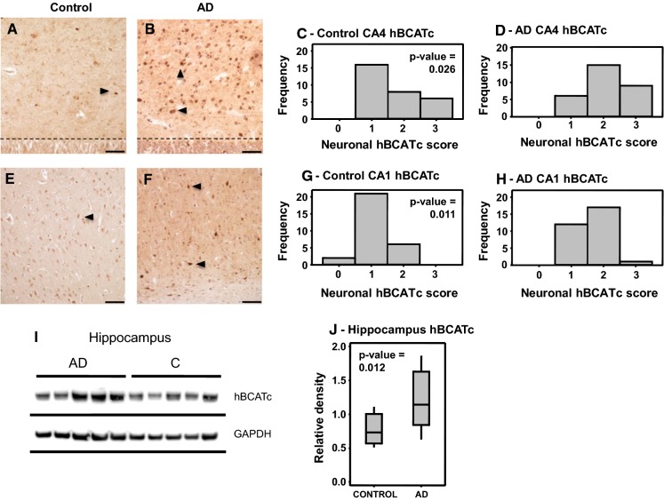
“Our focus was on the main regions of the brain affected by AD pathology where we found a regional increase in BCATc in the hippocampus (Hull et al. 2015a, b), specifically the CA1 and CA4 region, the area first targeted by AD pathology (Fig. 5).”
§ Alzheimer’s disease: targeting the glutamatergic system > Glutamate, BCAA toxicity and neurodegeneration > Regional increase of BCAT in AD brain—Glu toxicity.
What the sources say
- Not checked yetcites Hull et al. 2015a, b
plaincorrelational- Asserted: passes — The authors put it forward as true.
- Checkable: passes — It can be checked against evidence.
- Load-bearing: passes — The paper’s conclusions depend on it.
- Based: passes — The paper gives a reason why it holds.
A result the paper reports from its own data — it cites no other source for it, so there is none to check. The quote shows it is in the paper.
In the paper
“Here, BCATc-mediated autophagy was shown to be regulated through PKC-mediated phosphorylation, modulated by the redox state of BCATc.”
§ Alzheimer’s disease: targeting the glutamatergic system > Glutamate, BCAA toxicity and neurodegeneration > Moonlighting roles for BCAT
plaindirect- Asserted: passes — The authors put it forward as true.
- Checkable: passes — It can be checked against evidence.
- Load-bearing: passes — The paper’s conclusions depend on it.
- Based: passes — The paper gives a reason why it holds.
A result the paper reports from its own data — it cites no other source for it, so there is none to check. The quote shows it is in the paper.
In the paper
“Using the SH-SY5Y cell model, we showed that overexpression of BCATc, as observed in AD brain, resulted in Aβ accumulation through an increase in autophagosome synthesis.”
§ Alzheimer’s disease: targeting the glutamatergic system > Glutamate, BCAA toxicity and neurodegeneration > Moonlighting roles for BCAT
plaindirect- Asserted: passes — The authors put it forward as true.
- Checkable: passes — It can be checked against evidence.
- Load-bearing: passes — The paper’s conclusions depend on it.
- Based: passes — The paper gives a reason why it holds.
In the paper
“Neuronal loss has also been related to the dysregulation of the cholinergic system and glutamatergic system (Ferreira-Vieira et al. 2016).”
§ Alzheimer’s disease: targeting the glutamatergic system > Introduction
What the sources say
- Not checked yetcites Ferreira-Vieira et al. 2016 source
plaincorrelational- Asserted: passes — The authors put it forward as true.
- Checkable: passes — It can be checked against evidence.
- Load-bearing: passes — The paper’s conclusions depend on it.
- Based: passes — The paper gives a reason why it holds.
In the paper
“Currently, prescribed treatments for AD are based on cholinesterase inhibitors (galantamine, rivastigmine and donepezil) or glutamatergic targets (memantine) (Table 1).”
§ Alzheimer’s disease: targeting the glutamatergic system > Introduction
What the sources say
- Not checked yetcites Table 1
plain- Asserted: passes — The authors put it forward as true.
- Checkable: passes — It can be checked against evidence.
- Load-bearing: unclear — It is unclear whether the paper’s conclusions depend on it.
- Based: passes — The paper gives a reason why it holds.
In the paper
“N-methyl-d-aspartate receptor (NMDAR) activation and related excitotoxic events that lead to synaptic dysfunction has also been associated with AD pathology (Liu et al. 2019).”
§ Alzheimer’s disease: targeting the glutamatergic system > Introduction
What the sources say
- Not checked yetcites Liu et al. 2019 source
plaincorrelational- Asserted: passes — The authors put it forward as true.
- Checkable: passes — It can be checked against evidence.
- Load-bearing: passes — The paper’s conclusions depend on it.
- Based: passes — The paper gives a reason why it holds.
None of the sources cited for this claim have been checked yet.
In the paper
“A neuroprotective role for BCATc has been reported in related animal models of brain injury.”
§ Alzheimer’s disease: targeting the glutamatergic system > Glutamate, BCAA toxicity and neurodegeneration > Regional increase of BCAT in AD brain—Glu toxicity.
plaindirect- Asserted: passes — The authors put it forward as true.
- Checkable: passes — It can be checked against evidence.
- Load-bearing: passes — The paper’s conclusions depend on it.
- Based: passes — The paper gives a reason why it holds.
None of the sources cited for this claim have been checked yet.
In the paper
“Concentrations of Glu were also shown to be lower in the posterior cingulate cortex of AD patients, either when measured alone or as a combined measure of Glu+glutamine (Glx) (Antuono et al. 2001; Fayed et al. 2011; Rupsingh et al. 2011).”
§ Alzheimer’s disease: targeting the glutamatergic system > Brain glutamate > Glutamate in the brain, aging and AD-related changes
What the sources say
plaincorrelational- Asserted: passes — The authors put it forward as true.
- Checkable: passes — It can be checked against evidence.
- Load-bearing: passes — The paper’s conclusions depend on it.
- Based: passes — The paper gives a reason why it holds.
A conclusion the paper draws from its own results — it cites no other source for it, so there is none to check. The quote shows it is in the paper.
In the paper
“However, in disease conditions such as AD, Glu toxicity resulting in neuronal cell death indicates that there is a failure in this mechanism to effectively remove toxic levels of Glu.”
§ Alzheimer’s disease: targeting the glutamatergic system > Brain glutamate > The role of BCAT in brain metabolism.
hedgeddirect- Asserted: passes — The authors put it forward as true.
- Checkable: passes — It can be checked against evidence.
- Load-bearing: passes — The paper’s conclusions depend on it.
- Based: passes — The paper gives a reason why it holds.
None of the sources cited for this claim have been checked yet.
In the paper
“In addition, the anti-epileptic drug gabapentin, which inhibits BCATc, has proved successful in treating behavioural alterations in subjects with AD (Cooney et al. 2013; Supasitthumrong et al. 2019).”
§ Alzheimer’s disease: targeting the glutamatergic system > Glutamate, BCAA toxicity and neurodegeneration > Regional increase of BCAT in AD brain—Glu toxicity.
What the sources say
plaindirect- Asserted: passes — The authors put it forward as true.
- Checkable: passes — It can be checked against evidence.
- Load-bearing: unclear — It is unclear whether the paper’s conclusions depend on it.
- Based: passes — The paper gives a reason why it holds.
A conclusion the paper draws from its own results — it cites no other source for it, so there is none to check. The quote shows it is in the paper.
In the paper
“Therefore, it is possible that in AD too the upregulation of BCAT is a physiological response, as in general the neurons that were immunopositive for BCATc in the human brain tissue also appeared morphologically intact.”
§ Alzheimer’s disease: targeting the glutamatergic system > Glutamate, BCAA toxicity and neurodegeneration > Regional increase of BCAT in AD brain—Glu toxicity.
hedgedconditional- Asserted: passes — The authors put it forward as true.
- Checkable: passes — It can be checked against evidence.
- Load-bearing: unclear — It is unclear whether the paper’s conclusions depend on it.
- Based: passes — The paper gives a reason why it holds.
A conclusion the paper draws from its own results — it cites no other source for it, so there is none to check. The quote shows it is in the paper.
In the paper
“This study indicated that BCAT and glutamate hold promise as early indicators of disease pathology.”
§ Alzheimer’s disease: targeting the glutamatergic system > Glutamate, BCAA toxicity and neurodegeneration > Regional increase of BCAT in AD brain—Glu toxicity.
hedgedcorrelational- Asserted: passes — The authors put it forward as true.
- Checkable: passes — It can be checked against evidence.
- Load-bearing: passes — The paper’s conclusions depend on it.
- Based: passes — The paper gives a reason why it holds.
None of the sources cited for this claim have been checked yet.
In the paper
“A limiting factor in this reaction is the source of nitrogen, where the BCAAs, aspartate and more recently alanine serve as potential nitrogen donors (Hutson et al. 1998, 2001; Lieth et al. 2001; Bixel et al. 1997, 2001; Yudkoff et al. 1983, 1996; Kanamori et al. 1998).”
§ Alzheimer’s disease: targeting the glutamatergic system > Brain glutamate > Glutamate/glutamine cycle
What the sources say
- Not matchedcites Hutson et al. 1998, 2001
Couldn’t be matched to a paper in Crossref, so it can’t be checked.
- Not checked yetcites Lieth et al. 2001 source
- Not matchedcites Bixel et al. 1997, 2001
Couldn’t be matched to a paper in Crossref, so it can’t be checked.
plainconditional- Asserted: passes — The authors put it forward as true.
- Checkable: passes — It can be checked against evidence.
- Load-bearing: unclear — It is unclear whether the paper’s conclusions depend on it.
- Based: passes — The paper gives a reason why it holds.
- Not matched
A conclusion the paper draws from its own results — it cites no other source for it, so there is none to check. The quote shows it is in the paper.
In the paper
“An increase in the level of BCATc could be considered a compensatory response to expand the Glu pool in response to the reduced uptake of synaptic Glu by astrocytes.”
§ Alzheimer’s disease: targeting the glutamatergic system > Glutamate, BCAA toxicity and neurodegeneration > Regional increase of BCAT in AD brain—Glu toxicity.
hedgedconditional- Asserted: passes — The authors put it forward as true.
- Checkable: passes — It can be checked against evidence.
- Load-bearing: passes — The paper’s conclusions depend on it.
- Based: passes — The paper gives a reason why it holds.
In the paper
“This redox switch is unique to these aminotransferases and has been shown to be important in regulating metabolite shuttling between the BCKD complex and GDH (Islam et al. 2007).”
§ Alzheimer’s disease: targeting the glutamatergic system > Glutamate, BCAA toxicity and neurodegeneration > Moonlighting roles for BCAT
What the sources say
- Not checked yetcites Islam et al. 2007 source
plaindirect- Asserted: passes — The authors put it forward as true.
- Checkable: passes — It can be checked against evidence.
- Load-bearing: unclear — It is unclear whether the paper’s conclusions depend on it.
- Based: passes — The paper gives a reason why it holds.
A conclusion the paper draws from its own results — it cites no other source for it, so there is none to check. The quote shows it is in the paper.
In the paper
“However, as discussed, in human brain BCATm was found in endothelial cells indicating that an alternative model needs to be considered for human brain glutamate regulation.”
§ Alzheimer’s disease: targeting the glutamatergic system > Brain glutamate > The role of BCAT in brain metabolism.
hedgeddirect- Asserted: passes — The authors put it forward as true.
- Checkable: passes — It can be checked against evidence.
- Load-bearing: passes — The paper’s conclusions depend on it.
- Based: passes — The paper gives a reason why it holds.
In the paper
“We propose that the expression of BCATm, the BCKD complex and GDH offer an auxiliary mechanism to regulate brain Glu, in particular under conditions where the level of Glu is high, offering a neuroprotective role in the first instance (Hull et al. 2015a, b).”
§ Alzheimer’s disease: targeting the glutamatergic system > Brain glutamate > The role of BCAT in brain metabolism.
What the sources say
- Not checked yetcites Hull et al. 2015a, b
plaindirect- Asserted: passes — The authors put it forward as true.
- Checkable: passes — It can be checked against evidence.
- Load-bearing: passes — The paper’s conclusions depend on it.
- Based: passes — The paper gives a reason why it holds.
A conclusion the paper draws from its own results — it cites no other source for it, so there is none to check. The quote shows it is in the paper.
In the paper
“In human brain, we propose that in neuronal cells BCAAs are transaminated by BCATc generating Glu, which contributes both to the pool of neurotransmitters and the Glu required for release during excitation.”
§ Alzheimer’s disease: targeting the glutamatergic system > Brain glutamate > The role of BCAT in brain metabolism.
plaindirect- Asserted: passes — The authors put it forward as true.
- Checkable: passes — It can be checked against evidence.
- Load-bearing: passes — The paper’s conclusions depend on it.
- Based: unclear — It is unclear why the paper says it holds.
In the paper
“Damage to glutamatergic neurons, in particular cell bodies and neurites in layers III and IV of the neocortex, together with damage to glutamatergically-innervated cortical and hippocampal neurons, are particularly evident in AD brain (Albin and Greenamyre 1992).”
§ Alzheimer’s disease: targeting the glutamatergic system > Glutamate, BCAA toxicity and neurodegeneration > Glutamate excitotoxicity
What the sources say
- Not checked yetcites Albin and Greenamyre 1992 source
plaincorrelational- Asserted: passes — The authors put it forward as true.
- Checkable: passes — It can be checked against evidence.
- Load-bearing: passes — The paper’s conclusions depend on it.
- Based: passes — The paper gives a reason why it holds.
A conclusion the paper draws from its own results — it cites no other source for it, so there is none to check. The quote shows it is in the paper.
In the paper
“As a result, anaplerotic pathways must interface with the Glu-glutamine cycle to regenerate this ‘lost’ Glu necessary to sustain efficient neurotransmission.”
§ Alzheimer’s disease: targeting the glutamatergic system > Brain glutamate > Glutamate/glutamine cycle
plaindirect- Asserted: passes — The authors put it forward as true.
- Checkable: passes — It can be checked against evidence.
- Load-bearing: unclear — It is unclear whether the paper’s conclusions depend on it.
- Based: passes — The paper gives a reason why it holds.
None of the sources cited for this claim have been checked yet.
In the paper
“Targeted proteomic studies have indicated that the BCAT proteins have additional binding partners governed by their CXXC motif (Coles et al. 2009; Hindy and Conway 2019).”
§ Alzheimer’s disease: targeting the glutamatergic system > Glutamate, BCAA toxicity and neurodegeneration > Moonlighting roles for BCAT
What the sources say
- Not checked yetcites Coles et al. 2009
- Not checked yetcites Hindy and Conway 2019 source
hedgeddirect- Asserted: passes — The authors put it forward as true.
- Checkable: passes — It can be checked against evidence.
- Load-bearing: unclear — It is unclear whether the paper’s conclusions depend on it.
- Based: passes — The paper gives a reason why it holds.
In the paper
“Decades of clinical trials dedicated to target Aβ processing have failed to generate a treatment for AD (Tolar et al. 2019).”
§ Alzheimer’s disease: targeting the glutamatergic system > Introduction > The amyloid and Tau hypothesis
What the sources say
- Not checked yetcites Tolar et al. 2019 source
plain- Asserted: passes — The authors put it forward as true.
- Checkable: passes — It can be checked against evidence.
- Load-bearing: passes — The paper’s conclusions depend on it.
- Based: passes — The paper gives a reason why it holds.
In the paper
“A more recent study identified several additional BCAT2 mutations that ranged between homozygous or compound heterozygous BCAT2 mutations (Knerr et al. 2019).”
§ Alzheimer’s disease: targeting the glutamatergic system > Disorders of BCAA metabolism > Mutations of BCAT leads to mild cognitive impairment and brain lesions
What the sources say
- Not checked yetcites Knerr et al. 2019 source
plain- Asserted: passes — The authors put it forward as true.
- Checkable: passes — It can be checked against evidence.
- Load-bearing: unclear — It is unclear whether the paper’s conclusions depend on it.
- Based: passes — The paper gives a reason why it holds.
In the paper
“Moreover, oligomeric Aβ has been shown to interact with glutamate receptors contributing to glutamate excitotoxicity (Hascup and Hascup 2016).”
§ Alzheimer’s disease: targeting the glutamatergic system > Conclusion
What the sources say
- Not checked yetcites Hascup and Hascup 2016 source
plaindirect- Asserted: passes — The authors put it forward as true.
- Checkable: passes — It can be checked against evidence.
- Load-bearing: passes — The paper’s conclusions depend on it.
- Based: passes — The paper gives a reason why it holds.
A conclusion the paper draws from its own results — it cites no other source for it, so there is none to check. The quote shows it is in the paper.
In the paper
“In disease conditions it is clear that excess (MSUD and BCAT2) or limiting (TBI) amounts of these amino acids result in altered cognitive function.”
§ Alzheimer’s disease: targeting the glutamatergic system > Conclusion
boosteddirect- Asserted: passes — The authors put it forward as true.
- Checkable: passes — It can be checked against evidence.
- Load-bearing: passes — The paper’s conclusions depend on it.
- Based: passes — The paper gives a reason why it holds.
- Rests on an assumption: Excess or limiting dietary BCAAs cause neurological dysfunction, reducing cognitive function.
In the paper
“Although MRI imaging showed associated brain white matter lesions, a feature of MSUD (Jan et al. 2003; Schonberger et al. 2004), brain regions affected in MSUD patients are mainly in the mesencephalon and brain stem as well as thalamus and globus pallidus (Schonberger et al. 2004), whereas in this patient, supratentorial lesions were found without overt abnormalities in these regions.”
§ Alzheimer’s disease: targeting the glutamatergic system > Disorders of BCAA metabolism > Mutations of BCAT leads to mild cognitive impairment and brain lesions
What the sources say
- Not checked yetcites Schonberger et al. 2004 source
plain- Asserted: passes — The authors put it forward as true.
- Checkable: passes — It can be checked against evidence.
- Load-bearing: unclear — It is unclear whether the paper’s conclusions depend on it.
- Based: passes — The paper gives a reason why it holds.
A result the paper reports from its own data — it cites no other source for it, so there is none to check. The quote shows it is in the paper.
In the paper
“Although MRI imaging showed associated brain white matter lesions, a feature of MSUD (Jan et al. 2003; Schonberger et al. 2004), brain regions affected in MSUD patients are mainly in the mesencephalon and brain stem as well as thalamus and globus pallidus (Schonberger et al. 2004), whereas in this patient, supratentorial lesions were found without overt abnormalities in these regions.”
§ Alzheimer’s disease: targeting the glutamatergic system > Disorders of BCAA metabolism > Mutations of BCAT leads to mild cognitive impairment and brain lesions
plainSingle case- Asserted: passes — The authors put it forward as true.
- Checkable: passes — It can be checked against evidence.
- Load-bearing: passes — The paper’s conclusions depend on it.
- Based: passes — The paper gives a reason why it holds.
In the paper
“Moreover, treatment of the patient with vitamin B6 resulted in a sharp decline in valine levels compared with those of leucine and isoleucine.”
§ Alzheimer’s disease: targeting the glutamatergic system > Disorders of BCAA metabolism > Mutations of BCAT leads to mild cognitive impairment and brain lesions
What the sources say
- Not checked yetcites Wang et al. 2015a, b
plaindirectSingle case- Asserted: passes — The authors put it forward as true.
- Checkable: passes — It can be checked against evidence.
- Load-bearing: unclear — It is unclear whether the paper’s conclusions depend on it.
- Based: passes — The paper gives a reason why it holds.
In the paper
“Using an animal model of TBI, Cole et al. (2010) reported improved cognition in rats supplemented with BCAAs following lateral fluid percussion injury.”
§ Alzheimer’s disease: targeting the glutamatergic system > Disorders of BCAA metabolism > Serum BCAA indicators of brain injury
What the sources say
- Not checked yetcites Cole et al. 2010 source
plaindirect- Asserted: passes — The authors put it forward as true.
- Checkable: passes — It can be checked against evidence.
- Load-bearing: unclear — It is unclear whether the paper’s conclusions depend on it.
- Based: passes — The paper gives a reason why it holds.
None of the sources cited for this claim have been checked yet.
In the paper
“Although MRI imaging showed associated brain white matter lesions, a feature of MSUD (Jan et al. 2003; Schonberger et al. 2004), brain regions affected in MSUD patients are mainly in the mesencephalon and brain stem as well as thalamus and globus pallidus (Schonberger et al. 2004), whereas in this patient, supratentorial lesions were found without overt abnormalities in these regions.”
§ Alzheimer’s disease: targeting the glutamatergic system > Disorders of BCAA metabolism > Mutations of BCAT leads to mild cognitive impairment and brain lesions
What the sources say
plaincorrelational- Asserted: passes — The authors put it forward as true.
- Checkable: passes — It can be checked against evidence.
- Load-bearing: unclear — It is unclear whether the paper’s conclusions depend on it.
- Based: passes — The paper gives a reason why it holds.
In the paper
“These include proteins important in cell signalling and protein folding (e.g. BCAT was identified as a novel chaperone in protein folding through its thiol oxidoreductase activity, El Hindy et al. 2014).”
§ Alzheimer’s disease: targeting the glutamatergic system > Glutamate, BCAA toxicity and neurodegeneration > Moonlighting roles for BCAT
What the sources say
- Not checked yetcites El Hindy et al. 2014 source
plaindirect- Asserted: passes — The authors put it forward as true.
- Checkable: passes — It can be checked against evidence.
- Load-bearing: unclear — It is unclear whether the paper’s conclusions depend on it.
- Based: passes — The paper gives a reason why it holds.
A conclusion the paper draws from its own results — it cites no other source for it, so there is none to check. The quote shows it is in the paper.
In the paper
“Clearly, regulation of brain glutamate is important in AD pathology.”
§ Alzheimer’s disease: targeting the glutamatergic system > Conclusion
boostedcorrelational- Asserted: passes — The authors put it forward as true.
- Checkable: passes — It can be checked against evidence.
- Load-bearing: passes — The paper’s conclusions depend on it.
- Based: passes — The paper gives a reason why it holds.
A conclusion the paper draws from its own results — it cites no other source for it, so there is none to check. The quote shows it is in the paper.
In the paper
“Our data supports a novel mechanism by which Aβ accumulation may be altered by BCAAs such as leucine, but subject to a more in-depth understanding of nutritional signalling in aging and neurodegenerative conditions such as AD.”
§ Alzheimer’s disease: targeting the glutamatergic system > Glutamate, BCAA toxicity and neurodegeneration > Moonlighting roles for BCAT
hedgedconditional- Asserted: passes — The authors put it forward as true.
- Checkable: passes — It can be checked against evidence.
- Load-bearing: passes — The paper’s conclusions depend on it.
- Based: passes — The paper gives a reason why it holds.
A conclusion the paper draws from its own results — it cites no other source for it, so there is none to check. The quote shows it is in the paper.
In the paper
“Substantial evidence supports a role for ineffective astrocytic clearance of glutamate released during excitation.”
§ Alzheimer’s disease: targeting the glutamatergic system > Conclusion
boosteddirect- Asserted: passes — The authors put it forward as true.
- Checkable: passes — It can be checked against evidence.
- Load-bearing: passes — The paper’s conclusions depend on it.
- Based: passes — The paper gives a reason why it holds.
- It may state two separate claims as one. 38
- Probably not a substantial claim. 10
In the paper
“Our group subsequently mapped BCATc to the human brain, where it was found in all brain regions and consistent with the rat model was neuron-specific, with strongest labelling reported in the parietal cortex (Hull et al. 2012).”
§ Alzheimer’s disease: targeting the glutamatergic system > Brain glutamate > Cellular distribution of branched chain aminotransferases
What the sources say
- Not checked yetcites Hull et al. 2012
plain- Asserted: passes — The authors put it forward as true.
- Checkable: passes — It can be checked against evidence.
- Load-bearing: unclear — It is unclear whether the paper’s conclusions depend on it.
- Based: passes — The paper gives a reason why it holds.
It may state two separate claims as one.
A result the paper reports from its own data — it cites no other source for it, so there is none to check. The quote shows it is in the paper.
In the paper
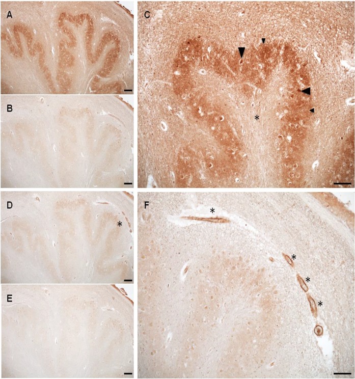
“What was clear was that wherever BCATc staining occurred BCATm was absent and vice versa (Fig. 4).”
§ Alzheimer’s disease: targeting the glutamatergic system > Brain glutamate > Cellular distribution of branched chain aminotransferases
boostedcorrelational- Asserted: passes — The authors put it forward as true.
- Checkable: passes — It can be checked against evidence.
- Load-bearing: unclear — It is unclear whether the paper’s conclusions depend on it.
- Based: passes — The paper gives a reason why it holds.
It may state two separate claims as one.
A result the paper reports from its own data — it cites no other source for it, so there is none to check. The quote shows it is in the paper.
In the paper
“Our model showed that BCAT and glutamate accurately distinguish between control and AD participants and in combination with the neurophysiological test Montreal Cognitive Assessment, improved the overall sensitivity to 1.00 and specificity to 0.978.”
§ Alzheimer’s disease: targeting the glutamatergic system > Glutamate, BCAA toxicity and neurodegeneration > Regional increase of BCAT in AD brain—Glu toxicity.
plaincorrelational- Asserted: passes — The authors put it forward as true.
- Checkable: passes — It can be checked against evidence.
- Load-bearing: passes — The paper’s conclusions depend on it.
- Based: passes — The paper gives a reason why it holds.
It may state two separate claims as one.
The paper states this without a source or data behind it here, so there is nothing to check it against. The quote shows it is in the paper.
In the paper
“The hippocampus showed intense immunoreactivity for BCATc of pyramidal cells (approximately 70% of neuronal cells) in the pyramidal cell layer, with moderate BCATc staining in the cell bodies of interneurons (GABAergic or glycine).”
§ Alzheimer’s disease: targeting the glutamatergic system > Brain glutamate > Cellular distribution of branched chain aminotransferases
plain- Asserted: passes — The authors put it forward as true.
- Checkable: passes — It can be checked against evidence.
- Load-bearing: unclear — It is unclear whether the paper’s conclusions depend on it.
- Based: passes — The paper gives a reason why it holds.
It may state two separate claims as one.
A result the paper reports from its own data — it cites no other source for it, so there is none to check. The quote shows it is in the paper.
In the paper
“Immunopositive staining for BCATm was vessel and capillary in nature. This staining was evident in all major anatomical regions of the brain assessed with the exception of the parietal lobe and medulla.”
§ Alzheimer’s disease: targeting the glutamatergic system > Brain glutamate > Cellular distribution of branched chain aminotransferases
plain- Asserted: passes — The authors put it forward as true.
- Checkable: passes — It can be checked against evidence.
- Load-bearing: unclear — It is unclear whether the paper’s conclusions depend on it.
- Based: passes — The paper gives a reason why it holds.
It may state two separate claims as one.
A result the paper reports from its own data — it cites no other source for it, so there is none to check. The quote shows it is in the paper.
In the paper
“Importantly, leucine, a key regulator of autophagic flux, significantly reduced BCATc-induced autophagy and consequently Aβ.”
§ Alzheimer’s disease: targeting the glutamatergic system > Glutamate, BCAA toxicity and neurodegeneration > Moonlighting roles for BCAT
plaindirect- Asserted: passes — The authors put it forward as true.
- Checkable: passes — It can be checked against evidence.
- Load-bearing: passes — The paper’s conclusions depend on it.
- Based: passes — The paper gives a reason why it holds.
It may state two separate claims as one.
In the paper
“Our most recent study offers evidence that these ‘moonlighting’ functions are fundamental in regulating autophagy and play a fundamental role in regulating Aβ (Harris et al. 2020).”
§ Alzheimer’s disease: targeting the glutamatergic system > Glutamate, BCAA toxicity and neurodegeneration > Moonlighting roles for BCAT
What the sources say
- Not checked yetcites Harris et al. 2020
boosteddirect- Asserted: passes — The authors put it forward as true.
- Checkable: passes — It can be checked against evidence.
- Load-bearing: passes — The paper’s conclusions depend on it.
- Based: passes — The paper gives a reason why it holds.
It may state two separate claims as one.
A conclusion the paper draws from its own results — it cites no other source for it, so there is none to check. The quote shows it is in the paper.
In the paper
“Whilst these treatments offer some reprive to symptoms, the improvement is modest and temporary, indicating that like Aβ targeted approaches, the heterogenity of the disease calls for a stratified approach for effective treatment.”
§ Alzheimer’s disease: targeting the glutamatergic system > Introduction
hedgeddirect- Asserted: passes — The authors put it forward as true.
- Checkable: passes — It can be checked against evidence.
- Load-bearing: passes — The paper’s conclusions depend on it.
- Based: passes — The paper gives a reason why it holds.
It may state two separate claims as one.
In the paper
“This is inhibited through oxidation that not only inactivates BCAT but also destabilises the metabolic complexes (Islam et al. 2010).”
§ Alzheimer’s disease: targeting the glutamatergic system > Glutamate, BCAA toxicity and neurodegeneration > Moonlighting roles for BCAT
What the sources say
- Not checked yetcites Islam et al. 2010
plaindirect- Asserted: passes — The authors put it forward as true.
- Checkable: passes — It can be checked against evidence.
- Load-bearing: passes — The paper’s conclusions depend on it.
- Based: passes — The paper gives a reason why it holds.
It may state two separate claims as one.
In the paper
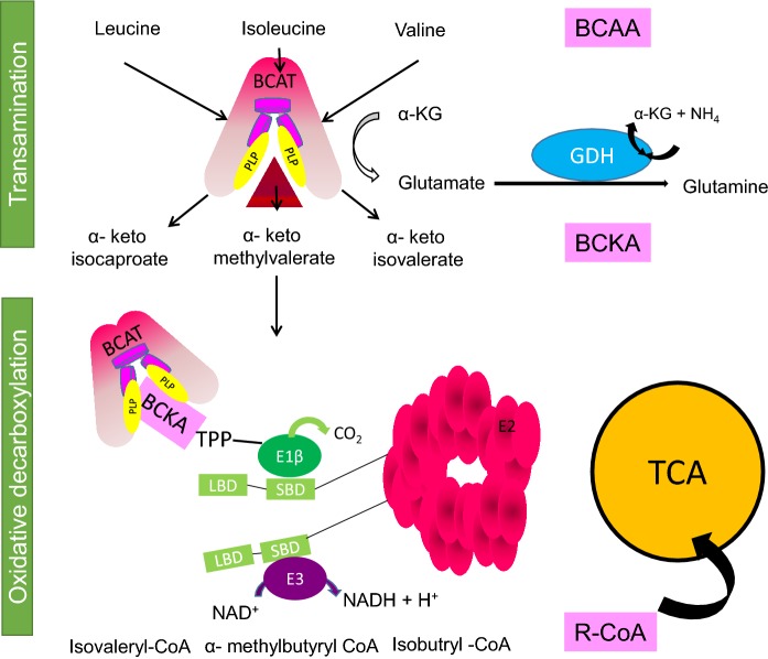
“The canonical role of the BCAT proteins is the transamination of the BCAAs, leucine, isoleucine and valine with α-keto glutarate to form Glu and their respective branched chain α-keto acids (BCKA) (α-ketoisocaproate (KIC), α-keto-β-methylvalerate (KMV) and α-ketoisovalerate (KIV)) regenerating the pyridoxal phosphate (PLP) form of the enzyme (Ichihara and Koyama 1966) (Fig. 3).”
§ Alzheimer’s disease: targeting the glutamatergic system > Brain glutamate > The branched chain aminotransferase proteins
What the sources say
- Not checked yetcites Ichihara and Koyama 1966
plaindirect- Asserted: passes — The authors put it forward as true.
- Checkable: passes — It can be checked against evidence.
- Load-bearing: fails — The paper’s conclusions may not depend on it.
- Based: passes — The paper gives a reason why it holds.
Probably not a substantial claim. The paper’s conclusions may not depend on it.
In the paper
“In AD brain, Aβ can interrupt effective glutamate uptake by astrocytes, which evokes a cascade of events that leads to neuronal swelling, destruction of membrane integrity and ultimately cell death.”
§ Alzheimer’s disease: targeting the glutamatergic system > Abstract
What the sources say
- Not checked yetcites Huang et al. 2018
plaindirect- Asserted: passes — The authors put it forward as true.
- Checkable: passes — It can be checked against evidence.
- Load-bearing: passes — The paper’s conclusions depend on it.
- Based: passes — The paper gives a reason why it holds.
It may state two separate claims as one.
None of the sources cited for this claim have been checked yet.
In the paper
“Whilst an important neurotransmitter, pathological accumulation of Glu results in this amino acid becoming a potent neurotoxin (Choi et al. 1987; Mattson and Chan 2003). This is in part due to the time-related exposure, overstimulating the post synaptic response causing an increase in the entry of calcium into neurons (Sattler and Tymianski 2000; Mattson and Chan 2003).”
§ Alzheimer’s disease: targeting the glutamatergic system > Glutamate, BCAA toxicity and neurodegeneration > Glutamate excitotoxicity
What the sources say
- Not checked yetcites Choi et al. 1987
- Not checked yetcites Mattson and Chan 2003
- Not checked yetcites Sattler and Tymianski 2000
plaindirect- Asserted: passes — The authors put it forward as true.
- Checkable: passes — It can be checked against evidence.
- Load-bearing: passes — The paper’s conclusions depend on it.
- Based: passes — The paper gives a reason why it holds.
It may state two separate claims as one.
A conclusion the paper draws from its own results — it cites no other source for it, so there is none to check. The quote shows it is in the paper.
In the paper
“Importantly, the immunohistochemistry data, showed that the intensity of BCATc labelling in both cell bodies and dendrites was most pronounced in the hippocampus, a region that is known to be particularly susceptible to excitotoxic neuronal injury at an early stage of AD, highlighting that sustained increase in BCATc may contribute to injury.”
§ Alzheimer’s disease: targeting the glutamatergic system > Glutamate, BCAA toxicity and neurodegeneration > Regional increase of BCAT in AD brain—Glu toxicity.
hedgedconditional- Asserted: passes — The authors put it forward as true.
- Checkable: passes — It can be checked against evidence.
- Load-bearing: passes — The paper’s conclusions depend on it.
- Based: passes — The paper gives a reason why it holds.
It may state two separate claims as one.
A conclusion the paper draws from its own results — it cites no other source for it, so there is none to check. The quote shows it is in the paper.
In the paper
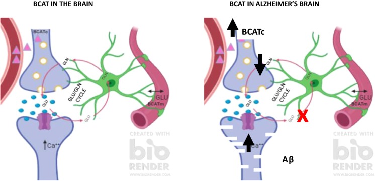
“We assigned a neuroprotective role to BCATc but proposed that should transamination in the direction of Glu synthesis persist it could exacerbate toxicity contributing to neuronal degeneration (Fig. 6).”
§ Alzheimer’s disease: targeting the glutamatergic system > Glutamate, BCAA toxicity and neurodegeneration > Regional increase of BCAT in AD brain—Glu toxicity.
hedgedconditional- Asserted: passes — The authors put it forward as true.
- Checkable: passes — It can be checked against evidence.
- Load-bearing: passes — The paper’s conclusions depend on it.
- Based: passes — The paper gives a reason why it holds.
It may state two separate claims as one.
None of the sources cited for this claim have been checked yet.
In the paper
“The regulation of brain Glu is important as uptake from the blood is minimal, however, precursors to its synthesis such as the BCAAs are easily taken up, in particular leucine (Oldendorf et al. 1977; Smith et al. 1987).”
§ Alzheimer’s disease: targeting the glutamatergic system > Brain glutamate > Glutamate/glutamine cycle
What the sources say
- Not checked yetcites Oldendorf et al. 1977
- Not checked yetcites Smith et al. 1987
plain- Asserted: passes — The authors put it forward as true.
- Checkable: passes — It can be checked against evidence.
- Load-bearing: unclear — It is unclear whether the paper’s conclusions depend on it.
- Based: passes — The paper gives a reason why it holds.
It may state two separate claims as one.
What the paper did — it cites no source for this, so there is none to check. The quote shows it is in the paper.
In the paper
“However, a limitation to this study was the timeframe, where a longitudinal study would better define the utility of BCAT and glutamate as biomarkers.”
§ Alzheimer’s disease: targeting the glutamatergic system > Glutamate, BCAA toxicity and neurodegeneration > Regional increase of BCAT in AD brain—Glu toxicity.
hedged- Asserted: passes — The authors put it forward as true.
- Checkable: passes — It can be checked against evidence.
- Load-bearing: fails — The paper’s conclusions may not depend on it.
- Based: passes — The paper gives a reason why it holds.
Probably not a substantial claim. The paper’s conclusions may not depend on it. It may state two separate claims as one.
None of the sources cited for this claim have been checked yet.
In the paper
“Although the canonical role for BCAT has traditionally been associated with transamination and glutamate regulation, it is important to note that it is also a redox-sensitive protein subject to oxidation (Conway et al. 2002), S-nitrosylation (Conway et al. 2004) and S-glutathionylation (Conway et al. 2008).”
§ Alzheimer’s disease: targeting the glutamatergic system > Glutamate, BCAA toxicity and neurodegeneration > Moonlighting roles for BCAT
What the sources say
- Not checked yetcites Conway et al. 2002
- Not checked yetcites Conway et al. 2004
- Not checked yetcites Conway et al. 2008
plain- Asserted: passes — The authors put it forward as true.
- Checkable: passes — It can be checked against evidence.
- Load-bearing: unclear — It is unclear whether the paper’s conclusions depend on it.
- Based: passes — The paper gives a reason why it holds.
It may state two separate claims as one.
None of the sources cited for this claim have been checked yet.
In the paper
“Whilst the glutamate/glutamine cycle is an efficient method to regenerate Glu, this recycling is not 100% effective. During its transition some Glu is ‘lost’ in astrocytes through metabolism, generating lactate (Sonnelwald et al. 1993), or in the production of purines and glutathione (Shank et al. 1985).”
§ Alzheimer’s disease: targeting the glutamatergic system > Brain glutamate > Glutamate/glutamine cycle
What the sources say
- Not checked yetcites Sonnelwald et al. 1993
- Not checked yetcites Shank et al. 1985
plaindirect- Asserted: passes — The authors put it forward as true.
- Checkable: passes — It can be checked against evidence.
- Load-bearing: unclear — It is unclear whether the paper’s conclusions depend on it.
- Based: passes — The paper gives a reason why it holds.
It may state two separate claims as one.
None of the sources cited for this claim have been checked yet.
In the paper
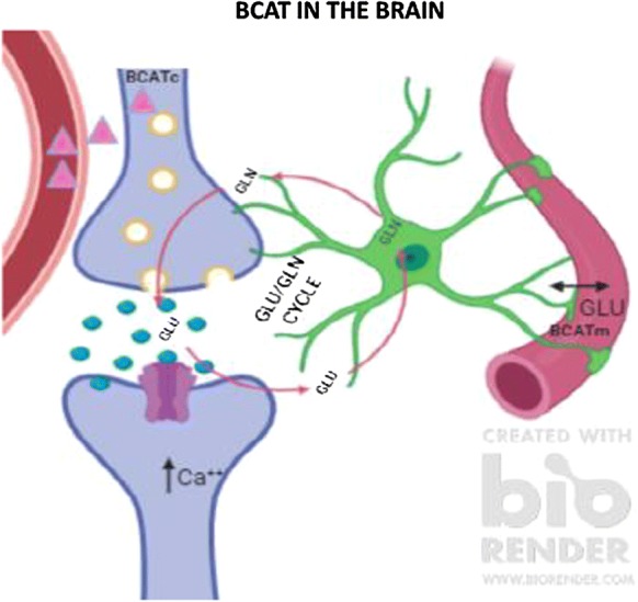
“Regulation of brain Glu is thought to be primarily governed through the Glu/glutamine cycle, where excess Glu remaining after excitation is taken up by astrocytes (Fig. 2) (reviewed in Conway and Hutson 2016; Yudkoff 2017).”
§ Alzheimer’s disease: targeting the glutamatergic system > Brain glutamate > Glutamate/glutamine cycle
What the sources say
- Not checked yetcites Conway and Hutson 2016
- Not checked yetcites Yudkoff 2017
hedgeddirect- Asserted: passes — The authors put it forward as true.
- Checkable: passes — It can be checked against evidence.
- Load-bearing: fails — The paper’s conclusions may not depend on it.
- Based: passes — The paper gives a reason why it holds.
Probably not a substantial claim. The paper’s conclusions may not depend on it. It may state two separate claims as one.
None of the sources cited for this claim have been checked yet.
In the paper
“Together, 1H MRS studies confirm that glutamatergic neurotransmission decreases as we age but is further perturbed in individuals with aMCI and AD, respectively and correlates with cognitive measures.”
§ Alzheimer’s disease: targeting the glutamatergic system > Brain glutamate > Glutamate in the brain, aging and AD-related changes
What the sources say
- Not checked yetcites Wang et al. 2015a, b
- Not checked yetcites Antuono et al. 2001
- Not checked yetcites Riese et al. 2015
boostedcorrelational- Asserted: passes — The authors put it forward as true.
- Checkable: passes — It can be checked against evidence.
- Load-bearing: passes — The paper’s conclusions depend on it.
- Based: passes — The paper gives a reason why it holds.
It may state two separate claims as one.
None of the sources cited for this claim have been checked yet.
In the paper
“Treatment using memantine has improved cognition, behaviour, global function but the degree of efficacy remains to be fully determined (Kishi et al. 2017; Matsunga et al. 2015).”
§ Alzheimer’s disease: targeting the glutamatergic system > Glutamate, BCAA toxicity and neurodegeneration > Glutamate excitotoxicity
What the sources say
- Not checked yetcites Kishi et al. 2017
- Not checked yetcites Matsunga et al. 2015
plaindirect- Asserted: passes — The authors put it forward as true.
- Checkable: passes — It can be checked against evidence.
- Load-bearing: unclear — It is unclear whether the paper’s conclusions depend on it.
- Based: passes — The paper gives a reason why it holds.
It may state two separate claims as one.
A conclusion the paper draws from its own results — it cites no other source for it, so there is none to check. The quote shows it is in the paper.
In the paper
“Similar to BCATc, we propose that increased levels of endothelial BCATm would serve in a neuroprotective capacity.”
§ Alzheimer’s disease: targeting the glutamatergic system > Glutamate, BCAA toxicity and neurodegeneration > Regional increase of BCAT in AD brain—Glu toxicity.
hedgeddirect- Asserted: passes — The authors put it forward as true.
- Checkable: passes — It can be checked against evidence.
- Load-bearing: passes — The paper’s conclusions depend on it.
- Based: passes — The paper gives a reason why it holds.
Probably not a substantial claim.
A conclusion the paper draws from its own results — it cites no other source for it, so there is none to check. The quote shows it is in the paper.
In the paper
“Collectively, these results support a role for BCAA in regulating brain Glu and can impact associated cognitive function.”
§ Alzheimer’s disease: targeting the glutamatergic system > Disorders of BCAA metabolism > Serum BCAA indicators of brain injury
plaindirect- Asserted: passes — The authors put it forward as true.
- Checkable: passes — It can be checked against evidence.
- Load-bearing: passes — The paper’s conclusions depend on it.
- Based: passes — The paper gives a reason why it holds.
It may state two separate claims as one.
- Rests on an assumption: BCAA are important nutrient signals in regulating brain Glu.
In the paper
“Following TBI, there is combined metabolic response including Glu toxicity, secondary brain oedema and increased cranial pressure (Vuille-Dit-bille et al. 2012).”
§ Alzheimer’s disease: targeting the glutamatergic system > Disorders of BCAA metabolism > Serum BCAA indicators of brain injury
What the sources say
- Not checked yetcites Vuille-Dit-bille et al. 2012
plaindirect- Asserted: passes — The authors put it forward as true.
- Checkable: passes — It can be checked against evidence.
- Load-bearing: unclear — It is unclear whether the paper’s conclusions depend on it.
- Based: passes — The paper gives a reason why it holds.
It may state two separate claims as one.
In the paper
“Interestingly, c.509G> A (p.Arg170Gln) was found in his father while c.790G>A (p.Glu264Lys) was found in his mother.”
§ Alzheimer’s disease: targeting the glutamatergic system > Disorders of BCAA metabolism > Mutations of BCAT leads to mild cognitive impairment and brain lesions
What the sources say
- Not checked yetcites Wang et al. 2015a, b
plainSingle case- Asserted: passes — The authors put it forward as true.
- Checkable: passes — It can be checked against evidence.
- Load-bearing: unclear — It is unclear whether the paper’s conclusions depend on it.
- Based: passes — The paper gives a reason why it holds.
It may state two separate claims as one.
A conclusion the paper draws from its own results — it cites no other source for it, so there is none to check. The quote shows it is in the paper.
In the paper
“As discussed the BCAA and aromatic amino acids are important sources of neurotransmitter synthesis.”
§ Alzheimer’s disease: targeting the glutamatergic system > Conclusion
plaindirect- Asserted: passes — The authors put it forward as true.
- Checkable: passes — It can be checked against evidence.
- Load-bearing: passes — The paper’s conclusions depend on it.
- Based: fails — The paper may give no reason why it holds.
Probably not a substantial claim. The paper may give no reason why it holds.
In the paper
“Sequencing analysis revealed two heterogeneous mutations including: c.509G> A (p.Arg170Gln) and c.790G>A (p.Glu264Lys).”
§ Alzheimer’s disease: targeting the glutamatergic system > Disorders of BCAA metabolism > Mutations of BCAT leads to mild cognitive impairment and brain lesions
What the sources say
- Not checked yetcites Wang et al. 2015a, b
plainSingle case- Asserted: passes — The authors put it forward as true.
- Checkable: passes — It can be checked against evidence.
- Load-bearing: passes — The paper’s conclusions depend on it.
- Based: passes — The paper gives a reason why it holds.
It may state two separate claims as one.
In the paper
“Wang et al. (2015a, b) first reported a case of a 22 year-old male who presented with MCI, lasting longer than 6 years, headaches and a striking blood profile of hypervalinaemia, hyperleucinemia and isoleucinemia, which aligned with a diagnosis of MSUD (Wang et al. 2015a, b).”
§ Alzheimer’s disease: targeting the glutamatergic system > Disorders of BCAA metabolism > Mutations of BCAT leads to mild cognitive impairment and brain lesions
What the sources say
- Not checked yetcites Wang et al. 2015a, b
plaincorrelationalSingle case- Asserted: passes — The authors put it forward as true.
- Checkable: passes — It can be checked against evidence.
- Load-bearing: passes — The paper’s conclusions depend on it.
- Based: passes — The paper gives a reason why it holds.
It may state two separate claims as one.
None of the sources cited for this claim have been checked yet.
In the paper
“The mechanisms underlying the brain damage observed in these patients are not well established but will most likely be related to the increased BCAA levels similar to that reported for MSUD (Yudkoff 1997; Barschak et al. 2009; Funchal et al. 2006; Sgaravatti et al. 2003).”
§ Alzheimer’s disease: targeting the glutamatergic system > Disorders of BCAA metabolism > Mutations of BCAT leads to mild cognitive impairment and brain lesions
What the sources say
- Not checked yetcites Yudkoff 1997 source
- Not checked yetcites Barschak et al. 2009
- Not checked yetcites Funchal et al. 2006
hedgedconditional- Asserted: passes — The authors put it forward as true.
- Checkable: passes — It can be checked against evidence.
- Load-bearing: unclear — It is unclear whether the paper’s conclusions depend on it.
- Based: passes — The paper gives a reason why it holds.
It may state two separate claims as one.
A result the paper reports from its own data — it cites no other source for it, so there is none to check. The quote shows it is in the paper.
In the paper
“Mutations linked with MSUD were not indicated nor did the patient present with the classical smell of maple syrup from his urine.”
§ Alzheimer’s disease: targeting the glutamatergic system > Disorders of BCAA metabolism > Mutations of BCAT leads to mild cognitive impairment and brain lesions
plainSingle case- Asserted: passes — The authors put it forward as true.
- Checkable: passes — It can be checked against evidence.
- Load-bearing: unclear — It is unclear whether the paper’s conclusions depend on it.
- Based: passes — The paper gives a reason why it holds.
It may state two separate claims as one.
In the paper
“In untreated cases, infants present with symptoms including neurological dysfunction, seizures and infant death (Chuang et al. 2006).”
§ Alzheimer’s disease: targeting the glutamatergic system > Disorders of BCAA metabolism > Maple syrup urine disease (MSUD)
What the sources say
- Not checked yetcites Chuang et al. 2006
plaindirect- Asserted: passes — The authors put it forward as true.
- Checkable: passes — It can be checked against evidence.
- Load-bearing: fails — The paper’s conclusions may not depend on it.
- Based: passes — The paper gives a reason why it holds.
Probably not a substantial claim. The paper’s conclusions may not depend on it. It may state two separate claims as one.
A conclusion the paper draws from its own results — it cites no other source for it, so there is none to check. The quote shows it is in the paper.
In the paper
“Clinically, a role for excitotoxicity in AD pathology is underpinned by the approval of memantine to treat moderate to severe confusion related to AD.”
§ Alzheimer’s disease: targeting the glutamatergic system > Conclusion
plaindirect- Asserted: passes — The authors put it forward as true.
- Checkable: passes — It can be checked against evidence.
- Load-bearing: passes — The paper’s conclusions depend on it.
- Based: passes — The paper gives a reason why it holds.
It may state two separate claims as one.
- Rests on an assumption: Memantine does not cure AD but can improve memory, awareness, and the ability to carry out day-to-day activities.
- Rests on an assumption: Memantine only targets the NMDA receptor; it reduces excitotoxicity but does not address other aspects of brain glutamate regulation.
None of the sources cited for this claim have been checked yet.
In the paper
“The autosomal recessive congenital disease, MSUD, is clinically characterised by an accumulation of both BCAAs and their respective α-keto-acid derivatives that are severely toxic to cells (Dancis et al. 1960; Menkes 1959).”
§ Alzheimer’s disease: targeting the glutamatergic system > Disorders of BCAA metabolism > Maple syrup urine disease (MSUD)
What the sources say
- Not checked yetcites Dancis et al. 1960
- Not checked yetcites Menkes 1959
plaindirect- Asserted: passes — The authors put it forward as true.
- Checkable: passes — It can be checked against evidence.
- Load-bearing: fails — The paper’s conclusions may not depend on it.
- Based: passes — The paper gives a reason why it holds.
Probably not a substantial claim. The paper’s conclusions may not depend on it.
None of the sources cited for this claim have been checked yet.
In the paper
“Interestingly, studies have reported a significant decrease in the plasma BCAAs, whereas the aromatic amino acids (phenylalanine, tryptophan and tyrosine) were increased (Vuille-Dit-bille et al. 2012; Jeter et al. 2013).”
§ Alzheimer’s disease: targeting the glutamatergic system > Disorders of BCAA metabolism > Serum BCAA indicators of brain injury
What the sources say
- Not checked yetcites Vuille-Dit-bille et al. 2012
- Not checked yetcites Jeter et al. 2013
plaindirect- Asserted: passes — The authors put it forward as true.
- Checkable: passes — It can be checked against evidence.
- Load-bearing: passes — The paper’s conclusions depend on it.
- Based: passes — The paper gives a reason why it holds.
It may state two separate claims as one.
In the paper
“Whilst leucine concentrations are usually the highest among the BCAA that are increased in MSUD, valine was shown to be the highest in this case.”
§ Alzheimer’s disease: targeting the glutamatergic system > Disorders of BCAA metabolism > Mutations of BCAT leads to mild cognitive impairment and brain lesions
What the sources say
- Not checked yetcites Wang et al. 2015a, b
plainSingle case- Asserted: passes — The authors put it forward as true.
- Checkable: passes — It can be checked against evidence.
- Load-bearing: unclear — It is unclear whether the paper’s conclusions depend on it.
- Based: passes — The paper gives a reason why it holds.
It may state two separate claims as one.
In the paper
“In patients with TBI, it was demonstrated that supplementation with BCAA improved cognition and has in some cases improved recovery from a vegetative or minimally conscious state (Aquilani et al. 2008a, 2008).”
§ Alzheimer’s disease: targeting the glutamatergic system > Disorders of BCAA metabolism > Serum BCAA indicators of brain injury
What the sources say
- Not checked yetcites Aquilani et al. 2008a, 2008
boosteddirect- Asserted: passes — The authors put it forward as true.
- Checkable: passes — It can be checked against evidence.
- Load-bearing: unclear — It is unclear whether the paper’s conclusions depend on it.
- Based: passes — The paper gives a reason why it holds.
It may state two separate claims as one.
A conclusion the paper draws from its own results — it cites no other source for it, so there is none to check. The quote shows it is in the paper.
In the paper
“Decreased transamination would also impact Glu regeneration potentially explaining the MCI presentation.”
§ Alzheimer’s disease: targeting the glutamatergic system > Disorders of BCAA metabolism > Mutations of BCAT leads to mild cognitive impairment and brain lesions
hedgedconditional- Asserted: passes — The authors put it forward as true.
- Checkable: passes — It can be checked against evidence.
- Load-bearing: fails — The paper’s conclusions may not depend on it.
- Based: fails — The paper may give no reason why it holds.
Probably not a substantial claim. The paper’s conclusions may not depend on it. The paper may give no reason why it holds. It may state two separate claims as one.
A conclusion the paper draws from its own results — it cites no other source for it, so there is none to check. The quote shows it is in the paper.
In the paper
“Treatment through Vitamin B supplementation (BCAT2), BCAA restriction (MSUD), or supplementation (TBI) has improved cognitive outcome.”
§ Alzheimer’s disease: targeting the glutamatergic system > Conclusion
plaindirect- Asserted: passes — The authors put it forward as true.
- Checkable: passes — It can be checked against evidence.
- Load-bearing: passes — The paper’s conclusions depend on it.
- Based: passes — The paper gives a reason why it holds.
It may state two separate claims as one.
In the paper
“The authors reported similar biochemical profiles i.e. raised plasma BCAA with wide-ranging cognitive challenges.”
§ Alzheimer’s disease: targeting the glutamatergic system > Disorders of BCAA metabolism > Mutations of BCAT leads to mild cognitive impairment and brain lesions
What the sources say
- Not checked yetcites Knerr et al. 2019
plaincorrelational- Asserted: passes — The authors put it forward as true.
- Checkable: passes — It can be checked against evidence.
- Load-bearing: unclear — It is unclear whether the paper’s conclusions depend on it.
- Based: passes — The paper gives a reason why it holds.
It may state two separate claims as one.
A conclusion the paper draws from its own results — it cites no other source for it, so there is none to check. The quote shows it is in the paper.
In the paper
“Moreover, mutations in key metabolic enzymes specific to BCAA metabolism, such as BCAT2 and the BCKD complex, result in blood profiles that show BCAAs or BCAA and α-keto acids accumulation, respectively.”
§ Alzheimer’s disease: targeting the glutamatergic system > Conclusion
plaindirect- Asserted: passes — The authors put it forward as true.
- Checkable: passes — It can be checked against evidence.
- Load-bearing: fails — The paper’s conclusions may not depend on it.
- Based: fails — The paper may give no reason why it holds.
Probably not a substantial claim. The paper’s conclusions may not depend on it. The paper may give no reason why it holds. It may state two separate claims as one.
None of the sources cited for this claim have been checked yet.
In the paper
“Should the levels of the BCAA decrease then uptake of the aromatic amino acids would exceed that of BCAAs, increasing the synthesis of serotonin and dopamine (Fernstrom and Fernstrom 2007; Scarna et al. 2005).”
§ Alzheimer’s disease: targeting the glutamatergic system > Disorders of BCAA metabolism > Serum BCAA indicators of brain injury
What the sources say
- Not checked yetcites Fernstrom and Fernstrom 2007 source
- Not checked yetcites Scarna et al. 2005
hedgeddirect- Asserted: passes — The authors put it forward as true.
- Checkable: passes — It can be checked against evidence.
- Load-bearing: unclear — It is unclear whether the paper’s conclusions depend on it.
- Based: passes — The paper gives a reason why it holds.
It may state two separate claims as one.
In the paper
“Other distinguishable features from MSUD such as low‐normal BCKAs with undetectable l‐allo‐isoleucine and absence of acute encephalopathy even with exceptionally high BCAA levels were evident.”
§ Alzheimer’s disease: targeting the glutamatergic system > Disorders of BCAA metabolism > Mutations of BCAT leads to mild cognitive impairment and brain lesions
What the sources say
- Not checked yetcites Knerr et al. 2019
plain- Asserted: passes — The authors put it forward as true.
- Checkable: passes — It can be checked against evidence.
- Load-bearing: unclear — It is unclear whether the paper’s conclusions depend on it.
- Based: passes — The paper gives a reason why it holds.
It may state two separate claims as one.
In the paper
“Five classifications of MSUD have been identified (varying from severe classic forms to mild variant types with an additional thiamine responsive form) based on the residual BCKD activity, the age of onset and the concentration of leucine in serum (Chuang and Chuang 2000).”
§ Alzheimer’s disease: targeting the glutamatergic system > Disorders of BCAA metabolism > Maple syrup urine disease (MSUD)
What the sources say
- Not checked yetcites Chuang and Chuang 2000
plaincorrelational- Asserted: passes — The authors put it forward as true.
- Checkable: passes — It can be checked against evidence.
- Load-bearing: fails — The paper’s conclusions may not depend on it.
- Based: passes — The paper gives a reason why it holds.
Probably not a substantial claim. The paper’s conclusions may not depend on it.
The paper relies on this without support.
There is little insight into how glutamate is regulated more broadly in the brain and the influence of anaplerotic pathways.
“Much work has focussed on the post-synaptic response with little insight into how glutamate is regulated more broadly in the brain and the influence of anaplerotic pathways that finely tune these mechanisms.”
The paper relies on this without support.
The number of plaques generated does not always reflect disease severity.
“Whilst defective amyloid precursor protein (APP) processing and Aβ aggregation more than likely contribute to AD pathology the number of plaques generated do not always reflect disease severity, indicating that other factors, such as genetics and environmental cues, have a role in disease progression.”
The paper relies on this without support.
Confirmation of AD can only be given at autopsy when morphological and histological examinations have been validated.
“Confirmation of AD can only be given at autopsy when morphological and histological examinations have been validated.”
The paper relies on this without support.
The aetiology of AD is not well understood.
“The aetiology of AD is not well understood, but is often characterised as early-onset familial AD (less than 65 years of age) and late onset AD (greater than 65 years of age) (Tellechea et al. 2018; Wattmo and Wallin 2017).”
The paper relies on this without support.
The redox switch is unique to these aminotransferases.
“This redox switch is unique to these aminotransferases and has been shown to be important in regulating metabolite shuttling between the BCKD complex and GDH (Islam et al. 2007).”
The paper relies on this without support.
Neuronal damage due to prolonged high brain BCAA concentrations was reported using MRS imaging.
“Neuronal damage due to prolonged high brain BCAA concentrations was reported using MRS imaging.”
The paper relies on this without support.
BCAAs and aromatic amino acids utilise the same transporters to access the brain.
“The BCAAs and aromatic amino acids utilise the same transporters to access the brain.”
The paper relies on this without support.
BCAA are important nutrient signals in regulating brain Glu.
“As the BCAA are important nutrient signals in regulating brain Glu, a decrease would signal a reduction in brain repair and dysregulation of glutamate metabolism.”
The paper relies on this without support.
Memantine does not cure AD but can improve memory, awareness, and the ability to carry out day-to-day activities.
“Although it does not cure AD it can improve memory, awareness and the ability to carry out day-to-day activities.”
The paper relies on this without support.
Memantine only targets the NMDA receptor; it reduces excitotoxicity but does not address other aspects of brain glutamate regulation.
“However, this medication only targets the NMDA receptor, which although reduces excitotoxicity, it does not address other aspects of brain glutamate regulation.”
The paper relies on this without support.
Excess or limiting dietary BCAAs cause neurological dysfunction, reducing cognitive function.
“Excess or limiting dietary BCAAs cause neurological dysfunction, reducing cognitive function.”
Claims other papers make about this one
Each is checked against this paper’s own text: does it say what the citing paper says it does?
In the citing paper
“The glutamatergic hypothesis posits that dysregulation of glutamate, the main excitatory neurotransmitter in the central nervous system, plays a critical role in the pathogenesis of AD.”
From Recent Advances in Therapeutics for the Treatment of Alzheimer’s Disease (2024) · cites this paper as [10]
Self-check
Percentages, sums and reported differences re-computed from the paper’s own numbers.
Each contradiction between two passages listed here was confirmed by an independent check.
Rated minor: it would not touch a result — a value no result rests on, a p-value that stays on the same side of the significance level, or a conflict that is clerical, resolved elsewhere in the paper, or outside its own findings.
In the paper
§ Alzheimer’s disease: targeting the glutamatergic system > Abstract
“Given that memantine, the glutamate receptor agonist, shows clinical relevance”
§ Alzheimer’s disease: targeting the glutamatergic system > Introduction
“| Memantine | Namenda | NMDA receptor antagonist | Mild |”
Rated minor: it would not touch a result — a value no result rests on, a p-value that stays on the same side of the significance level, or a conflict that is clerical, resolved elsewhere in the paper, or outside its own findings.
In the paper
§ Alzheimer’s disease: targeting the glutamatergic system > Brain glutamate > The branched chain aminotransferase proteins
“Glutamate can be further metabolised by glutamate dehydrogenase to generate glutamine.”
§ Alzheimer’s disease: targeting the glutamatergic system > Brain glutamate > The branched chain aminotransferase proteins
“Glutamate dehydrogenase (GDH), a mitochondrial matrix enzyme, is responsible for the oxidative deamination of Glu to α-KG and free NH4+, using either NAD+ or NADP+ as a co-factor.”
Rated minor: it would not touch a result — a value no result rests on, a p-value that stays on the same side of the significance level, or a conflict that is clerical, resolved elsewhere in the paper, or outside its own findings.
In the paper
§ Alzheimer’s disease: targeting the glutamatergic system > Introduction
“| Memantine | Namenda | NMDA receptor antagonist | Mild |”
§ Alzheimer’s disease: targeting the glutamatergic system > Conclusion
“Clinically, a role for excitotoxicity in AD pathology is underpinned by the approval of memantine to treat moderate to severe confusion related to AD.”
Rated minor: it would not touch a result — a value no result rests on, a p-value that stays on the same side of the significance level, or a conflict that is clerical, resolved elsewhere in the paper, or outside its own findings.
In the paper
§ Alzheimer’s disease: targeting the glutamatergic system > Brain glutamate > Cellular distribution of branched chain aminotransferases
“This staining was evident in all major anatomical regions of the brain assessed with the exception of the parietal lobe and medulla.”
§ Alzheimer’s disease: targeting the glutamatergic system > Brain glutamate > Cellular distribution of branched chain aminotransferases
“Figure: Fig. 4 Medulla BCATc and BCATm staining. a BCATc staining of the inferior olivary nucleus. b Antigen incubation of serial section, at 200× molar excess. c Increased magnification of the inferior olivary nucleus showing staining of small neurons (large arrow) and neuropil staining (small arrow) along with immunonegative hylum (*). d BCATm staining of the inferior olivary nucleus.”
Citations
Cites
2016 Alzheimer's disease facts and figures
Alzheimer's Association (2016) · Alzheimer's & Dementia · doi:10.1016/j.jalz.2016.03.001 · Cited by 5,069
Danbolt, N. C. (2001) · Progress in Neurobiology · doi:10.1016/s0301-0082(00)00067-8 · Cited by 4,227
Lane, C. A., Hardy, J., & Schott, J. M. (2018) · European Journal of Neurology · doi:10.1111/ene.13439 · Cited by 2,616
Amyloid Precursor Protein Processing and Alzheimer's Disease
O'Brien, R. J., & Wong, P. C. (2011) · Annual Review of Neuroscience · doi:10.1146/annurev-neuro-061010-113613 · Cited by 2,061
Epidemiology of Alzheimer disease
Reitz, C., Brayne, C., & Mayeux, R. (2011) · Nature Reviews Neurology · doi:10.1038/nrneurol.2011.2 · Cited by 1,675
Alzheimer's disease: Targeting the Cholinergic System
H. Ferreira-Vieira, T., M. Guimaraes, I., R. Silva, F., & M. Ribeiro, F. (2016) · Current Neuropharmacology · doi:10.2174/1570159x13666150716165726 · Cited by 1,551
Glutamate neurotoxicity in cortical cell culture
Choi, D. W., Maulucci-Gedde, M., & Kriegstein, A. (1987) · Journal of Neuroscience · doi:10.1523/jneurosci.07-02-00357.1987 · Cited by 1,506
Excitatory amino acid receptors in the vertebrate central nervous system.
Collingridge, G. L., & Lester, R. A. J. (1989) · Pharmacological Reviews · doi:10.1016/s0031-6997(25)00025-0 · Cited by 1,344
Patterns of Brain Activation in People at Risk for Alzheimer's Disease
Bookheimer, S. Y., Strojwas, M. H., Cohen, M. S., Saunders, A. M., Pericak-Vance, M. A., Mazziotta, J. C., & Small, G. W. (2000) · New England Journal of Medicine · doi:10.1056/nejm200008173430701 · Cited by 1,328
Alzheimer’s disease: pathogenesis, diagnostics, and therapeutics
Tiwari, S., Atluri, V., Kaushik, A., Yndart, A., & Nair, M. (2019) · International Journal of Nanomedicine · doi:10.2147/ijn.s200490 · Cited by 1,308
Cited by
Sorted by citations
Glutamate metabolism and recycling at the excitatory synapse in health and neurodegeneration
Andersen, J. V., Markussen, K. H., Jakobsen, E., Schousboe, A., Waagepetersen, H. S., Rosenberg, P. A., & Aldana, B. I. (2021) · Neuropharmacology · doi:10.1016/j.neuropharm.2021.108719 · Cited by 392
Dhillon, S. (2021) · Drugs · doi:10.1007/s40265-021-01569-z · Cited by 293
Bukke, V. N., Archana, M., Villani, R., Romano, A. D., Wawrzyniak, A., Balawender, K., Orkisz, S., Beggiato, S., Serviddio, G., & Cassano, T. (2020) · International Journal of Molecular Sciences · doi:10.3390/ijms21207452 · Cited by 161
Guo, J., Wang, Z., Liu, R., Huang, Y., Zhang, N., & Zhang, R. (2020) · Brain and Behavior · doi:10.1002/brb3.1831 · Cited by 165
Neurotransmitters in Prevention and Treatment of Alzheimer’s Disease
Yang, Z., Zou, Y., & Wang, L. (2023) · International Journal of Molecular Sciences · doi:10.3390/ijms24043841 · Cited by 138
Metadata
- Authors
- Conway, M. E.
- Affiliations
- University of the West of England (GB)
- Corresponding author
- Conway, Myra E.
- ORCID
- Conway, Myra E.
- Venue
- Biogerontology
- Published
- 2020-02-11
- Volume
- 21
- Issue
- 3
- Pages
- 257-274
- ISSN
- 1389-5729 (ISSN-L)
- 1573-6768
- PMC
- PMC7196085
- OpenAlex
- W3006557860
- Semantic Scholar
- 2de59f037abfd087bc036e020b3a584e8c7402c4
- Cited by
- 180
- References
- 144
- Citations by year
- 2026: 18 · 2025: 28 · 2024: 37 · 2023: 24 · 2022: 41 · 2021: 22 · 2020: 9
- FWCI
- 3.09
- Type
- journal article
- Language
- en
- Open access
- hybrid
- doi.org
- Locations
- Biogerontology (publishedVersion, cc-by)
- PubMed Central (publishedVersion, cc-by)
- PubMed (publishedVersion)
- Topics
- Life Sciences › Neuroscience › Cellular and Molecular Neuroscience › Alzheimer's disease research and treatments
- Life Sciences › Neuroscience › Cellular and Molecular Neuroscience › Neuroscience and Neuropharmacology Research
- Life Sciences › Biochemistry, Genetics and Molecular Biology › Biochemistry › Amino Acid Enzymes and Metabolism
- Keywords
- Alzheimer's disease, glutamatergic system, glutamate metabolism, amyloid-beta, astrocytes, glutamate uptake, anaplerotic pathway, branched-chain amino acids, branched-chain amino acid aminotransferase, BCAA supplementation, memantine, glutamate receptor agonists, neurodegeneration, cognitive decline
- MeSH terms
- Alzheimer Disease, Astrocytes, Humans, Amyloid beta-Peptides, Neurodegenerative Diseases, Endothelial Cells
About these checks
Registry, retraction, Cites and statistics results are computed, not judged, with one exception: a printed reference that no index lists is read once by a model before it counts as a citation error. Claims are extracted by a model and kept only when their quote appears verbatim in the text; each cited source is then checked, with a model’s help, for whether it says what the claim relies on. Those verdicts can be wrong — read the claim’s quote and the cited source before relying on one.
A language model drew the claims from the paper on 2026-10-10, under version 3 of the claim definition (model deepseek-v4.1-flash, prompt extract-v3).
A second model labelled each claim on 2026-10-10 (model jev-1.13-20260917, definition revision gold-guideline-v0.2).
Statistics and arithmetic are recomputed, not judged.
Confidence bands are shown only for fields calibrated on our annotated test set. None is calibrated yet, so no band is shown.
What counts as a claim ›How we measure confidence ›How we check this ›
Are you an author, or do you see a problem with this page? Request a review
