

The DOI matches its Crossref record; 95 of the 95 references it cites were found in a registry or an index.
Cell Types of the Human Retina and Its Organoids at Single-Cell Resolution
Open accessPublished version of a preprint
Abstract
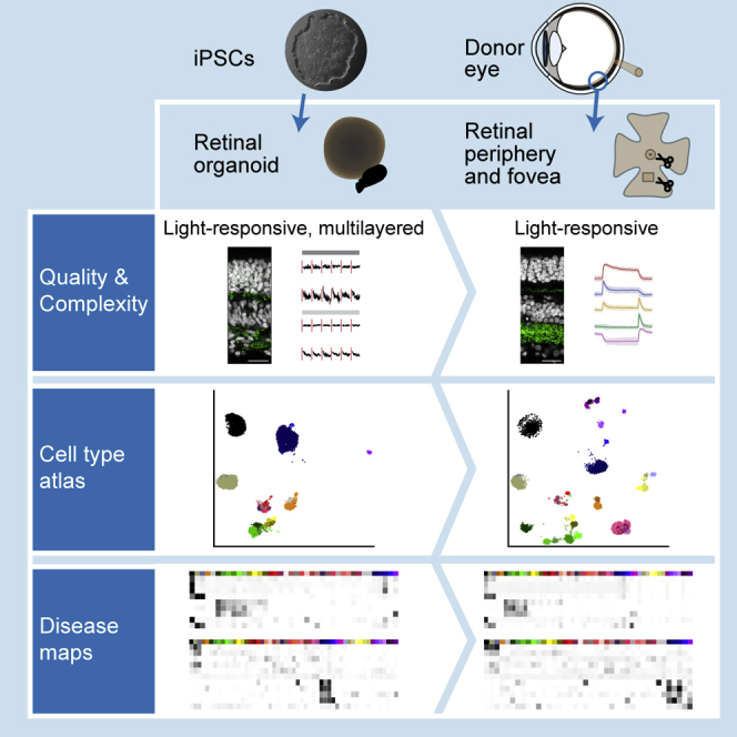
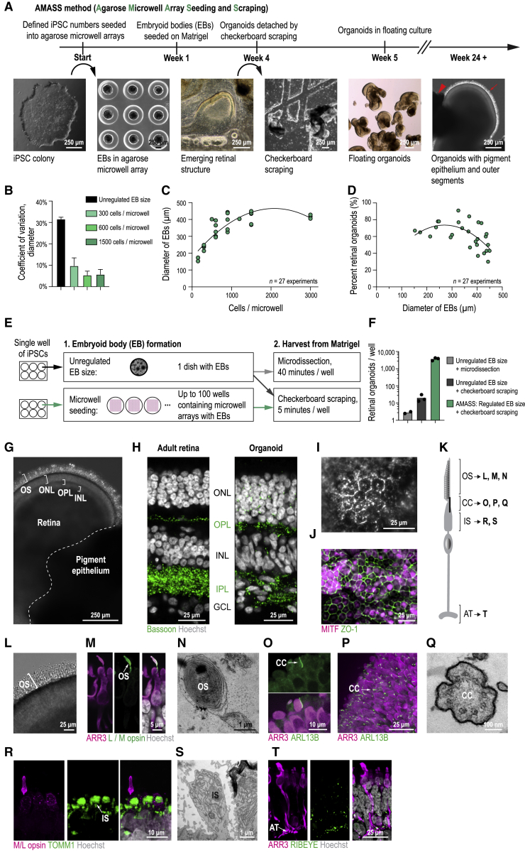
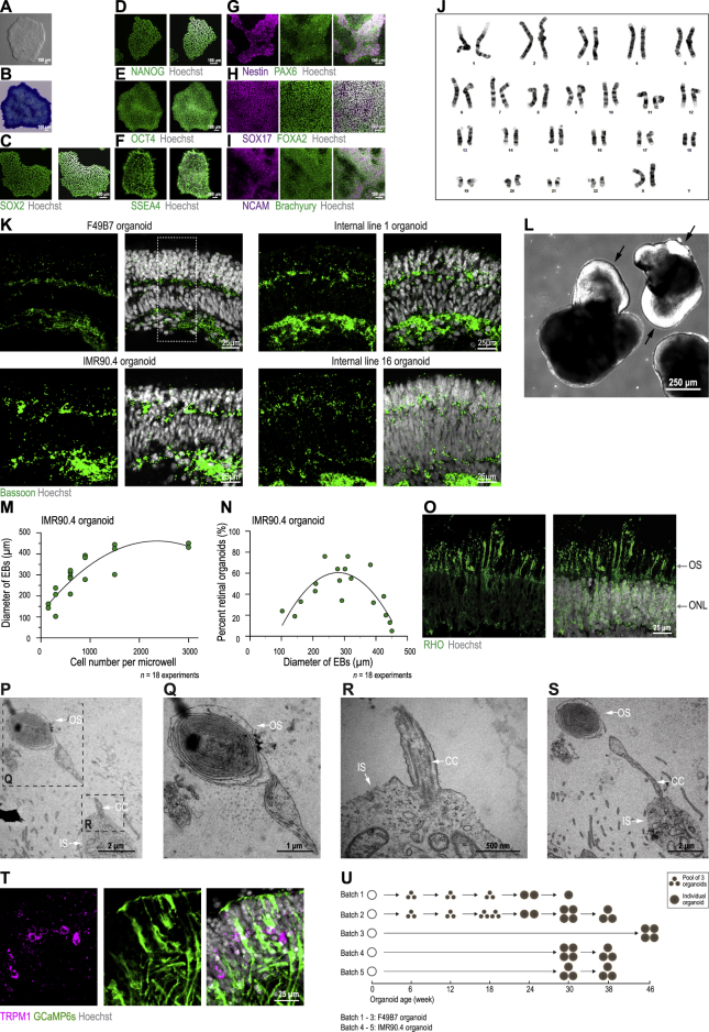
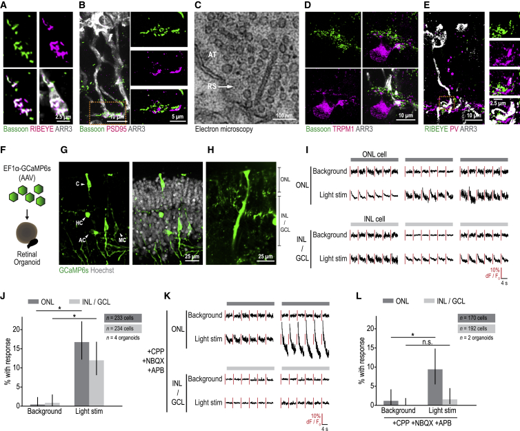
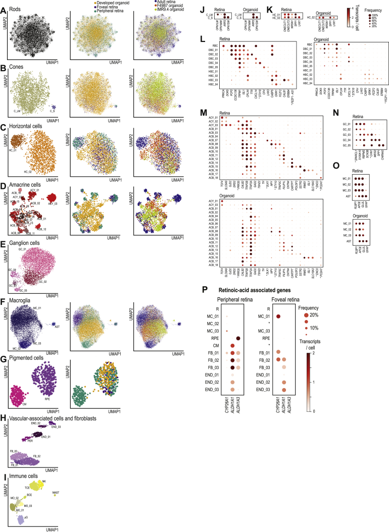
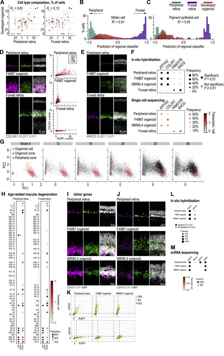
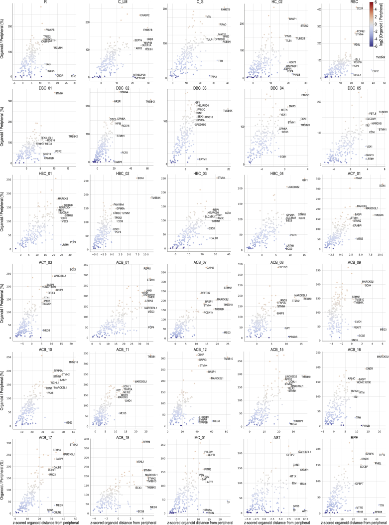
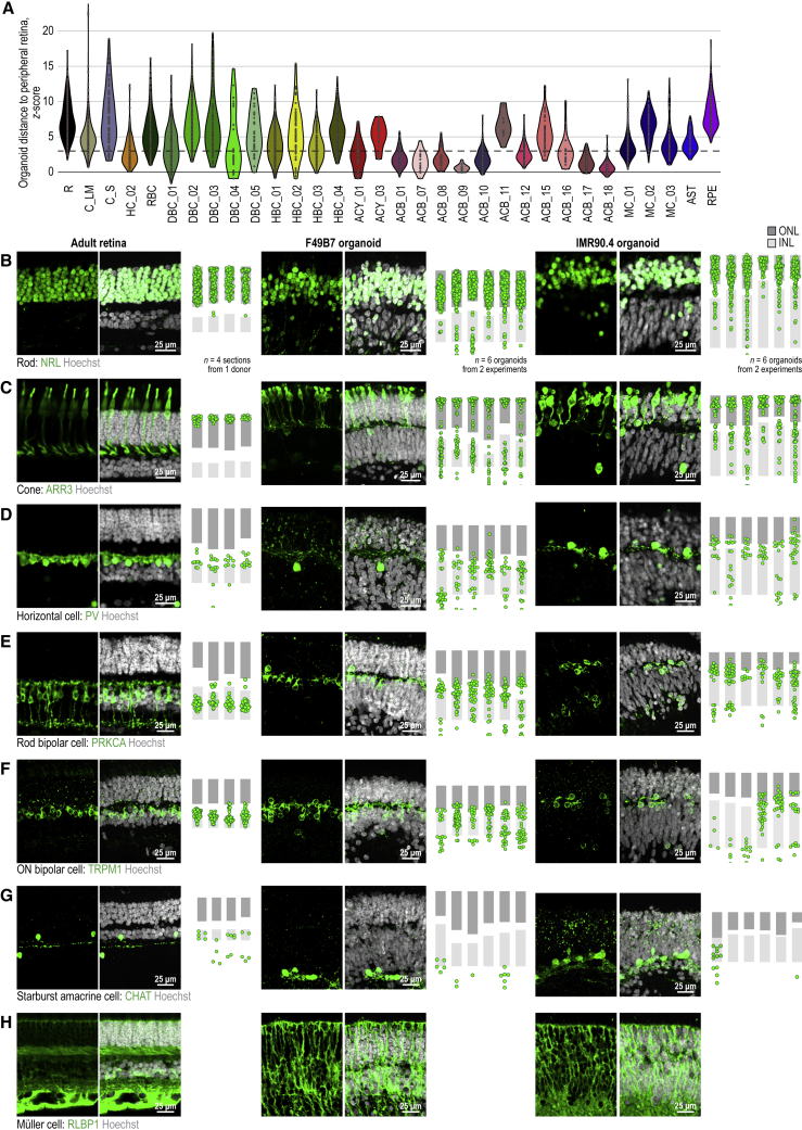
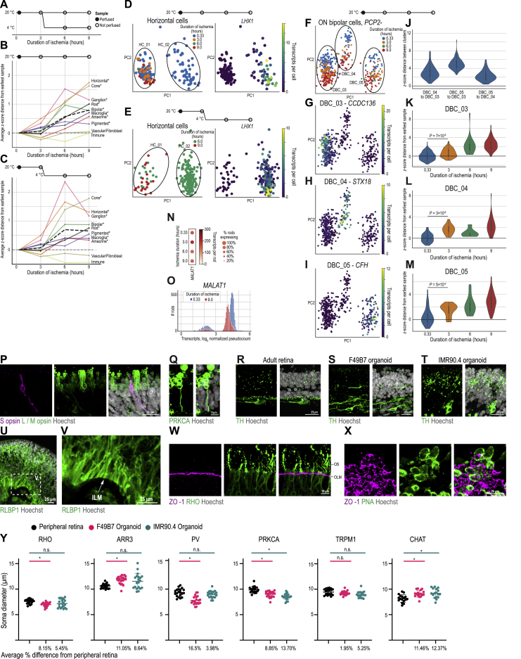
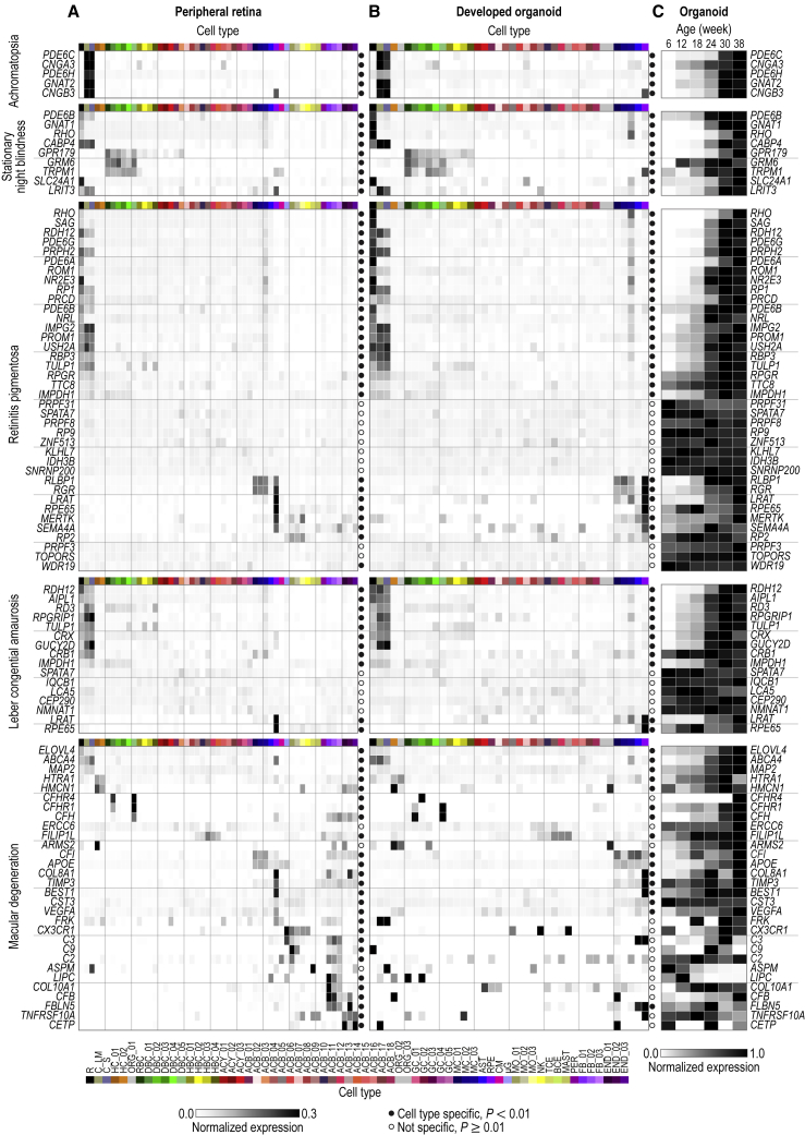
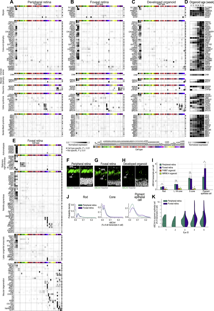
Human organoids recapitulating the cell-type diversity and function of their target organ are valuable for basic and translational research. We developed light-sensitive human retinal organoids with multiple nuclear and synaptic layers and functional synapses. We sequenced the RNA of 285,441 single cells from these organoids at seven developmental time points and from the periphery, fovea, pigment epithelium and choroid of light-responsive adult human retinas, and performed histochemistry. Cell types in organoids matured in vitro to a stable "developed" state at a rate similar to human retina development in vivo. Transcriptomes of organoid cell types converged toward the transcriptomes of adult peripheral retinal cell types. Expression of disease-associated genes was cell-type-specific in adult retina, and cell-type specificity was retained in organoids. We implicate unexpected cell types in diseases such as macular degeneration. This resource identifies cellular targets for studying disease mechanisms in organoids and for targeted repair in human retinas.
Overview
The claims and statistics of this paper haven’t been checked yet.
Run the check — free, no account needed. It reads the open-access text and checks the sources it cites.
Integrity signals
Registry
Cites
Statistics
Figures
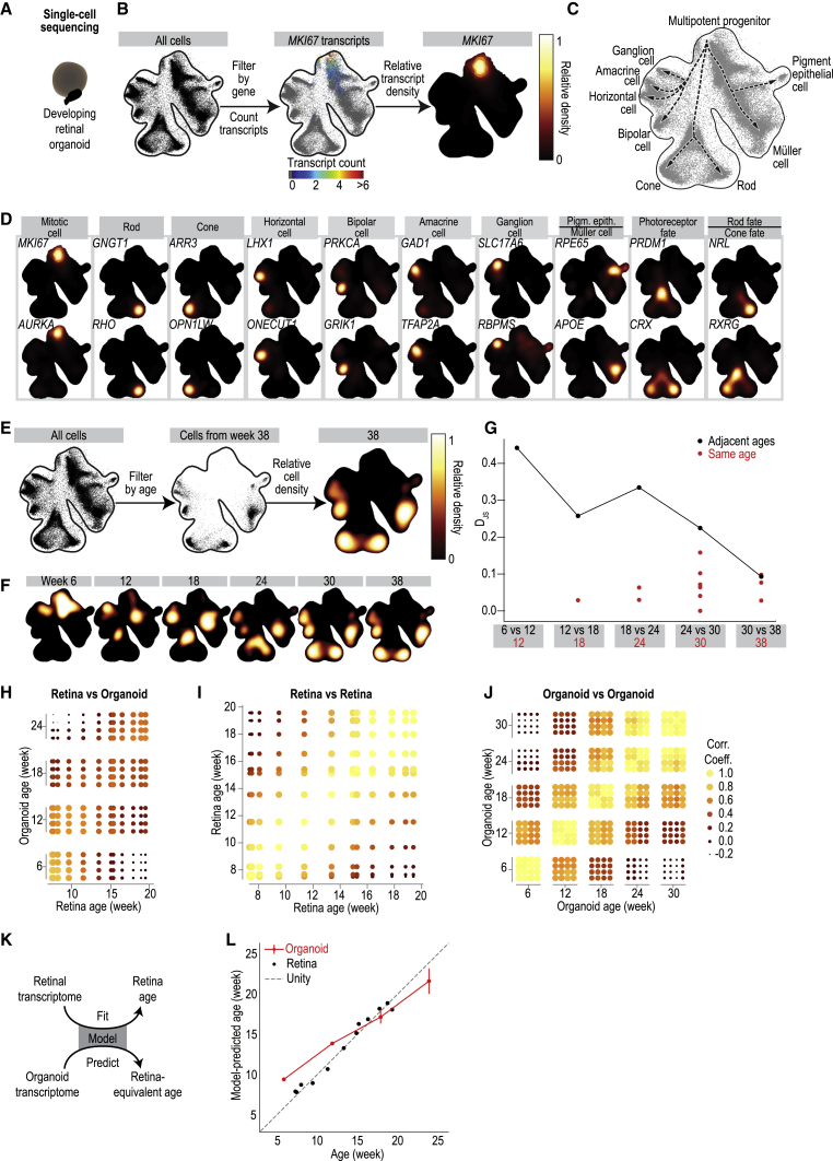
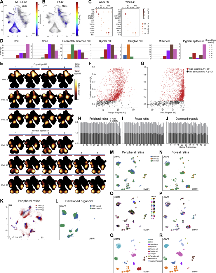
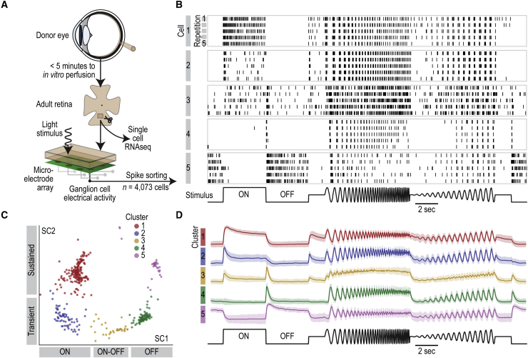
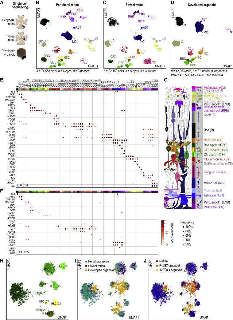
Verification
Claims the paper makes
The claims and statistics of this paper haven’t been checked yet.
Run the check — free, no account needed. It reads the open-access text and checks the sources it cites.
Claims other papers make about this one
Each is checked against this paper’s own text: does it say what the citing paper says it does?
The source backs part of the claim, not all of it — or backs it for a different population, condition or comparison.
In the citing paper
“ABCA4 is also present at extremely low levels in the retinal pigment epithelium (RPE) where it has been proposed to enhance endolysosomal function (2, 3).”
From Retinal gene therapy for Stargardt disease with dual AAV intein vectors is both safe and effective in large animal models (2025) · cites this paper as [2]
Experimental — accuracy not yet measuredIn the citing paper
“The physiological similarities between human and macaque retina (Cowan et al., 2019; Kling et al., 2020; Soto et al., 2020; Rodieck, 1998), including their responses to electrical stimulation (Madugula et al., 2022), suggest that the benefits of the present stimulation approach could translate from the ex vivo lab prototype with healthy macaque retina to an in vivo implant in the degenerated human”
From Precise control of neural activity using dynamically optimized electrical stimulation (2024) · cites this paper as Cowan et al., 2019
Self-check
Percentages, sums and reported differences re-computed from the paper’s own numbers.
The self-check needs the paper’s full text.
Citations
Cites
Scikit-learn: Machine Learning in Python
Pedregosa, F., Varoquaux, G., Gramfort, A., Michel, V., Thirion, B., Grisel, O., Blondel, M., Müller, A., Nothman, J., Louppe, G., et al. (2012) · arXiv · doi:10.48550/arxiv.1201.0490 · Cited by 71,016
Fiji: an open-source platform for biological-image analysis
Schindelin, J., Arganda-Carreras, I., Frise, E., Kaynig, V., Longair, M., Pietzsch, T., Preibisch, S., Rueden, C., Saalfeld, S., Schmid, B., et al. (2012) · Nature Methods · doi:10.1038/nmeth.2019 · Cited by 66,890
SciPy 1.0: fundamental algorithms for scientific computing in Python
Virtanen, P., Gommers, R., Oliphant, T. E., Haberland, M., Reddy, T., Cournapeau, D., Burovski, E., Peterson, P., Weckesser, W., Bright, J., et al. (2020) · Nature Methods · doi:10.1038/s41592-019-0686-2 · Cited by 38,430
Wickham, H., Averick, M., Bryan, J., Chang, W., McGowan, L., François, R., Grolemund, G., Hayes, A., Henry, L., Hester, J., et al. (2019) · Journal of Open Source Software · doi:10.21105/joss.01686 · Cited by 22,138
SciPy 1.0: fundamental algorithms for scientific computing in Python
Virtanen, P., Gommers, R., Oliphant, T. E., Haberland, M., Reddy, T., Cournapeau, D., Burovski, E., Peterson, P., Weckesser, W., Bright, J., et al. (2019) · Apollo - University of Cambridge Repository · doi:10.17863/cam.64134 · Cited by 10,460
The igraph software package for complex network research
Csárdi, G., & Nepusz, T. (2006) · Medical Entomology and Zoology · Cited by 10,078
The NumPy Array: A Structure for Efficient Numerical Computation
van der Walt, S., Colbert, S. C., & Varoquaux, G. (2011) · Computing in Science & Engineering · doi:10.1109/mcse.2011.37 · Cited by 9,381
SCANPY: large-scale single-cell gene expression data analysis
Wolf, F. A., Angerer, P., & Theis, F. J. (2018) · Genome Biology · doi:10.1186/s13059-017-1382-0 · Cited by 8,881
Highly parallel genome-wide expression profiling of individual cells using nanoliter droplets.
Macosko, E. Z., Basu, A., Satija, R., Nemesh, J., Shekhar, K., Goldman, M., Tirosh, I., Bialas, A. R., Kamitaki, N., Martersteck, E. M., et al. (2015) · Cell · doi:10.1016/j.cell.2015.05.002 · Cited by 7,110
UMAP: Uniform Manifold Approximation and Projection for Dimension Reduction
McInnes, L., Healy, J., & Melville, J. (2018) · arXiv · doi:10.48550/arxiv.1802.03426 · Cited by 7,101
Cited by
Sorted by citations
Items 401–500 of 665
Progress of iPSC-derived retinal organoids in the study of inherited retinal diseases
Liu, B., Liu, Y., Zhang, H., Huang, J., & Liu, G. (2025) · Orphanet Journal of Rare Diseases · doi:10.1186/s13023-025-04059-7 · Cited by 4
Li, R., Wu, Y., Zheng, Z., Zhou, F., Xu, K., & Su, J. (2026) · MedComm · doi:10.1002/mco2.70575 · Cited by 4
A cell fate specification and transition mechanism for human foveolar cone subtype patterning
Hussey, K. A., Eldred, K. C., Guy, B., Santiago, C. P., Zhang, J. S., Glass, I. A., Reh, T. A., Blackshaw, S., Goff, L. A., & Johnston, R. J. (2026) · Proceedings of the National Academy of Sciences · doi:10.1073/pnas.2510799123 · Cited by 4
Mitochondrial dysfunction in neurodegenerative disorders: mechanisms and therapeutic advances
Tong, Y., He, J. N., Zhou, L., Zhang, J., Ho, B. M., Du, L., Yip, Y. W. Y., Bisnauthsing, H., Chan, P. P. M., Tham, C. C. Y., et al. (2026) · Molecular Biomedicine · doi:10.1186/s43556-026-00480-x · Cited by 5
Eintracht, J., Owen, N., Harding, P., & Moosajee, M. (2024) · Stem Cell Reports · doi:10.1016/j.stemcr.2024.05.001 · Cited by 4
Assembling Retinal Organoids with Microglia
Xu, J., Yu, S., & Jin, Z. (2024) · Journal of Visualized Experiments · doi:10.3791/67016 · Cited by 4
Harding, P., Owen, N., Eintracht, J., Cunha, D. L., Chan, B. H. K., Rainger, J., & Moosajee, M. (2025) · Biochimica et Biophysica Acta (BBA) - Molecular Basis of Disease · doi:10.1016/j.bbadis.2025.167869 · Cited by 4
Development of Retinal Astroglia
Kay, J. N., Valdez‐Lopez, J. C., Dembla, E., & Miltner, A. M. (2025) · Annual Review of Vision Science · doi:10.1146/annurev-vision-121423-013153 · Cited by 4
Yang, L., Tao, Y., Pan, Q., Cai, T., Ye, Y., Liu, J., Zhou, Y., Shao, Y., Yi, Q., Lu, Z. H., et al. (2025) · Human Genomics · doi:10.1186/s40246-025-00796-9 · Cited by 4
Lemée, M. V., Loviglio, M. N., Ye, T., Tilly, P., Keime, C., Weber, C., Petrova, A., Klein, P., Morlet, B., Wendling, O., et al. (2025) · Nucleic Acids Research · doi:10.1093/nar/gkaf934 · Cited by 5
Single-Cell and Spatial Analysis of Emergent Organoid Platforms
Kumar, A., Cai, S., Allam, M., Henderson, S., Ozbeyler, M., Saiontz, L., & Coskun, A. F. (2023) · Methods in molecular biology · doi:10.1007/978-1-0716-3163-8_22 · Cited by 4
Pozo‐Valero, M. D., Almoallem, B., Rey, A. D., Mahieu, Q., Heetvelde, M. V., Jeddawi, L., Bauwens, M., & Baere, E. B. W. D. (2024) · Clinical Genetics · doi:10.1111/cge.14517 · Cited by 4
High-fat diet alters retinal lipid composition and gene expression networks in mice
Zou, R., Cai, J., Chen, T., Mo, W., Qian, H., Zhu, X., & Zhang, L. (2025) · BMC Biology · doi:10.1186/s12915-025-02212-z · Cited by 4
Single-cell RNA sequencing in the study of human retinal organoids
Chen, Y., Xu, H., Xiao, L., Zhang, M., & Yan, N. (2025) · Experimental Eye Research · doi:10.1016/j.exer.2025.110417 · Cited by 4
Jiawang, W., Liu, T., Zhang, X., Qu, C. F., Wu, J., Xu, S., Yang, J., & Xing, X. (2025) · Biofabrication · doi:10.1088/1758-5090/adde15 · Cited by 4
SUMOylation is a Translatable Target in Hypoxic MNPs Regulating Retinal Vasculopathy
Zhong, Z., Liang, G., Yu, H., Li, J., Wang, R., Ma, X., Zhou, Z., Zhao, Y., Sun, F., & Sun, X. (2025) · Advanced Science · doi:10.1002/advs.202503505 · Cited by 4
Yan, Y., Yang, H., Wei, Q., Yu, D., Gao, C., Diao, B., Song, Y., & Chen, X. (2025) · Experimental Eye Research · doi:10.1016/j.exer.2025.110508 · Cited by 4
Kelley, R. A., & Wu, Z. (2023) · Vision Research · doi:10.1016/j.visres.2023.108269 · Cited by 4
Kruczek, K., & Swaroop, A. (2023) · Current topics in developmental biology/Current Topics in Developmental Biology · doi:10.1016/bs.ctdb.2023.09.003 · Cited by 4
Mehra, D., & Le, P. H. (2019) · StatPearls · Cited by 4
[Study on the measurement of ambient ammonia in urban area based on open-path DOAS technique].
Wang, J., Xie, P.-H., Qin, M., Ling, L.-Y., Ye, C.-L., Liu, J., & Liu, W. (2012) · PubMed · Cited by 4
Gene Therapy for Rhodopsin MutationsPreprint
Lewin, A. S., & Smith, W. C. (2022) · Cold Spring Harbor Perspectives in Medicine · doi:10.1101/cshperspect.a041283 · Cited by 6
Human photoreceptor cell transplants integrate into human retina organoidsPreprint
Wagner, F., Carrera, R., Kurth, T., Michalakis, S., Naumann, R., Žužić, M., Neumann, K., Goureau, O., Busskamp, V., & Karl, M. (2022) · bioRxiv (Cold Spring Harbor Laboratory) · doi:10.1101/2022.08.09.500037 · Cited by 6
Monville, C., Goureau, O., & M’Barek, K. B. (2022) · Cold Spring Harbor Perspectives in Medicine · doi:10.1101/cshperspect.a041309 · Cited by 4
Hussey, K. A., Eldred, K. C., Guy, B., Santiago, C. P., Glass, I. A., Reh, T. A., Blackshaw, S., Goff, L. A., & Johnston, R. J. (2023) · bioRxiv (Cold Spring Harbor Laboratory) · doi:10.1101/2023.01.28.526051 · Cited by 4
A single-nucleus RNA sequencing atlas of the postnatal retina of the shark Scyliorhinus canicula
Vidal-Vázquez, N., Hernández‐Núñez, I., Carballo-Pacoret, P., Salisbury, S. J., Villamayor, P. R., Hervas-Sotomayor, F., Yuan, X., Lamanna, F., Schneider, C., Schmidt, J., et al. (2025) · Scientific Data · doi:10.1038/s41597-025-04547-2 · Cited by 4
Zhang, S., Dong, Z., Guo, J., Li, Z., Wu, H., Zhang, L., Min, F., & Zeng, T. (2025) · Journal of Neuroinflammation · doi:10.1186/s12974-025-03350-5 · Cited by 3
Edwards, K. L., Moore, B. M., Ganser, T.-S., Susaimanickam, P. J., Sovell, K., Martin, Y., Jager, L. D., Willes, A. M., Moyer, T. H., Bowar, L., et al. (2025) · Stem Cell Reports · doi:10.1016/j.stemcr.2025.102425 · Cited by 4
Podoliak, E., Guzman, G., & Busskamp, V. (2025) · Molecular Aspects of Medicine · doi:10.1016/j.mam.2025.101388 · Cited by 3
Engineering infrared light detection in blind human retina using ultrasensitive human TRPV1 channels
Chevalier, M., Fadel, F., Májer, T., Magda, D. P., Gerendás, L., Kilin, F., Nagy, Z. Z., Szabó, A., Roska, B., & Testa-Silva, G. (2025) · Trends in biotechnology · doi:10.1016/j.tibtech.2025.07.033 · Cited by 3
Self-citationWu, J. ‐., Fu, Y., Wang, X., Liu, T., Zhang, X., Qu, C., Zhang, Q., Xu, S., Yang, J., & Xing, X. (2025) · International Journal of Pharmaceutics · doi:10.1016/j.ijpharm.2025.126203 · Cited by 3
USH2A -Mutated Human Retinal Organoids Model Rod–Cone Dystrophy
Ashworth, K. E., Zhang, J., D’Amata, C., Héon, E., & Ballios, B. G. (2025) · Investigative Ophthalmology & Visual Science · doi:10.1167/iovs.66.14.2 · Cited by 3
A deep learning-based computational pipeline predicts developmental outcome in retinal organoids
Afting, C., Bhatti, N., Schlagheck, C., Salvador, E. S., Herrera, M., Agarwal, R., Suzuki, R., Hackert, N. S., Lorenz, H., Žílová, L., et al. (2026) · PLoS Biology · doi:10.1371/journal.pbio.3003597 · Cited by 3
Gene regulatory roles of growth and differentiation factors in retinal development
Luo, Z., Shah, S. H., Tanasă, B., Chang, K., & Goldberg, J. L. (2024) · iScience · doi:10.1016/j.isci.2024.110100 · Cited by 3
In vivo mechanism of the interaction between trimethylamine lyase expression and glycolytic pathways
Li, Q., Wu, D., Song, Y., Zhang, L., Wang, T., Chen, X., & Zhang, M. (2024) · Food & Function · doi:10.1039/d4fo03809f · Cited by 3
Lu, H., Liu, X., Zhou, Q., Li, R., Zuo, H., Hu, K., Tian, M., Wang, H., Li, J., Li, N., et al. (2025) · Genes & Diseases · doi:10.1016/j.gendis.2025.101610 · Cited by 3
Application of hiPSC as a Drug Tester Via Mimicking a Personalized Mini Heart
Wei, L., Xia, S., Li, Y., Qi, Y., Wang, Y., Zhang, D., Hua, Y., & Luo, S. (2022) · Frontiers in Genetics · doi:10.3389/fgene.2022.891159 · Cited by 3
Wu, Y., Qiu, J., Chen, S., Chen, X., Zhang, J., Zhuang, J., Liu, S., Yang, M., Zhou, P., Chen, H., et al. (2022) · International Journal of Molecular Sciences · doi:10.3390/ijms23137088 · Cited by 3
A minimal-complexity light-sheet microscope maps network activity in 3D neuronal systems
Wysmolek, P. M., Kiessler, F., Salbaum, K. A., Shelton, E., Sonntag, S. M., & Serwane, F. (2022) · Scientific Reports · doi:10.1038/s41598-022-24350-y · Cited by 5
Shen, W., Shao, A., Zhou, W., Lou, L., Grzybowski, A. E., Jin, K., & Ye, J. (2023) · Cell Transplantation · doi:10.1177/09636897231214321 · Cited by 4
RNA fusion in human retinal development
Wang, W., Zhang, X., Zhao, N., Xu, Z.-H., Jin, K., & Jin, Z. (2024) · eLife · doi:10.7554/elife.92523 · Cited by 3
In a century from agitated cells to human organoids
Layer, P. G. (2024) · Journal of Neuroscience Methods · doi:10.1016/j.jneumeth.2024.110083 · Cited by 3
Liu, J.-B., Yuan, H.-L., Zhang, G., & Ke, J.-B. (2024) · eNeuro · doi:10.1523/eneuro.0145-24.2024 · Cited by 3
Iso-Seq enables discovery of novel isoform variants in human retina at single cell resolutionPreprint
Wang, L., Urrutia-Cabrera, D., Hung, S. S. C., Hewitt, A. W., Lukowski, S. W., Foord, C., Wang, P., Tilgner, H., & Wong, R. C. B. (2024) · bioRxiv (Cold Spring Harbor Laboratory) · doi:10.1101/2024.08.08.607267 · Cited by 3
Uribe‐Salazar, J. M., Kaya, G., Weyenberg, K., Radke, B., Hino, K., Soto, D. C., Shiu, J.-L., Zhang, W., Ingamells, C., Haghani, N. K., et al. (2024) · bioRxiv (Cold Spring Harbor Laboratory) · doi:10.1101/2024.09.11.612570 · Cited by 3
Deep learning predicts tissue outcomes in retinal organoidsPreprint
Afting, C., Bhatti, N., Schlagheck, C., Salvador, E. S., Herrera, M., Agarwal, R., Suzuki, R., Hackert, N. S., Lorenz, H., Žílová, L., et al. (2025) · bioRxiv (Cold Spring Harbor Laboratory) · doi:10.1101/2025.02.19.639061 · Cited by 3
Connectome of a human foveal retinaPreprint
Kim, Y. J., PACKER, O. S., Macrina, T., Pollreisz, A., Curcio, C. A., Lee, K., Kemnitz, N., Ih, D., Nguyen, T., Lu, R., et al. (2025) · bioRxiv (Cold Spring Harbor Laboratory) · doi:10.1101/2025.04.05.647403 · Cited by 3
RetiGene, a comprehensive gene atlas for inherited retinal diseases (IRDs)Preprint
Quinodoz, M., Çelik, E., Kamdar, D., Cancellieri, F., Kamińska, K., Ullah, M., Barberán-Martínez, P., Bouckaert, M., Cortón, M., Delanote, E., et al. (2025) · bioRxiv (Cold Spring Harbor Laboratory) · doi:10.1101/2025.06.08.653722 · Cited by 3
Unified Generation of Regionalized Neural Organoids from Single-Lumen NeuroepitheliumPreprint
Rao, J., He, Z., Loskarn, S., Bender, A., Jo, Y., Lückel, J., Curcio, M., Cubela, I., Camp, J. G., Treutlein, B., et al. (2025) · bioRxiv (Cold Spring Harbor Laboratory) · doi:10.1101/2025.11.18.689013 · Cited by 4
Retinal cytoarchitecture is preserved in an organotypic perfused human and porcine eye model
Chan, D. W.-H., Zhang, J. W., Won, G.-J., & Sivak, J. M. (2024) · Acta Neuropathologica Communications · doi:10.1186/s40478-024-01892-y · Cited by 2
Corti, S., Kim, K. H., Chen, T., Botezatu, A., Cora, V., Ma, K., Pashkovskaia, N., Vergara, A. B., Sperlich, D., Dave, K., et al. (2025) · Cell Reports Medicine · doi:10.1016/j.xcrm.2025.102244 · Cited by 5
Xu, X., Zhang, Y., Geng, Y., Luo, Y., & Sun, X. (2025) · Frontiers in Pharmacology · doi:10.3389/fphar.2025.1650200 · Cited by 3
Organoids in Genetic Disorders: from Disease Modeling to Translational Applications
Zhu, Y., Lin, N., Li, J., Zhang, H., Zhang, P., Cheng, X., Yang, Q., & Liu, L. (2025) · Stem Cell Reviews and Reports · doi:10.1007/s12015-025-10973-x · Cited by 2
The genesis of neurons and glia in the developing retina
Kaplan, L., Wohlschlegel, J., & Reh, T. A. (2025) · Current topics in developmental biology/Current Topics in Developmental Biology · doi:10.1016/bs.ctdb.2025.05.001 · Cited by 2
Weber, V. J., Reschigna, A., Gerhardt, M. J., Heigl, T., Hinrichsmeyer, K. S., van den Engel, S., Otify, D. Y., Gavrilov, Z., Blaser, F., Meneau, I., et al. (2025) · JCI Insight · doi:10.1172/jci.insight.189615 · Cited by 2
Wang, C., Chen, M., Fang, Y., Fu, Y., Lv, Y., Zhang, X., Li, B., Bai, Y., Li, Q., Zeng, Y., et al. (2025) · Stem Cell Research & Therapy · doi:10.1186/s13287-025-04848-8 · Cited by 2
Ma, J. Y. W., Daniszewski, M., & Pébay, A. (2025) · Molecular Medicine · doi:10.1186/s10020-025-01405-0 · Cited by 3
Retinal organoids in age-related macular degeneration: Promise, pitfalls, and progress
Palakkan, A. A., & Kumar, G. S. (2025) · Experimental Eye Research · doi:10.1016/j.exer.2025.110831 · Cited by 2
Bellapianta, A., Qi, J., Giugliano, M., Ouaidat, S., Rawas, R. E., Bolz, M., & Salti, A. (2025) · Biomedicines · doi:10.3390/biomedicines14010045 · Cited by 2
Zhang, Y., Niu, L., Xia, H., & Wang, J. (2026) · Frontiers in Endocrinology · doi:10.3389/fendo.2025.1733368 · Cited by 2
Wang, Z., Chen, G., Chen, G., Zhao, C., Wang, C., Shi, J., & Chen, H. (2026) · Journal of Neuroinflammation · doi:10.1186/s12974-026-03693-7 · Cited by 3
Healing of ischemic injury in the retina
Becker, S., L'Ecuyer, Z., Allen, J., Saeid, S., Sturgis, L., Adderley, A., Koskelainen, A., & Vinberg, F. (2026) · Science Advances · doi:10.1126/sciadv.adx7204 · Cited by 2
Advances in Quality Control of Tumor Organoid Living Biobanks
Fan, R., Zhou, S., An, P., Ye, X., Yu, M., Wang, Y., & Wu, C. (2026) · Biopreservation and Biobanking · doi:10.1177/19475535261416482 · Cited by 2
Development of a 3D-printed microfluidic chip for retinal organoid–endothelial co-culture
Abdalkader, R., Kawakami, S., Takashima, Y., & Fujita, T. (2026) · Lab on a Chip · doi:10.1039/d5lc00939a · Cited by 5
Organoid–microglia system for modeling the immune microenvironment of the brain and retina
Yu, J., Tang, B., Gu, Z., Wang, G., & Liu, A. (2026) · Frontiers in Immunology · doi:10.3389/fimmu.2026.1747589 · Cited by 2
Protocol for quality control screening of brain organoid morphology
Chiaradia, I., Jérôme Boulanger, A., Elmkvist, S. B., Larsen, M. R., & Lancaster, M. A. (2026) · STAR Protocols · doi:10.1016/j.xpro.2026.104423 · Cited by 2
Organoid models: Regulations and prospects for the development of investigational new drugs
Li, H., Pan, Y., Zheng, Z., Lin, R., Pan, Y., Lin, L., Quan, M., Lu, H., Hu, Z., Zheng, M., et al. (2026) · Chinese Medical Journal · doi:10.1097/cm9.0000000000004067 · Cited by 4
Agarwal, R., Iezhitsa, I. N., Hombrebueno, J. R., & Agarwal, P. (2026) · International Journal of Retina and Vitreous · doi:10.1186/s40942-026-00846-x · Cited by 2
Revisiting retinal and macular degeneration in the genomics era
Swaroop, A., & Roska, B. (2026) · Nature Reviews Genetics · doi:10.1038/s41576-026-00960-4 · Cited by 2
Self-citationThe Analysis of Embryoid Body Formation and Its Role in Retinal Organoid Development
Berzal, A. H., Wagstaff, E. L., ten Asbroek, A. L. M. A., ten Brink, J. B., Bergen, A. A. B., & Boon, C. J. F. (2024) · International Journal of Molecular Sciences · doi:10.3390/ijms25031444 · Cited by 2
Zhang, Y., Yan, S., Mei, Z., Zhang, H., Ding, C., Zhang, S., & Wei, S. (2024) · Tissue Engineering and Regenerative Medicine · doi:10.1007/s13770-024-00632-6 · Cited by 2
Non-Canonical Wnt Pathway Expression in the Developing Mouse and Human Retina
Campos, R. C., Matsunaga, K., Reid, M. W., Fernández, G. E., Stepanian, K., Bharathan, S. P., Li, M., Thornton, M. E., Grubbs, B. H., & Nagiel, A. (2023) · Experimental Eye Research · doi:10.1016/j.exer.2024.109947 · Cited by 3
Morphogenetic Designs, and Disease Models in Central Nervous System Organoids
Bock, M., Hong, S. J., Zhang, S., Yu, Y., Lee, S., Shin, H.-E., Choi, B. H., & Han, I. (2024) · International Journal of Molecular Sciences · doi:10.3390/ijms25147750 · Cited by 3
Hu, X., Chen, J., Dai, W., Xiao, Y., Chen, X., Chen, Z., Zhang, S., & Hu, Y. (2024) · Cellular and Molecular Life Sciences · doi:10.1007/s00018-024-05279-z · Cited by 2
Advances in Organ-on-a-chip Technologies for Biomedical Research and Applications
Zhu, Q., Liao, M., Hu, Y., & Chai, R. (2025) · Materials Lab · doi:10.54227/mlab.20240006 · Cited by 2
3D Quantification of Viral Transduction Efficiency in Living Human Retinal Organoids
Rogler, T. S., Salbaum, K. A., Brinkop, A. T., Sonntag, S. M., James, R. E., Shelton, E., Thielen, A., Rose, R., Babutzka, S., Klopstock, T., et al. (2025) · Small Methods · doi:10.1002/smtd.202401050 · Cited by 2
Investigation of ABCA4 Missense Variants and Potential Small Molecule Rescue in Retinal Organoids
Piccolo, D., Sladen, P. E., Guarascio, R., Ziaka, K., & Cheetham, M. E. (2025) · Investigative Ophthalmology & Visual Science · doi:10.1167/iovs.66.9.58 · Cited by 2
SOX2-driven enhancer landscape defines the transcriptional architecture of retinogenesis
Bian, F., Golestaneh, K., Davis, E., Khan, A., Nicholson, B., Daghsni, M., Flohr, K., Liu, S., da Silva, S., Pennacchio, L. A., et al. (2025) · Development · doi:10.1242/dev.204986 · Cited by 3
Numb deficiency impairs retinal structure and visual function in mice
Yan, J., Li, Y., Zhang, T., & Shen, Y. (2022) · Experimental Eye Research · doi:10.1016/j.exer.2022.109066 · Cited by 2
Sharma, M., Jangra, S., Dhiman, S., Yangzes, S., Tiwari, A., Patro, S. K., Sharma, K., Rathore, S., Ramavat, A. S., Chhabra, S., et al. (2023) · Elsevier eBooks · doi:10.1016/b978-0-323-99987-8.00007-2 · Cited by 2
Modeling Retinitis Pigmentosa with Patient-Derived iPSCs
Leong, Y. C., & Sowden, J. C. (2023) · Advances in experimental medicine and biology · doi:10.1007/978-3-031-27681-1_81 · Cited by 2
Gasparini, S. J., Tessmer, K., Reh, M., Wieneke, S., Carido, M., Völkner, M., Borsch, O., Swiersy, A., Žužić, M., Goureau, O., et al. (2021) · bioRxiv (Cold Spring Harbor Laboratory) · doi:10.1101/2021.08.26.457641 · Cited by 2
Lyu, P., Hoang, T. V., Santiago, C. P., Thomas, E. D., Timms, A. E., Appel, H., Gimmen, M. Y., Le, N., Jiang, L., Kim, D. W., et al. (2021) · SSRN Electronic Journal · doi:10.2139/ssrn.3921283 · Cited by 3
Editorial: Hereditary Optic Neuropathies: A New Perspective
Carelli, V., Karanjia, R., & Morgia, C. L. (2021) · Frontiers in Neurology · doi:10.3389/fneur.2021.742484 · Cited by 2
Scale Invariance of Mechanical Properties in the Developing Mammalian RetinaPreprint
Shelton, E., Frischmann, M., Brinkop, A. T., James, R. E., Hüttl, L. M., & Serwane, F. (2024) · bioRxiv (Cold Spring Harbor Laboratory) · doi:10.1101/2024.10.21.619491 · Cited by 2
Maggi, K., Atac, D., Maggi, J., Feil, S., Koller, S., & Berger, W. (2024) · bioRxiv (Cold Spring Harbor Laboratory) · doi:10.1101/2024.11.15.623746 · Cited by 2
Developmental wave of programmed ganglion cell death in human retinal organoidsPreprint
Brooks, T., Park, Y. K., Vielle, A., Ha, M., Rio‐Tsonis, K. D., Robinson, M. L., & Vergara, M. N. (2025) · bioRxiv (Cold Spring Harbor Laboratory) · doi:10.1101/2025.07.25.666895 · Cited by 2
Rendón, J. M., Polvillo, R., González-Cid, Á., Corbacho, J., Naranjo, S., Manzo, S., Sousa‐Ortega, A., Fernández‐Miñán, A., Tena, J. J., & Martínez‐Morales, J. R. (2026) · bioRxiv (Cold Spring Harbor Laboratory) · doi:10.64898/2026.01.31.703039 · Cited by 2
Pan, X., Saunders, R. A., Replogle, J. M., Weissman, J. S., & Zhuang, X. (2026) · bioRxiv (Cold Spring Harbor Laboratory) · doi:10.64898/2026.05.16.725005 · Cited by 3
A great diversity of ROBO4 expression and regulations identified by data mining and transgene mice
Li, H., Xiao, H., Mai, X., Huang, S., Chen, J., & Xiao, X. (2024) · Gene Expression Patterns · doi:10.1016/j.gep.2024.119375 · Cited by 1
Generation of Retinal Organoids Using Human-Induced Pluripotent Stem Cells
Theodore, K., Imventarza, J. A., Tahir, S., da Costa, B. L., & Quinn, P. M. J. (2025) · Methods in molecular biology · doi:10.1007/7651_2024_600 · Cited by 1
“Armed in-vitro retina”-generating microglial retinal organoids, where are we now?
Liu, Y., Gao, L., Chen, W., Yan, Y., Ye, Z., & Li, Z. (2025) · Frontiers in Cell and Developmental Biology · doi:10.3389/fcell.2025.1574283 · Cited by 2
Han, T., Yu, Y., Wang, Y., Peng, X., Chen, Z., Lin, Q., & Zhou, X. (2025) · International Journal of Biological Macromolecules · doi:10.1016/j.ijbiomac.2025.147969 · Cited by 1
Bao, Y., Soh, S., Li, J., Zuo, Z. C., Ferdous, S., Qian, X., Li, J., Lu, J., Cheng, X., Matynia, A., et al. (2025) · Experimental Eye Research · doi:10.1016/j.exer.2025.110677 · Cited by 2
The association of ARR3 genetic polymorphisms with pathologic myopia in Northern Chinese population
Gao, W., Zhang, M., An, G., Yang, F., Li, L., Jin, X., & Du, L. (2025) · Experimental Eye Research · doi:10.1016/j.exer.2025.110691 · Cited by 1
Matsushita, T., Matsuyama, T., Kawasaki, T., & Uchiumi, F. (2025) · Organoids · doi:10.3390/organoids4040027 · Cited by 1
Electroporation-based gene delivery and whole-organoid imaging in human retinal organoids
Flohr, K., Janeček, M., Wang, L., Valle, V., Pi, S., Peixoto, R. T., & da Silva, S. (2025) · Developmental Biology · doi:10.1016/j.ydbio.2025.11.016 · Cited by 1
Busson, S. L., Naeem, A., Ferrara, S., Sarcar, S., Adefila-Ideozu, T., Wells, S., Alami, S. E., Boot, J. R., Sladen, P. E., Michaelides, M., et al. (2025) · International Journal of Molecular Sciences · doi:10.3390/ijms27010449 · Cited by 1
Endothelial stem cells of the retinal vasculature reside in the optic nerve
Sakimoto, S., Takigawa, T., Oguchi, A., Yoshihara, M., Sekito, S., Ueda, C., Shiraki, A., Shiki, K., Yamaguchi, K., Fukushima, Y., et al. (2026) · Nature Communications · doi:10.1038/s41467-025-68201-6 · Cited by 1
Tendon Organoids Enable Functional Tendon Rejuvenation Through ALKBH5‐Dependent RNA Demethylation
Qin, T., Pan, A., Miao, Z., Zhao, Y., Mo, X., Sun, H., Fan, C., Guo, J., Wu, B., Shen, W., et al. (2026) · Advanced Science · doi:10.1002/advs.202508376 · Cited by 1
Metadata
- Authors
- Cowan, C. S., Renner, M., De Gennaro, M., Gross-Scherf, B., Goldblum, D., Hou, Y., Munz, M., Rodrigues, T. M., Krol, J., Szikra, T., Cuttat, R., Waldt, A., Papasaikas, P., Diggelmann, R., Patino-Alvarez, C. P., Galliker, P., Spirig, S. E., Pavlinic, D., Gerber-Hollbach, N., Schuierer, S., Srdanovic, A., Balogh, M., Panero, R., Kusnyerik, A., Szabo, A., Stadler, M. B., Orgül, S., Picelli, S., Hasler, P. W., Hierlemann, A., Scholl, H. P. N., Roma, G., Nigsch, F., & Roska, B.
- Affiliations
- Institute of Molecular and Clinical Ophthalmology Basel (CH)
- University of Basel (CH)
- Novartis Institutes for BioMedical Research (CH)
- SIB Swiss Institute of Bioinformatics (CH)
- ETH Zurich (CH)
- Friedrich Miescher Institute (CH)
- Johns Hopkins University (US)
- Novartis (Switzerland) (CH)
- Corresponding author
- Roma, Guglielmo
- Nigsch, Florian
- Roska, Botond
- ORCID
- Cowan, Cameron S.
- Renner, Magdalena
- Goldblum, David
- Hou, Yanyan
- Munz, Martin
- Rodrigues, Tiago M.
- Cuttat, Rachel
- Waldt, Annick
- Papasaikas, Panagiotis
- Diggelmann, Roland
- Spirig, Stefan E.
- Schuierer, Sven
- Srdanovic, Aldin
- Panero, Riccardo
- Stadler, Michael B.
- Orgül, Selim
- Picelli, Simone
- Hasler, Pascal W.
- Hierlemann, Andreas
- Scholl, Hendrik P.N.
- Roma, Guglielmo
- Nigsch, Florian
- Roska, Botond
- Venue
- Cell
- Publisher
- Elsevier BV
- Published
- 2020-09
- Volume
- 182
- Issue
- 6
- Pages
- 1623-1640.e34
- ISSN
- 0092-8674 (ISSN-L)
- PMC
- PMC7505495
- OpenAlex
- W3087102910
- Cited by
- 654
- References
- 94
- Citations by year
- 2026: 79 · 2025: 118 · 2024: 118 · 2023: 113 · 2022: 123 · 2021: 74 · 2020: 23
- FWCI
- 26.98
- Type
- journal article
- Language
- en
- Open access
- hybrid
- doi.org
- Licence
- Locations
- Cell (publishedVersion, cc-by)
- bioRxiv (Cold Spring Harbor Laboratory) (acceptedVersion)
- PubMed Central (publishedVersion, cc-by)
- PubMed (publishedVersion)
- The Novartis Repository (Novartis) (submittedVersion, cc-by)
- Repository for Publications and Research Data (ETH Zurich) (submittedVersion, cc-by)
- ETH Zürich Research Collection (cc-by)
- Funders
- European Research Council
- Topics
- Health Sciences › Medicine › Ophthalmology › Retinal Development and Disorders
- Life Sciences › Biochemistry, Genetics and Molecular Biology › Molecular Biology › Single-cell and spatial transcriptomics
- Health Sciences › Medicine › Ophthalmology › Retinal Diseases and Treatments
- Keywords
- retinal cell types, single-cell transcriptomics, retinal development, photosensitivity, retinal pigment epithelium, choroid, fovea, macular degeneration, cell type specificity, retinal repair
- MeSH terms
- Cell Differentiation, Cell Line, Electrophysiology, Female, Multigene Family, Humans, Microscopy, Electron, Naphthoquinones, Organoids, Retina, Synapses, In Situ Hybridization, Gene Expression Regulation, Developmental, Cell Culture Techniques, Genetic Predisposition to Disease, Induced Pluripotent Stem Cells, Single-Cell Analysis, Transcriptome
About these checks
Registry, retraction, Cites and statistics results are computed, not judged, with one exception: a printed reference that no index lists is read once by a model before it counts as a citation error. Claims are extracted by a model and kept only when their quote appears verbatim in the text; each cited source is then checked, with a model’s help, for whether it says what the claim relies on. Those verdicts can be wrong — read the claim’s quote and the cited source before relying on one.
Statistics and arithmetic are recomputed, not judged.
Confidence bands are shown only for fields calibrated on our annotated test set. None is calibrated yet, so no band is shown.
What counts as a claim ›How we measure confidence ›How we check this ›
Are you an author, or do you see a problem with this page? Request a review
