Cited by 157

The DOI matches its Crossref record; 8 of the 8 references it cites were found in a registry or an index.

Giardia duodenalis induces pathogenic dysbiosis of human intestinal microbiota biofilms

Beatty, Jennifer K., Akierman, Sarah V., Motta, Jean-Paul, Muise, Stacy, Workentine, Matthew L., Harrison, Joe J., Bhargava, Amol, Beck, Paul L., Rioux, Kevin P., McKnight, Gordon Webb, Wallace, John L., Buret, Andre G.·2017·International Journal for Parasitology

Overview

The claims and statistics of this paper haven’t been checked yet.

Run the check — free, no account needed. It reads the open-access text and checks the sources it cites.

A few free checks a day · papers already verified open instantly · results are public

Integrity signals

Registry

Crossref · 10.1016/j.ijpara.2016.11.010

Cites

8 of 8 references verified0 citation errors · 0 not checkable

Statistics

Figures

Image 1
CC-BY-NC-ND · From the PDF
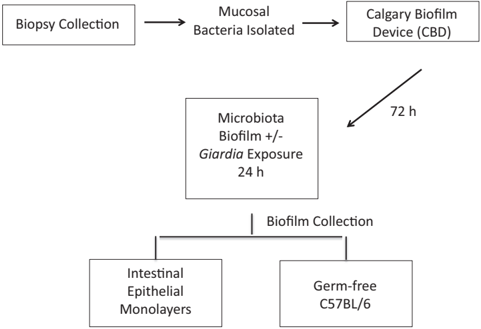
Fig. 1 Outline of experimental procedure. Human mucosal biopsies were collected from healthy donors (see Section 2.2), and mucosal bacteria were isolated and seeded into the Calgary Biofilm Device. Microbiota biofilms were cultivated on the Calgary Biofilm Device for 72 h and exposed to live Giardia duodenalis , spent medium, or control conditions for 24 h. Following exposure, biofilms were processed for microscopic analysis, intestinal epithelial monolayer challenge, or inoculation into germ-free C57BL/6 mice. Multiple aliquots of three biopsies, from a total of six donors, were used to seed biofilms used for all experiments.
Fig. 1 Outline of experimental procedure. Human mucosal biopsies were collected from healthy donors (see Section 2.2), and mucosal bacteria were isolated and seeded into the Calgary Biofilm Device. Microbiota biofilms were cultivated on the Calgary Biofilm Device for 72 h and exposed to live Giardia duodenalis , spent medium, or control conditions for 24 h. Following exposure, biofilms were processed for microscopic analysis, intestinal epithelial monolayer challenge, or inoculation into germ-free C57BL/6 mice. Multiple aliquots of three biopsies, from a total of six donors, were used to seed biofilms used for all experiments.CC-BY-NC-ND · From the PDF
log2 fold-change in Giardia relative to control
log2 fold-change in Giardia relative to controlCC-BY-NC-ND · From the PDF
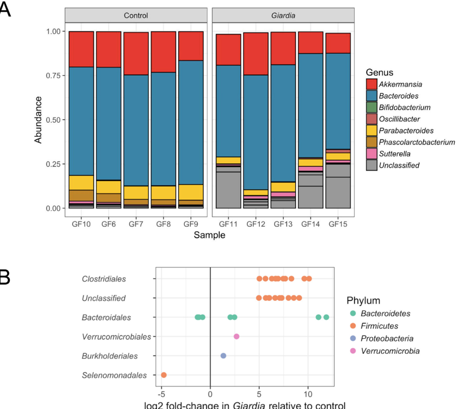
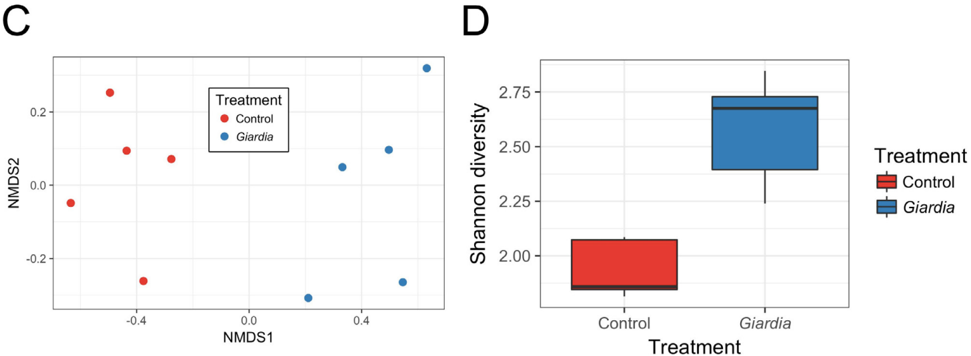
Fig. 2 Community profiles of faecal microbiota of germ-free mice reconstituted with human microbiota biofilms; mice were given either untreated microbiota (Control), or microbiota challenged with Giardia duodenalis ( Giardia ). Germ-free C57Bl6 mice were administered two rounds of human microbiota biofilm communities exposed to G. duodenalis , or control medium containing no parisites, and were euthanised after 14 days ( n = 5 per group). Faecal samples were collected from each animal and analysed via 16S amplicon sequencing of the V3-V4 rRNA region. (A) Operational taxomomic units in faeces of mice either given untreated microbiota (Control), or microbiota challenged with G. duodenalis ( Giardia ). (B) Changes in operational taxomomic unit abundance by phylum in the same samples. (C) Non-metric multidimensional scaling ordination plot of b -diversity using the Bray-Curtis metric in the same samples. (D) a -Diversity of the same samples as assessed using the Shannon diversity index.
Fig. 2 Community profiles of faecal microbiota of germ-free mice reconstituted with human microbiota biofilms; mice were given either untreated microbiota (Control), or microbiota challenged with Giardia duodenalis ( Giardia ). Germ-free C57Bl6 mice were administered two rounds of human microbiota biofilm communities exposed to G. duodenalis , or control medium containing no parisites, and were euthanised after 14 days ( n = 5 per group). Faecal samples were collected from each animal and analysed via 16S amplicon sequencing of the V3-V4 rRNA region. (A) Operational taxomomic units in faeces of mice either given untreated microbiota (Control), or microbiota challenged with G. duodenalis ( Giardia ). (B) Changes in operational taxomomic unit abundance by phylum in the same samples. (C) Non-metric multidimensional scaling ordination plot of b -diversity using the Bray-Curtis metric in the same samples. (D) a -Diversity of the same samples as assessed using the Shannon diversity index.CC-BY-NC-ND · From the PDF
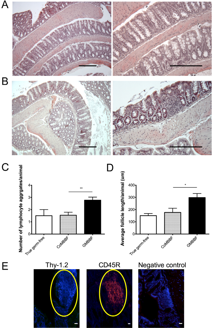
Fig. 3 Giardia -exposed microbiota biofilm communities promote B cell-containing lymphocyte follicles in the colonic epithelium of reconstituted germ-free mice. Sections of colon tissue from reconstituted germ-free C57Bl/6 mice. Animals were given intercolonic administration of two rounds of human microbiota biofilm communities exposed, or not, to Giardia and were euthanised after 14 days ( n = 5-10 per group). Representative pictures of colon tissue stained with H&E from mice receiving either control biofilms CoMBBF (A), or those exposed to Giardia GMBBF (B). The average number (C) and average size (D) (recorded blindly) of colonic lymphocyte follicles per microscopic field (200X) in groups of mice receiving control microbiota biofilms (CoMBBF), and mice receiving Giardia -exposed microbiota biofilms (GMBBF) (  four different fields were analysed per animal. True germ-free animals were neither exposed to CoMBBF nor GMBBF, but represented a measure of baseline colonisation following transfer from germfree facilities to the environment where experimentation took place. (E) Immunohistochemistry was performed on colonic sections of reconstituted germ-free mice. Representative images of animals receiving Giardia -exposed microbiota biofilms are presented as stained for T and B cell phenotypes. Host nuclei were coloured in blue (DAPI), Thy-1.2 (CD90-pan T cell marker), and CD45r-positive cells in red (rat polyclonal anti-CD45R coupled with alexa455). Lymphocyte aggregates were negative for Thy1.2 (CD90-pan T cell marker) staining. Negative control staining used rat IgG coupled with the same fluorochrome. Scale bars represent 50 m mfor E and represent the limit of intraepithelial lymphocyte follicles ( n  4 different fields were analysed per animal). / P < 0.05, // P < 0.01 (ANOVA and Tukey's multiple comparison test for data in C and D).
Fig. 3 Giardia -exposed microbiota biofilm communities promote B cell-containing lymphocyte follicles in the colonic epithelium of reconstituted germ-free mice. Sections of colon tissue from reconstituted germ-free C57Bl/6 mice. Animals were given intercolonic administration of two rounds of human microbiota biofilm communities exposed, or not, to Giardia and were euthanised after 14 days ( n = 5-10 per group). Representative pictures of colon tissue stained with H&E from mice receiving either control biofilms CoMBBF (A), or those exposed to Giardia GMBBF (B). The average number (C) and average size (D) (recorded blindly) of colonic lymphocyte follicles per microscopic field (200X) in groups of mice receiving control microbiota biofilms (CoMBBF), and mice receiving Giardia -exposed microbiota biofilms (GMBBF) (  four different fields were analysed per animal. True germ-free animals were neither exposed to CoMBBF nor GMBBF, but represented a measure of baseline colonisation following transfer from germfree facilities to the environment where experimentation took place. (E) Immunohistochemistry was performed on colonic sections of reconstituted germ-free mice. Representative images of animals receiving Giardia -exposed microbiota biofilms are presented as stained for T and B cell phenotypes. Host nuclei were coloured in blue (DAPI), Thy-1.2 (CD90-pan T cell marker), and CD45r-positive cells in red (rat polyclonal anti-CD45R coupled with alexa455). Lymphocyte aggregates were negative for Thy1.2 (CD90-pan T cell marker) staining. Negative control staining used rat IgG coupled with the same fluorochrome. Scale bars represent 50 m mfor E and represent the limit of intraepithelial lymphocyte follicles ( n  4 different fields were analysed per animal). / P < 0.05, // P < 0.01 (ANOVA and Tukey's multiple comparison test for data in C and D).CC-BY-NC-ND · From the PDF
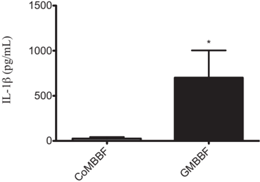
Fig. 4 Germ-free mice receiving Giardia -exposed microbiota biofilm communities (GMBBF) show increased colonic IL-1 b protein levels. Animals were given intracolonic administration of two rounds of human microbiota biofilm communities exposed, or not, to Giardia and were euthanised after 14 days ( n = 4-5 per group). Protein expression of IL-1 b was measured in colonic segments collected from germ-free C57Bl/6 mice reconstituted with differential microbiota biofilm communities using the multiplex technology of the Eve Technologies Luminex platform. Mice receiving Giardia -exposed microbiota biofilm communities exhibited greater expression of IL-1 b compared with those receiving control microbiota biofilms (CoMBBF). * P < 0.05 (Student's t -test).
Fig. 4 Germ-free mice receiving Giardia -exposed microbiota biofilm communities (GMBBF) show increased colonic IL-1 b protein levels. Animals were given intracolonic administration of two rounds of human microbiota biofilm communities exposed, or not, to Giardia and were euthanised after 14 days ( n = 4-5 per group). Protein expression of IL-1 b was measured in colonic segments collected from germ-free C57Bl/6 mice reconstituted with differential microbiota biofilm communities using the multiplex technology of the Eve Technologies Luminex platform. Mice receiving Giardia -exposed microbiota biofilm communities exhibited greater expression of IL-1 b compared with those receiving control microbiota biofilms (CoMBBF). * P < 0.05 (Student's t -test).CC-BY-NC-ND · From the PDF
Image 7
CC-BY-NC-ND · From the PDF
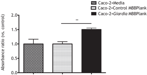
Fig. 6 Planktonic bacteria from human microbiota biofilms exposed to Giardia spent medium promote heightened rates of epithelial apoptosis. Human microbiota biofilm communities were cultivated on the Calgary Biofilm Device using colonic biopsies collected from healthy human donors. Following a 72 h growth period, microbiota biofilms were subjected to either control conditions (fresh sTSY medium) or exposure to Giardia spent medium. Caco-2 monolayers co-incubated, for 24 h, with planktonic constituents of microbiota biofilms exposed to Giardia spent medium (Caco-2 + Giardia MBBPlank) exhibited significantly higher rates of apoptosis compared with cells incubated with microbiota biofilm communities exposed to fresh sTSY (Caco-2 + Control MBBPlank). Caco-2 cells incubated with fresh cell medium served as baseline indicators of cell apoptosis. Values were calculated as absorbance ratios versus control: the mean of all control values was calculated and set at 1.0, and ratio changes were calculated for each measurement in each group against this calculated control mean. Values are mean ± S.E.M. n = 3/group. * P < 0.05 versus Caco-2 + Medium (ANOVA and Tukey's multiple comparison test).
Fig. 6 Planktonic bacteria from human microbiota biofilms exposed to Giardia spent medium promote heightened rates of epithelial apoptosis. Human microbiota biofilm communities were cultivated on the Calgary Biofilm Device using colonic biopsies collected from healthy human donors. Following a 72 h growth period, microbiota biofilms were subjected to either control conditions (fresh sTSY medium) or exposure to Giardia spent medium. Caco-2 monolayers co-incubated, for 24 h, with planktonic constituents of microbiota biofilms exposed to Giardia spent medium (Caco-2 + Giardia MBBPlank) exhibited significantly higher rates of apoptosis compared with cells incubated with microbiota biofilm communities exposed to fresh sTSY (Caco-2 + Control MBBPlank). Caco-2 cells incubated with fresh cell medium served as baseline indicators of cell apoptosis. Values were calculated as absorbance ratios versus control: the mean of all control values was calculated and set at 1.0, and ratio changes were calculated for each measurement in each group against this calculated control mean. Values are mean ± S.E.M. n = 3/group. * P < 0.05 versus Caco-2 + Medium (ANOVA and Tukey's multiple comparison test).CC-BY-NC-ND · From the PDF
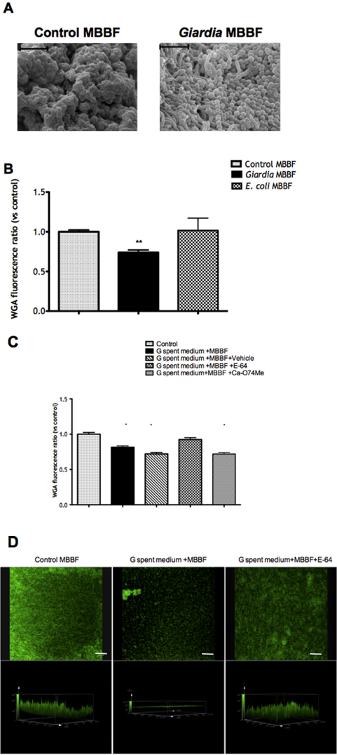
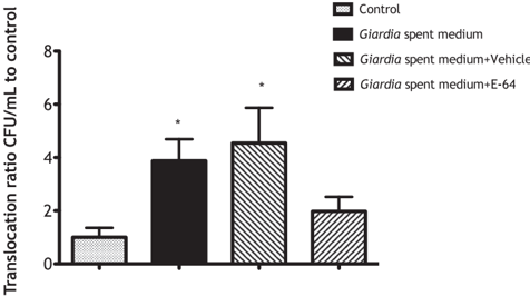
Fig. 7 Planktonic bacteria from human microbiota biofilms exposed to Giardia spent medium more readily translocate across epithelial monolayers. Human microbiota biofilm communities were cultivated on the Calgary Biofilm Device using colonic biopsies collected from healthy human donors. Following a 72 h growth period, microbiota biofilms were subjected to either control conditions (fresh sTSY medium), or exposure to Giardia spent medium. Bacteria released from human microbiota biofilm communities exposed to Giardia spent medium translocate at significantly higher levels than those exposed to fresh sTSY (Control), and this effect was inhibited by pre-treatment with E-64 ( Giardia spent medium + E-64). DMSO pre-treatment served as the E-64 vehicle control. Values were calculated as log colony forming unit (CFU)/mL ratios versus Control CFU/mL, arbitrarily set to 1.0. Values are mean ± S.E.M. n = 3/group. * P < 0.05 versus Control (ANOVA and Tukey's multiple comparison test).
Fig. 7 Planktonic bacteria from human microbiota biofilms exposed to Giardia spent medium more readily translocate across epithelial monolayers. Human microbiota biofilm communities were cultivated on the Calgary Biofilm Device using colonic biopsies collected from healthy human donors. Following a 72 h growth period, microbiota biofilms were subjected to either control conditions (fresh sTSY medium), or exposure to Giardia spent medium. Bacteria released from human microbiota biofilm communities exposed to Giardia spent medium translocate at significantly higher levels than those exposed to fresh sTSY (Control), and this effect was inhibited by pre-treatment with E-64 ( Giardia spent medium + E-64). DMSO pre-treatment served as the E-64 vehicle control. Values were calculated as log colony forming unit (CFU)/mL ratios versus Control CFU/mL, arbitrarily set to 1.0. Values are mean ± S.E.M. n = 3/group. * P < 0.05 versus Control (ANOVA and Tukey's multiple comparison test).CC-BY-NC-ND · From the PDF
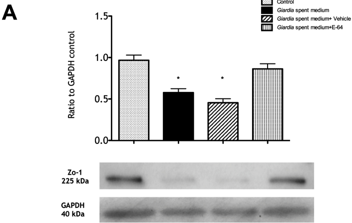
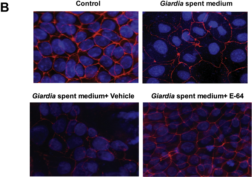
Fig. 8 Planktonic bacteria from human microbiota biofilms exposed to Giardia spent medium reduce zonula occludens (ZO)-1 protein and modify ZO-1 distribution in Caco-2 epithelial monolayers. Human microbiota biofilm communities were cultivated on the Calgary Biofilm Device using colonic biopsies collected from healthy human donors. Following a 72 h growth period, microbiota biofilms were subjected to either control conditions (fresh sTSY medium), or exposure to Giardia spent medium. Cell monolayers were challenged with planktonic constituents released from microbiota biofilms, either exposed to Giardia spent medium or fresh sTSY (Control) in the presence/absence of broad-spectrum, E-64, pre-treatment on trophozoites, for 24 h, and immunoblotted for ZO-1 (A). Western blots (densitometry and representative blots images) illustrate that Caco-2 monolayers co-incubated with planktonic constituents released from Giardia spent medium- exposed microbiota biofilm communities contain significantly decreased ZO-1 protein levels. Pre-treatment of trophozoites with E-64 inhibited this decrease. (B) Representative immunostaining for ZO-1 (red) reveals a mislocalization, and loss in stain intensity in Caco-2 monolayers co-incubated with planktonic constituents released from microbiota biofilm communities modified by exposure to Giardia spent medium. Densitometry data are presented as % glyceraldehyde 3 phosphate dehydrogenase. Values were calculated as % glyceraldehyde 3 phosphate dehydrogenase ratios versus control, arbitrarily set to 1.0 (A). Values are mean ± S.E.M. n = 3/group. * P < 0.05 versus Control (ANOVA and Tukey's multiple comparison test).
Fig. 8 Planktonic bacteria from human microbiota biofilms exposed to Giardia spent medium reduce zonula occludens (ZO)-1 protein and modify ZO-1 distribution in Caco-2 epithelial monolayers. Human microbiota biofilm communities were cultivated on the Calgary Biofilm Device using colonic biopsies collected from healthy human donors. Following a 72 h growth period, microbiota biofilms were subjected to either control conditions (fresh sTSY medium), or exposure to Giardia spent medium. Cell monolayers were challenged with planktonic constituents released from microbiota biofilms, either exposed to Giardia spent medium or fresh sTSY (Control) in the presence/absence of broad-spectrum, E-64, pre-treatment on trophozoites, for 24 h, and immunoblotted for ZO-1 (A). Western blots (densitometry and representative blots images) illustrate that Caco-2 monolayers co-incubated with planktonic constituents released from Giardia spent medium- exposed microbiota biofilm communities contain significantly decreased ZO-1 protein levels. Pre-treatment of trophozoites with E-64 inhibited this decrease. (B) Representative immunostaining for ZO-1 (red) reveals a mislocalization, and loss in stain intensity in Caco-2 monolayers co-incubated with planktonic constituents released from microbiota biofilm communities modified by exposure to Giardia spent medium. Densitometry data are presented as % glyceraldehyde 3 phosphate dehydrogenase. Values were calculated as % glyceraldehyde 3 phosphate dehydrogenase ratios versus control, arbitrarily set to 1.0 (A). Values are mean ± S.E.M. n = 3/group. * P < 0.05 versus Control (ANOVA and Tukey's multiple comparison test).CC-BY-NC-ND · From the PDF
Image 11
CC-BY-NC-ND · From the PDF
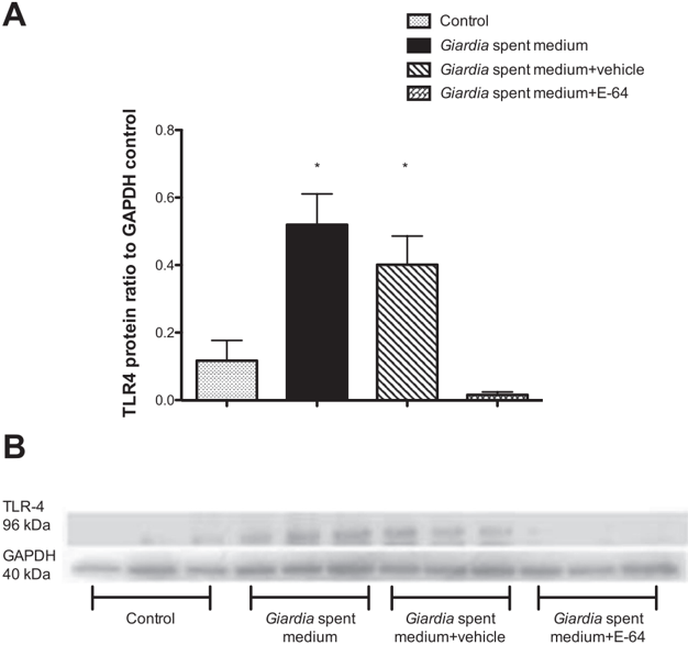
Fig. 9 Planktonic bacteria from human microbiota biofilms exposed to Giardia spent medium increase Toll-like receptor 4 expression in Caco-2 monolayers. Human microbiota biofilm communities were cultivated on the Calgary Biofilm Device using colonic biopsies collected from healthy human donors. Following a 72 h growth period, microbiota biofilms were subjected to either control conditions (fresh sTSY medium), and either control conditions (Control MBBPlank whereby no exposure to Giardia occured; or fresh sTSY medium), or exposure to Giardia spent medium (where trophozoites had been pre-treated with cysteine protease inhibitor E-64 or not), or exposure to the known pro-inflammatory cytokine IL1 beta. Data were also obtained from plain, untreated, CaCo-2 monolayers (Caco-2). (A) Caco-2 monolayers co-incubated with planktonic constituents released from human microbiota biofilms exposed to Giardia spent medium show a significant increase in TLR4 protein levels. Pre-treatment of trophozoites with E-64 prevented this increase of TLR4 protein levels in the monolayers. Densitometry data are presented as % glyceraldehyde 3 phosphate dehydrogenase. Values were calculated as % glyceraldehyde 3 phosphate dehydrogenase ratios versus control, arbitrarily set to 1.0. Values are mean ± S.E.M. n = 3/group. * P < 0.05 versus Control (ANOVA and Tukey's multiple comparison test). Numbers were representative of separate experiments conducted with microbiota biofilm cultures cultivated from colonic samples of multiple patients in order to ensure reproducibility. (B) Caco-2 monolayers co-incubated with planktonic constituents released from human microbiota biofilms exposed to Giardia spent medium show a significant increase in secreted CXCL-8. To further identify the mechanisms of epithelial pathophysiology induced by enteropathogen-modified MBBFs, we assessed the consequences of exposure to Giardia -modified microbiota constituents on the production of pro-inflammatory CXCL-8. Pre-treatment of trophozoites with E-64 inhibited this increase. Values were calculated as % control arbitrarily set to 1.0. Values are mean ± S.E.M. n = 3/group. / P < 0.05 versus Control (ANOVA and Tukey's multiple comparison test).
Fig. 9 Planktonic bacteria from human microbiota biofilms exposed to Giardia spent medium increase Toll-like receptor 4 expression in Caco-2 monolayers. Human microbiota biofilm communities were cultivated on the Calgary Biofilm Device using colonic biopsies collected from healthy human donors. Following a 72 h growth period, microbiota biofilms were subjected to either control conditions (fresh sTSY medium), and either control conditions (Control MBBPlank whereby no exposure to Giardia occured; or fresh sTSY medium), or exposure to Giardia spent medium (where trophozoites had been pre-treated with cysteine protease inhibitor E-64 or not), or exposure to the known pro-inflammatory cytokine IL1 beta. Data were also obtained from plain, untreated, CaCo-2 monolayers (Caco-2). (A) Caco-2 monolayers co-incubated with planktonic constituents released from human microbiota biofilms exposed to Giardia spent medium show a significant increase in TLR4 protein levels. Pre-treatment of trophozoites with E-64 prevented this increase of TLR4 protein levels in the monolayers. Densitometry data are presented as % glyceraldehyde 3 phosphate dehydrogenase. Values were calculated as % glyceraldehyde 3 phosphate dehydrogenase ratios versus control, arbitrarily set to 1.0. Values are mean ± S.E.M. n = 3/group. * P < 0.05 versus Control (ANOVA and Tukey's multiple comparison test). Numbers were representative of separate experiments conducted with microbiota biofilm cultures cultivated from colonic samples of multiple patients in order to ensure reproducibility. (B) Caco-2 monolayers co-incubated with planktonic constituents released from human microbiota biofilms exposed to Giardia spent medium show a significant increase in secreted CXCL-8. To further identify the mechanisms of epithelial pathophysiology induced by enteropathogen-modified MBBFs, we assessed the consequences of exposure to Giardia -modified microbiota constituents on the production of pro-inflammatory CXCL-8. Pre-treatment of trophozoites with E-64 inhibited this increase. Values were calculated as % control arbitrarily set to 1.0. Values are mean ± S.E.M. n = 3/group. / P < 0.05 versus Control (ANOVA and Tukey's multiple comparison test).CC-BY-NC-ND · From the PDF
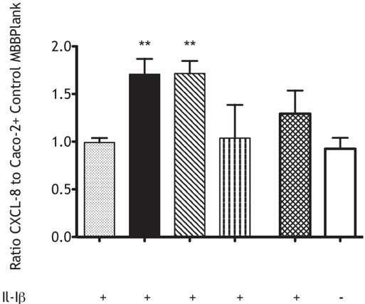
Fig. 10 Giardia spent medium-exposed microbiota biofilms planktonic constituents (MBBPlank) promote increased CXCL-8 protein levels secreted from Caco-2 monolayers. Human microbiota biofilm communities were cultivated on the Calgary Biofilm Device using colonic biopsies collected from healthy human donors. Following a 72 h growth period, microbiota biofilms were subjected to either control conditions (fresh sTSY medium), or exposure to Giardia spent medium. Caco-2 monolayers co-incubated with planktonic constituents released from Giardia spent medium-exposed microbiota biofilm communities showed a significant increase in secreted CXCL-8. Pre-treatment of trophozoites with cysteine protease inhibitor E-64 prevented increased secreted CXCL-8 levels in stimulated Caco-2 monolayers challenged with planktonic microbial constituents released from microbiota biofilms. Values were calculated as % control, arbitrarily set to 1.0. Values are mean ± S.E.M. n = 3/group. * P < 0.05, ** P < 0.01 versus Control (ANOVA and Tukey's multiple comparison test). Cells given IL-1 beta served as a positive ''pro-inflammatory'' control.
Fig. 10 Giardia spent medium-exposed microbiota biofilms planktonic constituents (MBBPlank) promote increased CXCL-8 protein levels secreted from Caco-2 monolayers. Human microbiota biofilm communities were cultivated on the Calgary Biofilm Device using colonic biopsies collected from healthy human donors. Following a 72 h growth period, microbiota biofilms were subjected to either control conditions (fresh sTSY medium), or exposure to Giardia spent medium. Caco-2 monolayers co-incubated with planktonic constituents released from Giardia spent medium-exposed microbiota biofilm communities showed a significant increase in secreted CXCL-8. Pre-treatment of trophozoites with cysteine protease inhibitor E-64 prevented increased secreted CXCL-8 levels in stimulated Caco-2 monolayers challenged with planktonic microbial constituents released from microbiota biofilms. Values were calculated as % control, arbitrarily set to 1.0. Values are mean ± S.E.M. n = 3/group. * P < 0.05, ** P < 0.01 versus Control (ANOVA and Tukey's multiple comparison test). Cells given IL-1 beta served as a positive ''pro-inflammatory'' control.CC-BY-NC-ND · From the PDF

Verification

Claims the paper makes

The claims and statistics of this paper haven’t been checked yet.

Run the check — free, no account needed. It reads the open-access text and checks the sources it cites.

A few free checks a day · papers already verified open instantly · results are public

Claims other papers make about this one

Each is checked against this paper’s own text: does it say what the citing paper says it does?

Self-check

Percentages, sums and reported differences re-computed from the paper’s own numbers.

The self-check needs the paper’s full text.

Citations

Cites

Show all 8 references

Research works printed but not linked: 76

  1. Abreu, 2010, Toll-like receptor signalling in the intestinal epithelium: how bacterial recognition shapes intestinal function, Nature Rev. Immunol., 10, 131, 10.1038/nri2707 · DOI · Paper page
  2. Amat, 2015, Giardia duodenalis depletes goblet cell mucins and degrades MUC2, Facilitating bacterial translocation, FASEB J., 29
  3. Abreu, 2002, TLR4 and MD-2 expression is regulated by immune-mediated signals in human intestinal epithelial cells, J. Biol. Chem., 277, 20431, 10.1074/jbc.M110333200 · DOI
  4. Barbara, 2002, A role for inflammation in irritable bowel syndrome?, Gut, 51, i41, 10.1136/gut.51.suppl_1.i41 · DOI
  5. Barrett, 1980, Fluorimetric assays for cathepsin B and cathepsin H with methylcoumarylamide substrates, Biochem, J187, 909, 10.1042/bj1870909 · DOI
  6. Bhargava, 2015, Giardia duodenalis Surface Cysteine Proteases Induce Cleavage of the Intestinal Epithelial Cytoskeletal Protein Villin via Myosin Light Chain Kinase, PLoS ONE, 10, e0136102, 10.1371/journal.pone.0136102 · DOI
  7. Buret, 2007, Mechanisms of epithelial dysfunction in giardiasis, Gut, 56, 316, 10.1136/gut.2006.107771 · DOI
  8. Buret, 2014, Modulatory mechanisms of enterocyte apoptosis by viral, bacterial and parasitic pathogens, Crit. Rev. Microbiol., 40, 1, 10.3109/1040841X.2012.746952 · DOI
  9. Cario, 2000, Differential alteration in intestinal epithelial cell expression of toll-like receptor 3 (TLR3) and TLR4 in inflammatory bowel disease, Infect. Immun., 68, 7010, 10.1128/IAI.68.12.7010-7017.2000 · DOI
  10. Carroll, 2011, Molecular analysis of the luminal- and mucosal-associated intestinal microbiota in diarrhea-predominant irritable bowel syndrome, Am. J. Physiol. Gastrointest. Liver Physiol., 301, G799, 10.1152/ajpgi.00154.2011 · DOI · Paper page
  11. Ceri, 1999, The Calgary Biofilm Device: new technology for rapid determination of antibiotic susceptibilities of bacterial biofilms, J. Clin. Microbiol., 37, 1771, 10.1128/JCM.37.6.1771-1776.1999 · DOI · Paper page
  12. Chadwick, 2002, Activation of the mucosal immune system in irritable bowel syndrome, Gastroenterology, 122, 1778, 10.1053/gast.2002.33579 · DOI · Paper page
  13. Chen, 2009, Altered expression of inflammatory cytokine receptors in response to LPS challenge through interaction between intestinal epithelial cells and lymphocytes of Peyer's patch, Cell Biol. Int., 33, 369, 10.1016/j.cellbi.2009.01.004 · DOI
  14. Chua, 2014, Dispersed cells represent a distinct stage in the transition from bacterial biofilm to planktonic lifestyles, Nat. Commun., 5, 4462, 10.1038/ncomms5462 · DOI
  15. Cotton, 2015, Disruptions of Host Immunity and Inflammation by Giardia Duodenalis: Potential Consequences for Co-Infections in the Gastro-Intestinal Tract, Pathog, 4, 764, 10.3390/pathogens4040764 · DOI
  16. Cotton, 2014, Giardia duodenalis cathepsin B proteases degrade intestinal epithelial interleukin-8 and attenuate interleukin-8-induced neutrophil chemotaxis, Infect. Immun., 82, 2772, 10.1128/IAI.01771-14 · DOI
  17. Cremon, 2009, Mucosal immune activation in irritable bowel syndrome: gender-dependence and association with digestive symptoms, Am. J. Gastroenterol., 104, 392, 10.1038/ajg.2008.94 · DOI · Paper page
  18. Cremon, 2014, Salmonella gastroenteritis during childhood is a risk factor for irritable bowel syndrome in adulthood, Gastroenterology, 147, 69, 10.1053/j.gastro.2014.03.013 · DOI
  19. David, 2012, Characterisation of protease activity in extracellular products secreted by Giardia duodenalis trophozoites treated with propolis, Nat. Prod. Res., 26, 370, 10.1080/14786419.2010.515547 · DOI
  20. Dongari-Bagtzoglou, 2008, Pathogenesis of mucosal biofilm infections: challenges and progress, Expert Rev. Anti Infect. Ther., 6, 201, 10.1586/14787210.6.2.201 · DOI
  21. DuBois, 2006, Giardia lamblia cysteine proteases, Parasitol. Res., 99, 313, 10.1007/s00436-006-0149-4 · DOI
  22. Edgar, 2013, UPARSE: highly accurate OTU sequences from microbial amplicon reads, Nat. Methods, 10, 996, 10.1038/nmeth.2604 · DOI
  23. Fahlgren, 2003, Increased expression of antimicrobial peptides and lysozyme in colonic epithelial cells of patients with ulcerative colitis, Clin. Exp. Immunol., 131, 90, 10.1046/j.1365-2249.2003.02035.x · DOI
  24. Gerbaba, 2015, Giardia duodenalis-induced alterations of commensal bacteria kill Caenorhabditis elegans: a new model to study microbial-microbial interactions in the gut, Am. J. Physiol. Gastrointest. Liver Physiol., 308, G550, 10.1152/ajpgi.00335.2014 · DOI
  25. Halliez, 2013, Extra-intestinal and long term consequences of Giardia duodenalis infections, World J. Gastroenterol., 19, 8974, 10.3748/wjg.v19.i47.8974 · DOI
  26. Halliez, 2016, Giardia duodenalis induces para-cellular bacterial translocation and causes post-infectious visceral hypersensitivity, Am. J. Physiol. Gastrointest. Liver Physiol., 00144, 02015
  27. Hanevik, 2009, Development of functional gastrointestinal disorders after Giardia lamblia infection, BMC Gastroenterol., 9, 27, 10.1186/1471-230X-9-27 · DOI
  28. Hare, 1989, Giardia lamblia: characterization of proteinase activity in trophozoites, Exp. Parasitol., 68, 168, 10.1016/0014-4894(89)90094-5 · DOI
  29. Harrison, 2006, The use of microscopy and three-dimensional visualization to evaluate the structure of microbial biofilms cultivated in the Calgary Biofilm Device, Biol. Proc. Online, 8, 194, 10.1251/bpo127 · DOI
  30. Harrison, 2010, Microtiter susceptibility testing of microbes growing on peg lids: a miniaturized biofilm model for high-throughput screening, Nat. Protoc., 5, 1236, 10.1038/nprot.2010.71 · DOI
  31. Kalischuk, 2009, Campylobacter jejuni induces transcellular translocation of commensal bacteria via lipid rafts, Gut Pathog, 1, 2, 10.1186/1757-4749-1-2 · DOI
  32. Kalischuk, 2010, Campylobacter jejuni induces transcytosis of commensal bacteria across the intestinal epithelium through M-like cells, Gut Pathog, 2, 14, 10.1186/1757-4749-2-14 · DOI
  33. Keselman, 2016, The microbiota contributes to CD8+ T cell activation and nutrient malabsorption following intestinal infection with giardia duodenalis, Infect. Immun., 84, 2853, 10.1128/IAI.00348-16 · DOI
  34. Kleessen, 2005, Modulation of gut mucosal biofilms, T Brit J Nutr, 93, S35, 10.1079/BJN20041346 · DOI
  35. Konig, 2014, Alteration of the intestinal microbiota as a cause of and a potential therapeutic option in irritable bowel syndrome, Beneficial Microb., 3, 1
  36. Koster, 2012, Snakemake—a scalable bioinformatics workflow engine, Bioinformatics, 28, 2520, 10.1093/bioinformatics/bts480 · DOI · Paper page
  37. Kozlowski, 2013, An entirely automated method to score DSS-induced colitis in mice by digital image analysis of pathology slides, Dis. Models Mech., 6, 855, 10.1242/dmm.011759 · DOI
  38. Lapointe, 2012, Interleukin-18 facilitates neutrophil transmigration via myosin light chain kinase-dependent disruption of occludin, without altering epithelial permeability, Am. J. Physiol. Gastrointest. Liver Physiol., 302, G343, 10.1152/ajpgi.00202.2011 · DOI
  39. Liebregts, 2007, Immune activation in patients with irritable bowel syndrome, Gastroenterology, 132, 913, 10.1053/j.gastro.2007.01.046 · DOI · Paper page
  40. Love, 2014, Moderated estimation of fold change and dispersion for RNA-seq data with DESeq2, Genome Biol., 15, 550, 10.1186/s13059-014-0550-8 · DOI · Paper page
  41. Macfarlane, 1997, Human colonic microbiota: ecology, physiology and metabolic potential of intestinal bacteria, Scand. J. Gastroenterol. Suppl., 222, 3, 10.1080/00365521.1997.11720708 · DOI
  42. Macfarlane, 2005, Colonization of mucin by human intestinal bacteria and establishment of biofilm communities in a two-stage continuous culture system, Appl. Environ. Microbiol., 71, 7483, 10.1128/AEM.71.11.7483-7492.2005 · DOI
  43. Marshall, 2010, Eight year prognosis of postinfectious irritable bowel syndrome following waterborne bacterial dysentery, Gut, 59, 605, 10.1136/gut.2009.202234 · DOI · Paper page
  44. Mason, 2011, From commensal to pathogen: translocation of Enterococcus faecalis from the midgut to the hemocoel of Manduca sexta, mBio, 2, 10.1128/mBio.00065-11 · DOI
  45. McMurdie, 2013, Phyloseq: an R package for reproducible interactive analysis and graphics of microbiome census data, PLoS ONE, 8, e61217, 10.1371/journal.pone.0061217 · DOI
  46. McMurdie, 2014, Waste not, want not: why rarefying microbiome data is inadmissible, PLoS Comput. Biol., 10, e1003531, 10.1371/journal.pcbi.1003531 · DOI
  47. Noronha, 2009, Hyperactivated B cells in human inflammatory bowel disease, J. Leuk. Biol., 86, 1007, 10.1189/jlb.0309203 · DOI
  48. O'Hara, 2008, Mechanisms of intestinal tight junctional disruption during infection, Front. Biosci., 13, 7008
  49. O'Neil, 1999, Expression and regulation of the human beta-defensins hBD-1 and hBD-2 in intestinal epithelium, J. Immunol., 163, 6718, 10.4049/jimmunol.163.12.6718 · DOI
  50. Perez-Mendoza, 2011, N-Acetylglucosamine-dependent biofilm formation in Pectobacterium atrosepticum is cryptic and activated by elevated c-di-GMP levels, Microbiology, 157, 3340, 10.1099/mic.0.050450-0 · DOI
  51. Rajilic-Stojanovic, 2011, Global and deep molecular analysis of microbiota signatures in fecal samples from patients with irritable bowel syndrome, Gastroenterology, 141, 1792, 10.1053/j.gastro.2011.07.043 · DOI · Paper page
  52. Ramachandran, 2000, Apoptosis in the intestinal epithelium: its relevance in normal and pathophysiological conditions, J. Gastroenterol. Hepatol., 15, 109, 10.1046/j.1440-1746.2000.02059.x · DOI
  53. Reti, 2015, Campylobacter jejuni increases flagellar expression and adhesion of noninvasive Escherichia coli: effects on enterocytic Toll-like receptor 4 and CXCL-8 expression, Infect. Immun., 83, 4571, 10.1128/IAI.00970-15 · DOI
  54. Roberts-Thomson, 1976, Giardiasis in the mouse: an animal model, Gastroenterology, 71, 57, 10.1016/S0016-5085(76)80097-2 · DOI
  55. Rodriguez-Fuentes, 2006, Giardia duodenalis: analysis of secreted proteases upon trophozoite-epithelial cell interaction in vitro, Mem. Inst. Oswaldo Cruz, 101, 693, 10.1590/S0074-02762006000600020 · DOI
  56. Roslev, 1993, Application of a tetrazolium salt with a water-soluble formazan as an indicator of viability in respiring bacteria, Appl. Environ. Microbiol., 59, 2891, 10.1128/AEM.59.9.2891-2896.1993 · DOI
  57. Sambuy, 2005, The Caco-2 cell line as a model of the intestinal barrier: influence of cell and culture-related factors on Caco-2 cell functional characteristics, Cell Biol. Toxicol., 21, 1, 10.1007/s10565-005-0085-6 · DOI
  58. Sawada, 2013, Tight junction-related human diseases, Pathol. Int., 63, 1, 10.1111/pin.12021 · DOI
  59. Sepehri, 2007, Microbial diversity of inflamed and noninflamed gut biopsy tissues in inflammatory bowel disease, Inflamm. Bowel Dis., 13, 675, 10.1002/ibd.20101 · DOI
  60. Spiller, 2006, Post-infectious irritable bowel syndrome, Curr. Opin. Gastroenterol., 22, 13, 10.1097/01.mog.0000194792.36466.5c · DOI
  61. Spiller, 2000, Increased rectal mucosal enteroendocrine cells, T lymphocytes, and increased gut permeability following acute Campylobacter enteritis and in post-dysenteric irritable bowel syndrome, Gut, 47, 804, 10.1136/gut.47.6.804 · DOI · Paper page
  62. Sproule-Willoughby, 2010, In vitro anaerobic biofilms of human colonic microbiota, J. Microbiol. Methods, 83, 296, 10.1016/j.mimet.2010.09.020 · DOI
  63. Stevens, 1978, Animal model of human disease: giardiasis, Am. J. Pathol., 90, 529
  64. Sutherland, 2001, The biofilm matrix–an immobilized but dynamic microbial environment, Trends Microbiol., 9, 222, 10.1016/S0966-842X(01)02012-1 · DOI
  65. Tchoupe, 1991, Photometric or fluorometric assay of cathepsin B, L and H and papain using substrates with an aminotrifluoromethylcoumarin leaving group, Biochim. Biophys. Acta, 1076, 149, 10.1016/0167-4838(91)90232-O · DOI
  66. Teoh, 2000, Giardia lamblia rearranges F-actin and alpha-actinin in human colonic and duodenal monolayers and reduces transepithelial electrical resistance, J. Parasitol., 86, 800
  67. Thabane, 2009, Post-infectious irritable bowel syndrome, World J. Gastroenterol., 15, 3591, 10.3748/wjg.15.3591 · DOI
  68. Tornblom, 2002, Full-thickness biopsy of the jejunum reveals inflammation and enteric neuropathy in irritable bowel syndrome, Gastroenterology, 123, 1972, 10.1053/gast.2002.37059 · DOI · Paper page
  69. Toyofuku, 2012, Identification of proteins associated with the Pseudomonas aeruginosa biofilm extracellular matrix, J. Proteo Res., 11, 4906, 10.1021/pr300395j · DOI
  70. von Rosenvinge, 2013, Microbial biofilms and gastrointestinal diseases, Pathog Dis., 67, 25, 10.1111/2049-632X.12020 · DOI
  71. Vora, 2004, Beta-defensin-2 expression is regulated by TLR signaling in intestinal epithelial cells, J. Immunol., 173, 5398, 10.4049/jimmunol.173.9.5398 · DOI
  72. Wang, 2009, The role of chemokines in intestinal inflammation and cancer, Curr. Opin. Pharmacol., 9, 688, 10.1016/j.coph.2009.08.003 · DOI
  73. Wensaas, 2012, Irritable bowel syndrome and chronic fatigue 3 years after acute giardiasis: historic cohort study, Gut, 61, 214, 10.1136/gutjnl-2011-300220 · DOI
  74. Williams, 1995, Multiple protease activities in Giardia intestinalis trophozoites, Int. J. Parasitol., 25, 771, 10.1016/0020-7519(94)00201-X · DOI
  75. Yu, 2005, SGLT-1-mediated glucose uptake protects intestinal epithelial cells against LPS-induced apoptosis and barrier defects: a novel cellular rescue mechanism?, FASEB J., 19, 1822, 10.1096/fj.05-4226com · DOI
  76. Zoetendal, 2008, High-throughput diversity and functionality analysis of the gastrointestinal tract microbiota, Gut, 57, 1605, 10.1136/gut.2007.133603 · DOI

Cited by

Sorted by citations

See all 157

Metadata

Authors
Beatty, J. K., Akierman, S. V., Motta, J.-P., Muise, S., Workentine, M. L., Harrison, J. J., Bhargava, A., Beck, P. L., Rioux, K. P., McKnight, G. W., Wallace, J. L., & Buret, A. G.
Venue
International Journal for Parasitology
Publisher
Elsevier BV
Published
2017-05
Volume
47
Issue
6
Pages
311-326
ISSN
0020-7519
Cited by
157
References
8
Language
en

About these checks

Registry, retraction, Cites and statistics results are computed, not judged, with one exception: a printed reference that no index lists is read once by a model before it counts as a citation error. Claims are extracted by a model and kept only when their quote appears verbatim in the text; each cited source is then checked, with a model’s help, for whether it says what the claim relies on. Those verdicts can be wrong — read the claim’s quote and the cited source before relying on one.

Statistics and arithmetic are recomputed, not judged.

Confidence bands are shown only for fields calibrated on our annotated test set. None is calibrated yet, so no band is shown.

What counts as a claim ›How we measure confidence ›How we check this ›

Are you an author, or do you see a problem with this page? Request a review

How to cite

Cite CoChat for the checks on this page, and the paper for its own findings.

This page
CoChat. (2026). Citation check: Giardia duodenalis induces pathogenic dysbiosis of human intestinal microbiota biofilms. https://app.cochat.ai/papers/10.1016/j.ijpara.2016.11.010
The paper
Beatty, J. K., Akierman, S. V., Motta, J.-P., Muise, S., Workentine, M. L., Harrison, J. J., Bhargava, A., Beck, P. L., Rioux, K. P., McKnight, G. W., Wallace, J. L., & Buret, A. G. (2017). Giardia duodenalis induces pathogenic dysbiosis of human intestinal microbiota biofilms. International Journal for Parasitology. https://doi.org/10.1016/j.ijpara.2016.11.010