

The DOI matches its Crossref record; 60 of the 60 references it cites were found in a registry or an index.
Corticospinal Neurons in Macaque Ventral Premotor Cortex with Mirror Properties: A Potential Mechanism for Action Suppression?
Open accessAbstract
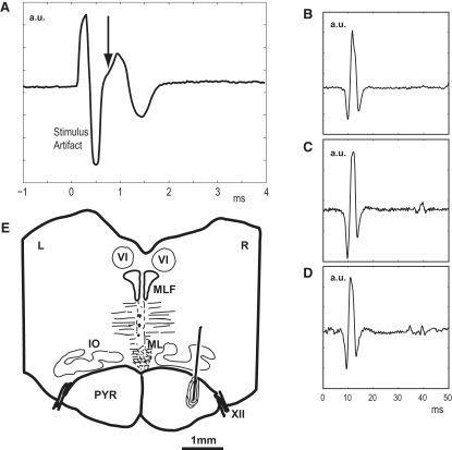
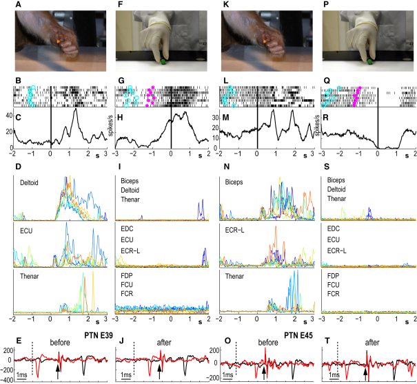
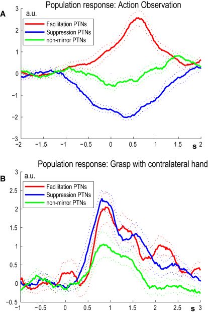
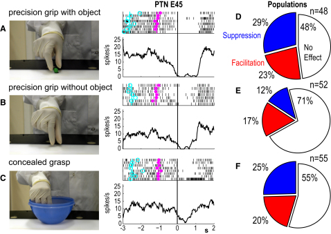
The discovery of "mirror neurons" in area F5 of the ventral premotor cortex has prompted many theories as to their possible function. However, the identity of mirror neurons remains unknown. Here, we investigated whether identified pyramidal tract neurons (PTNs) in area F5 of two adult macaques exhibited "mirror-like" activity. About half of the 64 PTNs tested showed significant modulation of their activity while monkeys observed precision grip of an object carried out by an experimenter, with somewhat fewer showing modulation during precision grip without an object or grasping concealed from the monkey. Therefore, mirror-like activity can be transmitted directly to the spinal cord via PTNs. A novel finding is that many PTNs (17/64) showed complete suppression of discharge during action observation, while firing actively when the monkey grasped food rewards. We speculate that this suppression of PTN discharge might be involved in the inhibition of self-movement during action observation.
Overview
The claims and statistics of this paper haven’t been checked yet.
Run the check — free, no account needed. It reads the open-access text and checks the sources it cites.
Integrity signals
Registry
Cites
Statistics
Figures
Verification
Claims the paper makes
The claims and statistics of this paper haven’t been checked yet.
Run the check — free, no account needed. It reads the open-access text and checks the sources it cites.
Claims other papers make about this one
Each is checked against this paper’s own text: does it say what the citing paper says it does?
The cited source says what the claim attributes to it.
In the citing paper
“subsequent studies confirmed that sizable proportions, perhaps the majority, of MNs exhibit robust responses to the observation of object-free body movement.”
From Mirror neurons: from origin to function (2014) · cites this paper as Kraskov et al., 2009
The check wasn’t confident either way, so it gives no verdict — never disputed.
In the citing paper
“MN responses start around 250ms after observed movement onset”
From Mirror neurons: from origin to function (2014) · cites this paper as Kraskov et al., 2009
Experimental — accuracy not yet measured
Self-check
Percentages, sums and reported differences re-computed from the paper’s own numbers.
The self-check needs the paper’s full text.
Citations
Cites
Action recognition in the premotor cortex
Gallese, V., Fadiga, L., Fogassi, L., & Rizzolatti, G. (1996) · Brain · doi:10.1093/brain/119.2.593 · Cited by 4,968
Premotor cortex and the recognition of motor actions
Rizzolatti, G., Fadiga, L., Gallese, V., & Fogassi, L. (1996) · Cognitive Brain Research · doi:10.1016/0926-6410(95)00038-0 · Cited by 4,870
Understanding motor events: a neurophysiological study
di Pellegrino, G., Fadiga, L., Fogassi, L., Gallese, V., & Rizzolatti, G. (1992) · Experimental Brain Research · doi:10.1007/bf00230027 · Cited by 3,539
Motor facilitation during action observation: a magnetic stimulation study
Fadiga, L., Fogassi, L., Pavesi, G., & Rizzolatti, G. (1995) · Journal of Neurophysiology · doi:10.1152/jn.1995.73.6.2608 · Cited by 2,297
Unsupervised spike detection and sorting with wavelets and superparamagnetic clustering
Quiroga, R. Q., Nadasdy, Z., & Ben-Shaul, Y. (2004) · Neural Computation · doi:10.1162/089976604774201631 · Cited by 2,119
Parietal Lobe: From Action Organization to Intention Understanding
Fogassi, L., Ferrari, P. F., Gesierich, B., Rozzi, S., Chersi, F., & Rizzolatti, G. (2005) · Science · doi:10.1126/science.1106138 · Cited by 1,990
Hearing Sounds, Understanding Actions: Action Representation in Mirror Neurons
Kohler, E., Keysers, C., Umiltà, M. A., Fogassi, L., Gallese, V., & Rizzolatti, G. (2002) · Science · doi:10.1126/science.1070311 · Cited by 1,801
Imitation, empathy, and mirror neurons
Iacoboni, M. (2009) · Annual Review of Psychology · doi:10.1146/annurev.psych.60.110707.163604 · Cited by 1,606
Descending Pathways in Motor Control
Lemon, R. N. (2008) · Annual Review of Neuroscience · doi:10.1146/annurev.neuro.31.060407.125547 · Cited by 1,599
Self-citationFunctional organization of inferior area 6 in the macaque monkey
Rizzolatti, G., Camarda, R., Fogassi, L., Gentilucci, M., Luppino, G., & Matelli, M. (1988) · Experimental Brain Research · doi:10.1007/bf00248742 · Cited by 1,550
Cited by
Sorted by citations
The functional role of the parieto-frontal mirror circuit: interpretations and misinterpretations
Rizzolatti, G., & Sinigaglia, C. (2010) · Nature Reviews Neuroscience · doi:10.1038/nrn2805 · Cited by 1,680
Single-Neuron Responses in Humans during Execution and Observation of Actions
Mukamel, R., Ekstrom, A. D., Kaplan, J., Iacoboni, M., & Fried, I. (2010) · Current Biology · doi:10.1016/j.cub.2010.02.045 · Cited by 1,178
What is so special about embodied simulation?
Gallese, V., & Sinigaglia, C. (2011) · Trends in Cognitive Sciences · doi:10.1016/j.tics.2011.09.003 · Cited by 728
Action-based language: A theory of language acquisition, comprehension, and production
Glenberg, A. M., & Gallese, V. (2011) · Cortex · doi:10.1016/j.cortex.2011.04.010 · Cited by 717
Mirror neurons: from origin to function
Cook, R., Bird, G., Catmur, C., Press, C., & Heyes, C. (2014) · Behavioral and Brain Sciences · doi:10.1017/s0140525x13000903 · Cited by 647
Rizzolatti, G., Cattaneo, L., Fabbri‐Destro, M., & Rozzi, S. (2014) · Physiological Reviews · doi:10.1152/physrev.00009.2013 · Cited by 523
The mirror mechanism: a basic principle of brain function
Rizzolatti, G., & Sinigaglia, C. (2016) · Nature reviews. Neuroscience · doi:10.1038/nrn.2016.135 · Cited by 521
What We Know Currently about Mirror Neurons
Kilner, J. M., & Lemon, R. (2013) · Current Biology · doi:10.1016/j.cub.2013.10.051 · Cited by 438
Self-citationMore than one pathway to action understanding
Kilner, J. M. (2011) · Trends in Cognitive Sciences · doi:10.1016/j.tics.2011.06.005 · Cited by 399
Mirror neurons 30 years later: implications and applications
Bonini, L., Rotunno, C., Arcuri, E., & Gallese, V. (2022) · Trends in Cognitive Sciences · doi:10.1016/j.tics.2022.06.003 · Cited by 373
The mirror mechanism: recent findings and perspectives
Rizzolatti, G., & Fogassi, L. (2014) · Philosophical Transactions of the Royal Society B Biological Sciences · doi:10.1098/rstb.2013.0420 · Cited by 312
Decoding a Perceptual Decision Process across Cortex
Hernández, A., Nácher, V., Luna, R., Zainos, A., Lemus, L., Álvarez, M., Vázquez, Y., Camarillo, L., & Romo, R. (2010) · Neuron · doi:10.1016/j.neuron.2010.03.031 · Cited by 278
Reflections on agranular architecture: predictive coding in the motor cortex
Shipp, S., Adams, R. A., & Friston, K. J. (2013) · Trends in Neurosciences · doi:10.1016/j.tins.2013.09.004 · Cited by 239
Embodied Cognition and Mirror Neurons: A Critical Assessment
Caramazza, A., Anzellotti, S., Strnad, L., & Lingnau, A. (2014) · Annual Review of Neuroscience · doi:10.1146/annurev-neuro-071013-013950 · Cited by 265
M1 Corticospinal Mirror Neurons and Their Role in Movement Suppression during Action Observation
Vigneswaran, G., Philipp, R., Lemon, R., & Kraskov, A. (2013) · Current Biology · doi:10.1016/j.cub.2012.12.006 · Cited by 226
Self-citationA Functional Role for the Motor System in Language Understanding
Willems, R. M., Labruna, L., D’Esposito, M., Ivry, R. B., & Casasanto, D. J. (2011) · Psychological Science · doi:10.1177/0956797611412387 · Cited by 213
Naish, K. R., Houston‐Price, C., Bremner, A. J., & Holmes, N. P. (2014) · Neuropsychologia · doi:10.1016/j.neuropsychologia.2014.09.034 · Cited by 212
Representation of Others' Action by Neurons in Monkey Medial Frontal Cortex
Yoshida, K., Saito, N., Iriki, A., & Isoda, M. (2011) · Current Biology · doi:10.1016/j.cub.2011.01.004 · Cited by 200
Conversion of sensory signals into perceptual decisions
Romo, R., & de Lafuente, V. (2012) · Progress in Neurobiology · doi:10.1016/j.pneurobio.2012.03.007 · Cited by 191
Gallese, V., Gernsbacher, M. A., Heyes, C. M., Hickok, G. S., & Iacoboni, M. (2011) · Perspectives on Psychological Science · doi:10.1177/1745691611413392 · Cited by 188
Vigneswaran, G., Kraskov, A., & Lemon, R. (2011) · Journal of Neuroscience · doi:10.1523/jneurosci.3142-11.2011 · Cited by 179
Self-citationA word in the hand: action, gesture and mental representation in humans and non-human primates
Cartmill, E. A., Beilock, S. L., & Susan Goldin-meadow, A. (2011) · Philosophical Transactions of the Royal Society B Biological Sciences · doi:10.1098/rstb.2011.0162 · Cited by 186
A touch with words: Dynamic synergies between manual actions and language
García, A. M., & Ibáñez, A. M. (2016) · Neuroscience & Biobehavioral Reviews · doi:10.1016/j.neubiorev.2016.04.022 · Cited by 157
Learning to move machines with the mind
Green, A. M., & Kalaska, J. F. (2010) · Trends in Neurosciences · doi:10.1016/j.tins.2010.11.003 · Cited by 157
Aping expressions? Chimpanzees produce distinct laugh types when responding to laughter of others.
Davila‐Ross, M., Allcock, B., Thomas, C., & Bard, K. A. (2011) · Emotion · doi:10.1037/a0022594 · Cited by 170
Making Mirrors: Premotor Cortex Stimulation Enhances Mirror and Counter-mirror Motor Facilitation
Catmur, C., Mars, R. B., Rushworth, M. F., & Heyes, C. (2011) · Journal of Cognitive Neuroscience · doi:10.1162/jocn.2010.21590 · Cited by 152
Urgesi, C., Candidi, M., & Avenanti, A. (2014) · Frontiers in Human Neuroscience · doi:10.3389/fnhum.2014.00344 · Cited by 157
Babiloni, C., Percio, C. D., Vecchio, F., Sebastiano, F., Gennaro, G. D., Quarato, P. P., Morace, R., Pavone, L., Soricelli, A., Noce, G., et al. (2015) · Clinical Neurophysiology · doi:10.1016/j.clinph.2015.04.068 · Cited by 150
Avenanti, A., Annella, L., Candidi, M., Urgesi, C., & Aglioti, S. M. (2012) · Cerebral Cortex · doi:10.1093/cercor/bhs040 · Cited by 145
The macaque lateral grasping network: A neural substrate for generating purposeful hand actions
Borra, E., Gerbella, M., Rozzi, S., & Luppino, G. (2017) · Neuroscience & Biobehavioral Reviews · doi:10.1016/j.neubiorev.2017.01.017 · Cited by 149
Species-dependent posttranscriptional regulation of NOS1 by FMRP in the developing cerebral cortex
Kwan, K. Y., Lam, M. M. S., Johnson, M. B., Dube, U., Shim, S., Rašin, M.-R., Sousa, A. M. M., Fertuzinhos, S., Chen, J.-G., Arellano, J. I., et al. (2012) · Cell · doi:10.1016/j.cell.2012.02.060 · Cited by 137
Mirror neurons: Functions, mechanisms and models
Öztop, E., Kawato, M., & Arbib, M. A. (2012) · Neuroscience Letters · doi:10.1016/j.neulet.2012.10.005 · Cited by 145
Two different mirror neuron networks: The sensorimotor (hand) and limbic (face) pathways
Ferrari, P. F., Gerbella, M., Coudé, G., & Rozzi, S. (2017) · Neuroscience · doi:10.1016/j.neuroscience.2017.06.052 · Cited by 137
Hanakawa, T. (2015) · Neuroscience Research · doi:10.1016/j.neures.2015.11.003 · Cited by 134
Motor system evolution and the emergence of high cognitive functions
Mendoza, G., & Merchant, H. (2014) · Progress in Neurobiology · doi:10.1016/j.pneurobio.2014.09.001 · Cited by 131
Bonini, L., Maranesi, M., Livi, A., Fogassi, L., & Rizzolatti, G. (2014) · The Journal of Neuroscience · doi:10.1523/jneurosci.4187-13.2014 · Cited by 121
Long- and short-term plastic modeling of action prediction abilities in volleyball
Urgesi, C., Savonitto, M. M., Fabbro, F., & Aglioti, S. M. (2011) · Psychological Research · doi:10.1007/s00426-011-0383-y · Cited by 129
Koch, G., Versace, V., Bonnì, S., Lupo, F., Gerfo, E. L., Oliveri, M., & Caltagirone, C. (2010) · Neuropsychologia · doi:10.1016/j.neuropsychologia.2010.07.037 · Cited by 119
The Extended Mirror Neuron Network
Bonini, L. (2016) · The Neuroscientist · doi:10.1177/1073858415626400 · Cited by 125
Borra, E., Belmalih, A., Gerbella, M., Rozzi, S., & Luppino, G. (2010) · The Journal of Comparative Neurology · doi:10.1002/cne.22353 · Cited by 120
Casile, A., Caggiano, V., & Ferrari, P. F. (2011) · The Neuroscientist · doi:10.1177/1073858410392239 · Cited by 119
Mirror neurons encode the subjective value of an observed action
Caggiano, V., Fogassi, L., Rizzolatti, G., Casile, A., Giese, M. A., & Thier, P. (2012) · Proceedings of the National Academy of Sciences · doi:10.1073/pnas.1205553109 · Cited by 120
Axon diameters and conduction velocities in the macaque pyramidal tract
Firmin, L., Field, P. M., Maier, M. A., Kraskov, A., Kirkwood, P., Nakajima, K., Lemon, R., & Glickstein, M. (2014) · Journal of Neurophysiology · doi:10.1152/jn.00720.2013 · Cited by 118
Self-citationRapid Facial Mimicry In Geladas
Mancini, G., Ferrari, P. F., & Palagi, E. (2013) · Scientific Reports · doi:10.1038/srep01527 · Cited by 113
Observed, Executed, and Imagined Action Representations can be Decoded From Ventral and Dorsal Areas
Filimon, F., Rieth, C. A., Sereno, M. I., & Cottrell, G. W. (2014) · Cerebral Cortex · doi:10.1093/cercor/bhu110 · Cited by 113
Mapping the flow of information within the putative mirror neuron system during gesture observation
Schippers, M. B., & Keysers, C. (2011) · NeuroImage · doi:10.1016/j.neuroimage.2011.02.018 · Cited by 103
Hogeveen, J., Obhi, S. S., Banissy, M. J., Santiesteban, I., Press, C., Catmur, C., & Bird, G. (2015) · Social Cognitive and Affective Neuroscience · doi:10.1093/scan/nsu148 · Cited by 100
Cattaneo, L., Barchiesi, G., Tabarelli, D., Arfeller, C., Sato, M., & Glenberg, A. M. (2011) · Social Cognitive and Affective Neuroscience · doi:10.1093/scan/nsq099 · Cited by 99
Inferring subjective states through the observation of actions
Patel, D., Fleming, S. M., & Kilner, J. M. (2012) · Proceedings of the Royal Society B Biological Sciences · doi:10.1098/rspb.2012.1847 · Cited by 102
Visuomotor Functions in the Frontal Lobe
Schall, J. D. (2015) · Annual Review of Vision Science · doi:10.1146/annurev-vision-082114-035317 · Cited by 92
A probabilistic map of negative motor areas of the upper limb and face: a brain stimulation study
Rech, F., Herbet, G., Gaudeau, Y., Wantz-Mézières, S., Moureau, J.-M., Moritz‐Gasser, S., & Duffau, H. (2019) · Brain · doi:10.1093/brain/awz021 · Cited by 91
Evolution of mirror systems: a simple mechanism for complex cognitive functions
Bonini, L., & Ferrari, P. F. (2011) · Annals of the New York Academy of Sciences · doi:10.1111/j.1749-6632.2011.06002.x · Cited by 89
Jeon, H., & Lee, S. (2018) · Clinical Psychopharmacology and Neuroscience · doi:10.9758/cpn.2018.16.1.18 · Cited by 109
Kraskov, A., Philipp, R., Waldert, S., Vigneswaran, G., Quallo, M. M., & Lemon, R. N. (2014) · Philosophical Transactions of the Royal Society B: Biological Sciences · doi:10.1098/rstb.2013.0174 · Cited by 82
Self-citationBruni, S., Gerbella, M., Bonini, L., Borra, E., Coudé, G., Ferrari, P. F., Fogassi, L., Maranesi, M., Rodà, F., Simone, L., et al. (2017) · Brain Structure and Function · doi:10.1007/s00429-017-1582-0 · Cited by 94
Reznik, D., Henkin, Y., Schadel, N., & Mukamel, R. (2014) · Nature Communications · doi:10.1038/ncomms5059 · Cited by 90
Varley, T. F., Sporns, O., Schaffelhofer, S., Scherberger, H., & Dann, B. (2023) · Proceedings of the National Academy of Sciences · doi:10.1073/pnas.2207677120 · Cited by 84
Catmur, C. (2015) · Consciousness and Cognition · doi:10.1016/j.concog.2015.03.012 · Cited by 89
Dann, B., Michaels, J. A., Schaffelhofer, S., & Scherberger, H. (2016) · eLife · doi:10.7554/elife.15719 · Cited by 87
Relating Anatomical and Social Connectivity: White Matter Microstructure Predicts Emotional Empathy
Parkinson, C. M., & Wheatley, T. P. (2012) · Cerebral Cortex · doi:10.1093/cercor/bhs347 · Cited by 90
Predicting Reaction Time from the Neural State Space of the Premotor and Parietal Grasping Network
Michaels, J. A., Dann, B., Intveld, R. W., & Scherberger, H. (2015) · Journal of Neuroscience · doi:10.1523/jneurosci.1714-15.2015 · Cited by 82
Ventral Premotor Neurons Encoding Representations of Action during Self and Others’ Inaction
Bonini, L., Maranesi, M., Livi, A., Fogassi, L., & Rizzolatti, G. (2014) · Current Biology · doi:10.1016/j.cub.2014.05.047 · Cited by 77
Frenkel‐Toledo, S., Bentin, S., Perry, A., Liebermann, D. G., & Soroker, N. (2013) · Brain Research · doi:10.1016/j.brainres.2013.03.004 · Cited by 81
Giese, M. A., & Rizzolatti, G. (2015) · Neuron · doi:10.1016/j.neuron.2015.09.040 · Cited by 81
Differences in self- and other-induced Mu suppression are correlated with empathic abilities
Woodruff, C. C., Martin, T., & Bilyk, N. (2011) · Brain Research · doi:10.1016/j.brainres.2011.05.046 · Cited by 79
Philipp, R., & Hoffmann, K. (2014) · Journal of Neuroscience · doi:10.1523/jneurosci.0443-13.2014 · Cited by 74
Frenkel‐Toledo, S., Bentin, S., Perry, A., Liebermann, D. G., & Soroker, N. (2013) · NeuroImage · doi:10.1016/j.neuroimage.2013.10.019 · Cited by 75
Timecourse of mirror and counter-mirror effects measured with transcranial magnetic stimulation
Cavallo, A., Heyes, C. M., Becchio, C., Bird, G., & Catmur, C. (2013) · Social Cognitive and Affective Neuroscience · doi:10.1093/scan/nst085 · Cited by 78
Action simulation in the human brain: Twelve questions
Pezzulo, G., Candidi, M., Dindo, H., & Barca, L. (2013) · New Ideas in Psychology · doi:10.1016/j.newideapsych.2013.01.004 · Cited by 78
Top-down modulation of the perception of other people in schizophrenia and autism
Cook, J. L., Barbalat, G., & Blakemore, S. (2012) · Frontiers in Human Neuroscience · doi:10.3389/fnhum.2012.00175 · Cited by 86
The Body Action Coding System I: Muscle activations during the perception and expression of emotion
Veld, E. M. J. H. I. ’., Boxtel, G. J. M. V., & de Gelder, B. (2014) · Social Neuroscience · doi:10.1080/17470919.2014.890668 · Cited by 78
Neurons in the Macaque Dorsal Premotor Cortex Respond to Execution and Observation of Actions
Papadourakis, V., & Raos, V. (2018) · Cerebral Cortex · doi:10.1093/cercor/bhy304 · Cited by 67
Neuroscience and Biobehavioral Reviews
Fitzgibbon, B. M., Enticott, P. G., Rich, A. N., Giummarra, M. J., Georgiou‐Karistianis, N., & Bradshaw, J. L. (2011) · Cited by 72
Lesion evidence for a human mirror neuron system
Binder, E., Dovern, A., Hesse, M. D., Ebke, M., Karbe, H., Saliger, J., Fink, G. R., & Weiss, P. H. (2017) · Cortex · doi:10.1016/j.cortex.2017.02.008 · Cited by 73
Romo, R., & Rossi-Pool, R. (2020) · Neuron · doi:10.1016/j.neuron.2019.11.033 · Cited by 72
Maranesi, M., Rodà, F., Bonini, L., Rozzi, S., Ferrari, P. F., Fogassi, L., & Coudé, G. (2012) · European Journal of Neuroscience · doi:10.1111/j.1460-9568.2012.08252.x · Cited by 72
Dissociating motor cortex from the motor
Schieber, M. H. (2011) · The Journal of Physiology · doi:10.1113/jphysiol.2011.215814 · Cited by 65
Grasping synergies: A motor-control approach to the mirror neuron mechanism
D’Ausilio, A., Bartoli, E., & Maffongelli, L. (2014) · Physics of Life Reviews · doi:10.1016/j.plrev.2014.11.002 · Cited by 63
Mirror neurons (and beyond) in the macaque brain: An overview of 20 years of research
Casile, A. (2012) · Neuroscience Letters · doi:10.1016/j.neulet.2012.11.003 · Cited by 61
Soma, S., Saiki, A., Yoshida, J., Ríos, A., Kawabata, M., Sakai, Y., & Isomura, Y. (2017) · Journal of Neuroscience · doi:10.1523/jneurosci.1188-17.2017 · Cited by 62
Mirror Neurons and Social Cognition
Spaulding, S. (2013) · Mind & Language · doi:10.1111/mila.12017 · Cited by 60
More than an imitation game: Top-down modulation of the human mirror system
Campbell, M. E. J., & Cunnington, R. (2017) · Neuroscience & Biobehavioral Reviews · doi:10.1016/j.neubiorev.2017.01.035 · Cited by 61
Murakami, T., Restle, J., & Ziemann, U. (2011) · Neuropsychologia · doi:10.1016/j.neuropsychologia.2011.03.034 · Cited by 57
Cowles, T. L., Clark, A., Mares, K., Peryer, G., Stuck, R. A., & Pomeroy, V. M. (2012) · Neurorehabilitation and neural repair · doi:10.1177/1545968312452470 · Cited by 57
Neuronal Chains for Actions in the Parietal Lobe: A Computational Model
Chersi, F., Ferrari, P. F., & Fogassi, L. (2011) · PLoS ONE · doi:10.1371/journal.pone.0027652 · Cited by 55
Mazurek, K. A., Rouse, A. G., & Schieber, M. H. (2018) · Journal of Neuroscience · doi:10.1523/jneurosci.3481-17.2018 · Cited by 58
Local and system mechanisms for action execution and observation in parietal and premotor cortices
Ferroni, C. G., Albertini, D., Lanzilotto, M., Livi, A., Maranesi, M., & Bonini, L. (2021) · Current Biology · doi:10.1016/j.cub.2021.04.034 · Cited by 52
Battaglia‐Mayer, A., Buiatti, T., Caminiti, R., Ferraina, S., Lacquaniti, F., & Shallice, T. (2014) · Neuroscience & Biobehavioral Reviews · doi:10.1016/j.neubiorev.2014.03.002 · Cited by 56
Mirror-sensory synaesthesia: Exploring ‘shared’ sensory experiences as synaesthesia
Fitzgibbon, B. M., Enticott, P. G., Rich, A. N., Giummarra, M. J., Georgiou‐Karistianis, N., & Bradshaw, J. L. (2011) · Neuroscience & Biobehavioral Reviews · doi:10.1016/j.neubiorev.2011.09.006 · Cited by 57
The Activity of Primary Motor Cortex Corticospinal Neurons during Tool Use by Macaque Monkeys
Quallo, M. M., Kraskov, A., & Lemon, R. (2012) · Journal of Neuroscience · doi:10.1523/jneurosci.1009-12.2012 · Cited by 54
Self-citationfMRI Evidence of ‘Mirror’ Responses to Geometric Shapes
Press, C., Catmur, C., Cook, R., Widmann, H., Heyes, C. M., & Bird, G. (2012) · PLoS ONE · doi:10.1371/journal.pone.0051934 · Cited by 51
Agent-based representations of objects and actions in the monkey pre-supplementary motor area
Livi, A., Lanzilotto, M., Maranesi, M., Fogassi, L., Rizzolatti, G., & Bonini, L. (2019) · Proceedings of the National Academy of Sciences · doi:10.1073/pnas.1810890116 · Cited by 51
Yoshie, M., Nagai, Y., Critchley, H. D., & Harrison, N. A. (2016) · Scientific Reports · doi:10.1038/srep19305 · Cited by 50
Kraskov, A., Prabhu, G., Quallo, M. M., Lemon, R., & Brochier, T. G. (2011) · Journal of Neuroscience · doi:10.1523/jneurosci.0525-11.2011 · Cited by 49
Self-citationMontgomery, L. R., Herbert, W. J., & Buford, J. A. (2013) · Experimental Brain Research · doi:10.1007/s00221-013-3639-5 · Cited by 49
Physiologically Inspired Model for the Visual Recognition of Transitive Hand Actions
Fleischer, F., Caggiano, V., Thier, P., & Giese, M. A. (2013) · Journal of Neuroscience · doi:10.1523/jneurosci.4129-12.2013 · Cited by 47
Mirror Neurons in a New World Monkey, Common Marmoset
Suzuki, W., Banno, T., Miyakawa, N., Abe, H., Goda, N., & Ichinohe, N. (2015) · Frontiers in Neuroscience · doi:10.3389/fnins.2015.00459 · Cited by 47
Sensorimotor learning and the ontogeny of the mirror neuron system
Catmur, C. (2012) · Neuroscience Letters · doi:10.1016/j.neulet.2012.10.001 · Cited by 49
The Body Action Coding System II: muscle activations during the perception and expression of emotion
Veld, E. M. J. H. I. ’., Boxtel, G. J. M. V., & de Gelder, B. (2014) · Frontiers in Behavioral Neuroscience · doi:10.3389/fnbeh.2014.00330 · Cited by 48
Hétu, S., Mercier, C., Eugène, F., Michon, P., & Jackson, P. L. (2011) · PLoS ONE · doi:10.1371/journal.pone.0024728 · Cited by 49
Metadata
- Authors
- Kraskov, A., Dancause, N., Quallo, M. M., Shepherd, S., & Lemon, R. N.
- Affiliations
- UCL Queen Square Institute of Neurology (GB)
- Corresponding author
- Lemon, Roger N.
- Venue
- Neuron
- Publisher
- Elsevier BV
- Published
- 2009-12
- Volume
- 64
- Issue
- 6
- Pages
- 922-930
- ISSN
- 0896-6273 (ISSN-L)
- PMC
- PMC2862290
- OpenAlex
- W1970012077
- Cited by
- 298
- References
- 60
- Citations by year
- 2026: 5 · 2025: 10 · 2024: 10 · 2023: 21 · 2022: 12 · 2021: 13 · 2020: 8 · 2019: 12 · 2018: 20 · 2017: 21 · 2016: 16 · 2015: 18 · 2014: 37 · 2013: 28 · 2012: 26
- FWCI
- 29.16
- Type
- journal article
- Language
- en
- Open access
- hybrid
- doi.org
- Licence
- Locations
- Neuron (publishedVersion, cc-by)
- PubMed Central (publishedVersion, cc-by)
- PubMed (publishedVersion)
- UCL Discovery (University College London) (submittedVersion)
- Funders
- Wellcome Trust (073920)
- Topics
- Social Sciences › Psychology › Social Psychology › Action Observation and Synchronization
- Life Sciences › Neuroscience › Cognitive Neuroscience › Motor Control and Adaptation
- Life Sciences › Agricultural and Biological Sciences › Ecology, Evolution, Behavior and Systematics › Primate Behavior and Ecology
- Keywords
- mirror neurons, ventral premotor cortex, pyramidal tract neurons, action observation, precision grip, action suppression, corticospinal neurons, motor inhibition, macaque
- MeSH terms
- Action Potentials, Animals, Behavior, Animal, Female, Frontal Lobe, Imitative Behavior, Macaca mulatta, Male, Motor Cortex, Movement, Neural Inhibition, Neurons, Photic Stimulation, Pyramidal Tracts, Reward, Visual Perception, Hand Strength
About these checks
Registry, retraction, Cites and statistics results are computed, not judged, with one exception: a printed reference that no index lists is read once by a model before it counts as a citation error. Claims are extracted by a model and kept only when their quote appears verbatim in the text; each cited source is then checked, with a model’s help, for whether it says what the claim relies on. Those verdicts can be wrong — read the claim’s quote and the cited source before relying on one.
Statistics and arithmetic are recomputed, not judged.
Confidence bands are shown only for fields calibrated on our annotated test set. None is calibrated yet, so no band is shown.
What counts as a claim ›How we measure confidence ›How we check this ›
Are you an author, or do you see a problem with this page? Request a review
