

The DOI matches its Crossref record; 66 of the 67 references it cites were found in a registry or an index.
Bassoon Speeds Vesicle Reloading at a Central Excitatory Synapse
Open accessAbstract
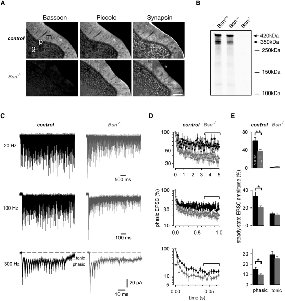
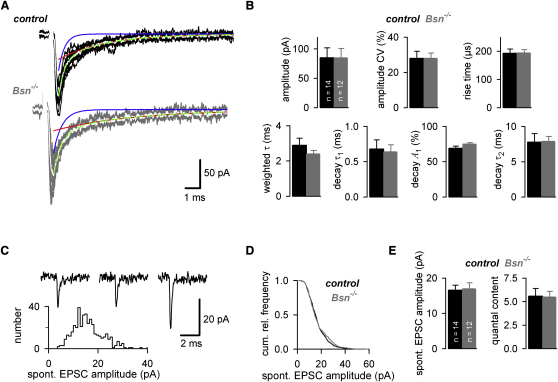
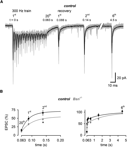
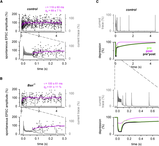
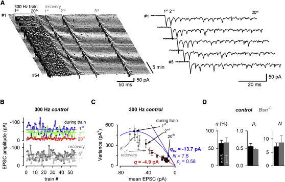
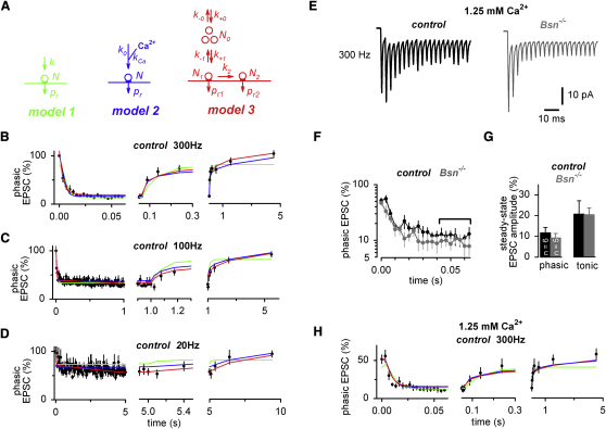
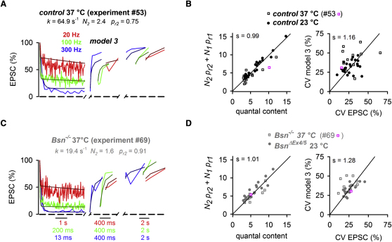
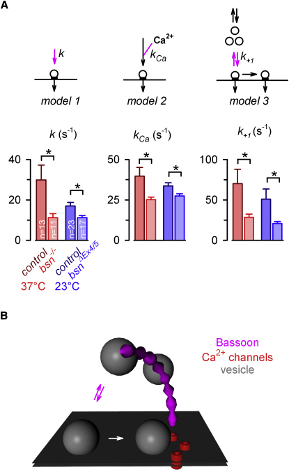
Sustained rate-coded signals encode many types of sensory modalities. Some sensory synapses possess specialized ribbon structures, which tether vesicles, to enable high-frequency signaling. However, central synapses lack these structures, yet some can maintain signaling over a wide bandwidth. To analyze the underlying molecular mechanisms, we investigated the function of the active zone core component Bassoon in cerebellar mossy fiber to granule cell synapses. We show that short-term synaptic depression is enhanced in Bassoon knockout mice during sustained high-frequency trains but basal synaptic transmission is unaffected. Fluctuation and quantal analysis as well as quantification with constrained short-term plasticity models revealed that the vesicle reloading rate was halved in the absence of Bassoon. Thus, our data show that the cytomatrix protein Bassoon speeds the reloading of vesicles to release sites at a central excitatory synapse.
Overview
The claims and statistics of this paper haven’t been checked yet.
Run the check — free, no account needed. It reads the open-access text and checks the sources it cites.
Integrity signals
Registry
Cites
Statistics
Figures
Verification
Claims the paper makes
The claims and statistics of this paper haven’t been checked yet.
Run the check — free, no account needed. It reads the open-access text and checks the sources it cites.
Claims other papers make about this one
Each is checked against this paper’s own text: does it say what the citing paper says it does?
The source backs part of the claim, not all of it — or backs it for a different population, condition or comparison.
In the citing paper
“Piccolo and bassoon are large proteins specific to vertebrates whose major function appears to be to guide synaptic vesicles from the backfield of the synapse to the active zone (Mukherjee et al., 2010; Hallermann et al., 2010).”
From The Presynaptic Active Zone (2012) · cites this paper as Hallermann et al., 2010
Experimental — accuracy not yet measuredThe cited source says what the claim attributes to it.
In the citing paper
“Recovery at MF–GC synapses follows a bi‐exponential time course (Hallermann et al., 2010).”
From MicroRNA‐138‐5p suppresses excitatory synaptic strength at the cerebellar input layer (2025) · cites this paper as Hallermann et al., 2010
The check wasn’t confident either way, so it gives no verdict — never disputed.
In the citing paper
“a deletion of the 17 C-terminal residues of bruchpilot (which are part of the plectin-homology region) impaired attachment of synaptic vesicles to t bars in Drosophila synapses and altered synaptic transmission”
From The Presynaptic Active Zone (2012) · cites this paper as Hallermann et al., 2010
Experimental — accuracy not yet measuredThe check wasn’t confident either way, so it gives no verdict — never disputed.
In the citing paper
“suggesting that a major contribution to bruchpilot function is derived from the plectin-homology region (Hallermann et al., 2010).”
From The Presynaptic Active Zone (2012) · cites this paper as Hallermann et al., 2010
Experimental — accuracy not yet measured
Self-check
Percentages, sums and reported differences re-computed from the paper’s own numbers.
The self-check needs the paper’s full text.
Citations
Cites
Südhof, T. C. (2004) · Annual Review of Neuroscience · doi:10.1146/annurev.neuro.26.041002.131412 · Cited by 2,491
Quantal components of the end‐plate potential
del Castillo, J., & Katz, B. (1954) · The Journal of Physiology · doi:10.1113/jphysiol.1954.sp005129 · Cited by 2,201
Synaptic depression and cortical gain control
Abbott, L. F., Varela, J. A., Sen, K., & Nelson, S. B. (1997) · Science · doi:10.1126/science.275.5297.221 · Cited by 1,690
Edwards, F. A., Konnerth, A., Sakmann, B. J., & Takahashi, T. (1989) · Pflügers Archiv - European Journal of Physiology · doi:10.1007/bf00580998 · Cited by 1,282
Differential signaling via the same axon of neocortical pyramidal neurons
Markram, H., Wang, Y., & Tsodyks, M. (1998) · Proceedings of the National Academy of Sciences · doi:10.1073/pnas.95.9.5323 · Cited by 1,102
Multiple Roles of Calcium Ions in the Regulation of Neurotransmitter Release
Neher, E., & Sakaba, T. (2008) · Neuron · doi:10.1016/j.neuron.2008.08.019 · Cited by 966
Hair cell synaptic ribbons are essential for synchronous auditory signalling
Khimich, D., Nouvian, R., Pujol, R., Dieck, S. T., Egner, A., Gundelfinger, E. D., & Moser, T. (2005) · Nature · doi:10.1038/nature03418 · Cited by 583
Self-citationDieck, S., Sanmartí-Vila, L., Langnaese, K., Richter, K., Kindler, S., Soyke, A., Wex, H., Smalla, K.-H., Kämpf, U., Fränzer, J.-T., et al. (1998) · The Journal of Cell Biology · doi:10.1083/jcb.142.2.499 · Cited by 482
Self-citationDick, O., Dieck, S. T., Altrock, W. D., Ammermüller, J., Weiler, R., Garner, C., Gundelfinger, E., & Brandstätter, J. (2003) · Neuron · doi:10.1016/s0896-6273(03)00086-2 · Cited by 453
Self-citationDittman, J. S., & Regehr, W. G. (1998) · Journal of Neuroscience · doi:10.1523/jneurosci.18-16-06147.1998 · Cited by 410
Cited by
Sorted by citations
Südhof, T. C. (2012) · Neuron · doi:10.1016/j.neuron.2012.06.012 · Cited by 1,062
Short-Term Presynaptic Plasticity
Regehr, W. G. (2012) · Cold Spring Harbor Perspectives in Biology · doi:10.1101/cshperspect.a005702 · Cited by 482
Sterling, P., & Laughlin, S. (2015) · doi:10.7551/mitpress/9780262028707.001.0001 · Cited by 482
Short-term forms of presynaptic plasticity
Fioravante, D., & Regehr, W. G. (2011) · Current Opinion in Neurobiology · doi:10.1016/j.conb.2011.02.003 · Cited by 375
Synaptic vesicle recycling: steps and principles
Rizzoli, S. O. (2014) · The EMBO Journal · doi:10.1002/embj.201386357 · Cited by 317
Metadata
- Authors
- Hallermann, S., Fejtova, A., Schmidt, H., Weyhersmüller, A., Silver, R. A., Gundelfinger, E. D., & Eilers, J.
- Affiliations
- Leipzig University (DE)
- University College London (GB)
- Leibniz Institute for Neurobiology (DE)
- Corresponding author
- Hallermann, Stefan
- Venue
- Neuron
- Publisher
- Elsevier BV
- Published
- 2010-11
- Volume
- 68
- Issue
- 4
- Pages
- 710-723
- ISSN
- 0896-6273 (ISSN-L)
- PMC
- PMC3004039
- OpenAlex
- W2050878615
- Cited by
- 223
- References
- 67
- Citations by year
- 2026: 7 · 2025: 13 · 2024: 7 · 2023: 12 · 2022: 15 · 2021: 11 · 2020: 16 · 2019: 17 · 2018: 13 · 2017: 16 · 2016: 17 · 2015: 13 · 2014: 17 · 2013: 15 · 2012: 16
- FWCI
- 6.34
- Type
- journal article
- Language
- en
- Open access
- hybrid
- doi.org
- Licence
- Locations
- Neuron (publishedVersion, cc-by)
- PubMed Central (publishedVersion, cc-by)
- PubMed (publishedVersion)
- UCL Discovery (University College London) (submittedVersion)
- Funders
- Wellcome Trust
- Topics
- Life Sciences › Neuroscience › Cellular and Molecular Neuroscience › Neuroscience and Neuropharmacology Research
- Life Sciences › Biochemistry, Genetics and Molecular Biology › Cell Biology › Cellular transport and secretion
- Life Sciences › Biochemistry, Genetics and Molecular Biology › Genetics › Genetics and Neurodevelopmental Disorders
- Keywords
- bassoon, short-term synaptic depression, cerebellum, active zone, quantal analysis, rate coding
- MeSH terms
- Animals, Cells, Cultured, Cerebellum, Hippocampus, Mice, Inbred C57BL, Mice, Transgenic, Nerve Tissue Proteins, Neural Inhibition, Reaction Time, Synapses, Synaptic Vesicles, Time Factors, Mice, Knockout, Excitatory Postsynaptic Potentials, Mice, Mice, 129 Strain
About these checks
Registry, retraction, Cites and statistics results are computed, not judged, with one exception: a printed reference that no index lists is read once by a model before it counts as a citation error. Claims are extracted by a model and kept only when their quote appears verbatim in the text; each cited source is then checked, with a model’s help, for whether it says what the claim relies on. Those verdicts can be wrong — read the claim’s quote and the cited source before relying on one.
Statistics and arithmetic are recomputed, not judged.
Confidence bands are shown only for fields calibrated on our annotated test set. None is calibrated yet, so no band is shown.
What counts as a claim ›How we measure confidence ›How we check this ›
Are you an author, or do you see a problem with this page? Request a review
