

The DOI matches its Crossref record; 4 of the 4 references it cites were found in a registry or an index.
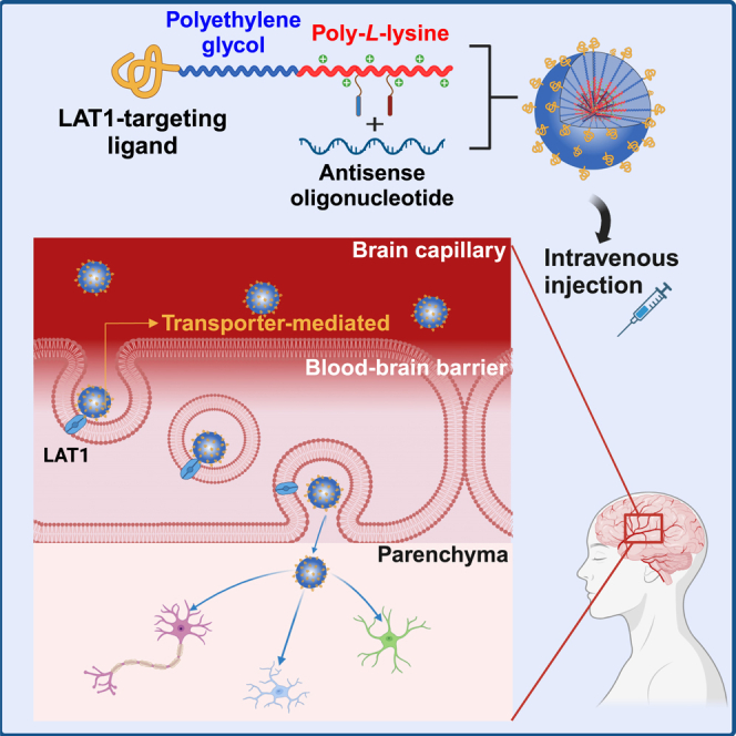
l-Type amino acid transporter 1-targeting nanoparticles for antisense oligonucleotide delivery to the CNS
Overview
The claims and statistics of this paper haven’t been checked yet.
Run the check — free, no account needed. It reads the open-access text and checks the sources it cites.
Integrity signals
Registry
Cites
Statistics
Figures
Verification
Claims the paper makes
The claims and statistics of this paper haven’t been checked yet.
Run the check — free, no account needed. It reads the open-access text and checks the sources it cites.
Claims other papers make about this one
Each is checked against this paper’s own text: does it say what the citing paper says it does?
In the citing paper
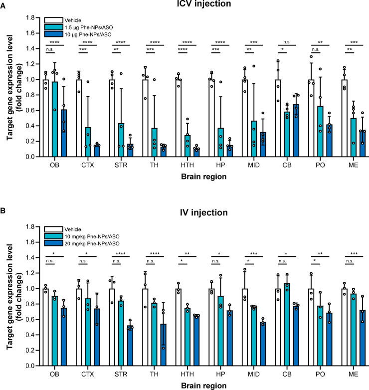
“The dose of BMD-001S (20 mg/kg) was determined based on our previous study using the same ASO-loaded nanoparticles, in which intravenous administration at this dose achieved efficient target knockdown [58].”
From Modulation of the miR-485-3p/PGC-1α Pathway by ASO-Loaded Nanoparticles Attenuates ALS Pathogenesis (2026) · cites this paper as [58]
In the citing paper
“was prepared using a protocol adapted from our previous study, in which this formulation strategy was validated [58]”
From Modulation of the miR-485-3p/PGC-1α Pathway by ASO-Loaded Nanoparticles Attenuates ALS Pathogenesis (2026) · cites this paper as [58]
Self-check
Percentages, sums and reported differences re-computed from the paper’s own numbers.
The self-check needs the paper’s full text.
Citations
Cites
The blood–brain barrier: Structure, regulation and drug delivery
Wu, D., Chen, Q., Chen, X., Han, F., Chen, Z., & Wang, Y. (2023) · Signal Transduction and Targeted Therapy · doi:10.1038/s41392-023-01481-w · Cited by 1,906
Endosomal escape pathways for delivery of biologicals
Varkouhi, A. K., Scholte, M., Storm, G., & Haisma, H. J. (2011) · Journal of Controlled Release · doi:10.1016/j.jconrel.2010.11.004 · Cited by 1,561
Strategies for delivering therapeutics across the blood–brain barrier
Terstappen, G. C., Meyer, A. H., Bell, R. D., & Zhang, W. (2021) · Nature Reviews Drug Discovery · doi:10.1038/s41573-021-00139-y · Cited by 1,138
Antisense Oligonucleotide Therapies for Neurodegenerative Diseases
Bennett, C. F., Krainer, A. R., & Cleveland, D. W. (2019) · Annual Review of Neuroscience · doi:10.1146/annurev-neuro-070918-050501 · Cited by 353
Research works printed but not linked: 69
- Mendonca, 2021, Advances in the Design of (Nano)Formulations for Delivery of Antisense Oligonucleotides and Small Interfering RNA: Focus on the Central Nervous System, Mol. Pharm., 18, 1491, 10.1021/acs.molpharmaceut.0c01238 · DOI
- Monine, 2021, A physiologically-based pharmacokinetic model to describe antisense oligonucleotide distribution after intrathecal administration, J. Pharmacokinet. Pharmacodyn., 48, 639, 10.1007/s10928-021-09761-0 · DOI
- MacCannell, 2022, Restoration of Nusinersen Levels Following Treatment Interruption in People With Spinal Muscular Atrophy: Simulations Based on a Population Pharmacokinetic Model, CNS Drugs, 36, 181, 10.1007/s40263-022-00899-0 · DOI
- Holm, 2022, Clinical advances of RNA therapeutics for treatment of neurological and neuromuscular diseases, RNA Biol., 19, 594, 10.1080/15476286.2022.2066334 · DOI
- Zhao, 2020, Therapeutic TVs for Crossing Barriers in the Brain, Cell, 182, 267, 10.1016/j.cell.2020.06.041 · DOI
- Zuchero, 2016, Discovery of Novel Blood-Brain Barrier Targets to Enhance Brain Uptake of Therapeutic Antibodies, Neuron, 89, 70, 10.1016/j.neuron.2015.11.024 · DOI
- Pinheiro, 2021, Nanoparticles for Targeted Brain Drug Delivery: What Do We Know?, Int. J. Mol. Sci., 22, 10.3390/ijms222111654 · DOI
- Jones, 2007, Blood-brain barrier transport of therapeutics via receptor-mediation, Pharm. Res. (N.Y.), 24, 1759, 10.1007/s11095-007-9379-0 · DOI · Paper page
- Chew, 2023, CD98hc is a target for brain delivery of biotherapeutics, Nat. Commun., 14, 5053, 10.1038/s41467-023-40681-4 · DOI
- Li, 2015, Choline Derivate-Modified Doxorubicin Loaded Micelle for Glioma Therapy, ACS Appl. Mater. Interfaces, 7, 21589, 10.1021/acsami.5b07045 · DOI
- Su, 2019, Emerging transporter-targeted nanoparticulate drug delivery systems, Acta Pharm. Sin. B, 9, 49, 10.1016/j.apsb.2018.10.005 · DOI
- Kou, 2018, L-Carnitine-conjugated nanoparticles to promote permeation across blood-brain barrier and to target glioma cells for drug delivery via the novel organic cation/carnitine transporter OCTN2, Artif. Cells, Nanomed. Biotechnol., 46, 1605
- Abdul Razzak, 2019, Approaches to CNS Drug Delivery with a Focus on Transporter-Mediated Transcytosis, Int. J. Mol. Sci., 20, 10.3390/ijms20123108 · DOI
- Mazumdar, 2021, Exploration and insights into the cellular internalization and intracellular fate of amphiphilic polymeric nanocarriers, Acta Pharm. Sin. B, 11, 903, 10.1016/j.apsb.2021.02.019 · DOI
- Gyimesi, 2023, Transporter-Mediated Drug Delivery, Molecules, 28, 10.3390/molecules28031151 · DOI
- Abbott, 2006, Astrocyte-endothelial interactions at the blood-brain barrier, Nat. Rev. Neurosci., 7, 41, 10.1038/nrn1824 · DOI · Paper page
- Chen, 2012, Modern methods for delivery of drugs across the blood-brain barrier, Adv. Drug Deliv. Rev., 64, 640, 10.1016/j.addr.2011.11.010 · DOI
- Min, 2020, Systemic Brain Delivery of Antisense Oligonucleotides across the Blood-Brain Barrier with a Glucose-Coated Polymeric Nanocarrier, Angew. Chem., Int. Ed. Engl., 59, 8173, 10.1002/anie.201914751 · DOI
- Somani, 2014, Transferrin-bearing polypropylenimine dendrimer for targeted gene delivery to the brain, J. Contr. Release, 188, 78, 10.1016/j.jconrel.2014.06.006 · DOI
- Zhang, 2003, Global non-viral gene transfer to the primate brain following intravenous administration, Mol. Ther., 7, 11, 10.1016/S1525-0016(02)00018-7 · DOI
- Pandit, 2020, The blood-brain barrier: Physiology and strategies for drug delivery, Adv. Drug Deliv. Rev., 165–166, 1, 10.1016/j.addr.2019.11.009 · DOI · Paper page
- Patra, 2018, Nano based drug delivery systems: recent developments and future prospects, J. Nanobiotechnol., 16, 71, 10.1186/s12951-018-0392-8 · DOI
- Banks, 2016, From blood-brain barrier to blood-brain interface: new opportunities for CNS drug delivery, Nat. Rev. Drug Discov., 15, 275, 10.1038/nrd.2015.21 · DOI · Paper page
- Ni, 2015, Poly(lactic-co-glycolic acid) nanoparticles conjugated with CD133 aptamers for targeted salinomycin delivery to CD133+ osteosarcoma cancer stem cells, Int. J. Nanomed., 10, 2537
- Shi, 2023, Active targeting schemes for nano-drug delivery systems in osteosarcoma therapeutics, J. Nanobiotechnol., 21, 103, 10.1186/s12951-023-01826-1 · DOI
- Ikotun, 2013, Imaging the L-type amino acid transporter-1 (LAT1) with Zr-89 immunoPET, PLoS One, 8, 10.1371/journal.pone.0077476 · DOI
- Scalise, 2018, The Human SLC7A5 (LAT1): The Intriguing Histidine/Large Neutral Amino Acid Transporter and Its Relevance to Human Health, Front. Chem., 6, 243, 10.3389/fchem.2018.00243 · DOI
- Barar, 2016, Blood-brain barrier transport machineries and targeted therapy of brain diseases, Bioimpacts, 6, 225, 10.15171/bi.2016.30 · DOI
- Yan, 2021, Mechanism of substrate transport and inhibition of the human LAT1-4F2hc amino acid transporter, Cell Discov., 7, 16, 10.1038/s41421-021-00247-4 · DOI
- Kanai, 2022, Amino acid transporter LAT1 (SLC7A5) as a molecular target for cancer diagnosis and therapeutics, Pharmacol. Ther., 230, 10.1016/j.pharmthera.2021.107964 · DOI
- Pardridge, 2022, A Historical Review of Brain Drug Delivery, Pharmaceutics, 14, 10.3390/pharmaceutics14061283 · DOI · Paper page
- Yanagida, 2001, Human L-type amino acid transporter 1 (LAT1): characterization of function and expression in tumor cell lines, Biochim. Biophys. Acta, 1514, 291, 10.1016/S0005-2736(01)00384-4 · DOI · Paper page
- Barthelemy, 2019, Ubiquitylation and endocytosis of the human LAT1/SLC7A5 amino acid transporter, Sci. Rep., 9, 10.1038/s41598-019-53065-w · DOI
- Zhang, 2020, Review of the Correlation of LAT1 With Diseases: Mechanism and Treatment, Front. Chem., 8, 10.3389/fchem.2020.564809 · DOI
- Huttunen, 2019, L-Type Amino Acid Transporter 1 (LAT1/Lat1)-Utilizing Prodrugs Can Improve the Delivery of Drugs into Neurons, Astrocytes and Microglia, Sci. Rep., 9, 10.1038/s41598-019-49009-z · DOI
- Puris, 2023, Protein Expression of Amino Acid Transporters Is Altered in Isolated Cerebral Microvessels of 5xFAD Mouse Model of Alzheimer's Disease, Mol. Neurobiol., 60, 732, 10.1007/s12035-022-03111-y · DOI
- Yan, 2019, Structure of the human LAT1-4F2hc heteromeric amino acid transporter complex, Nature, 568, 127, 10.1038/s41586-019-1011-z · DOI · Paper page
- Zhang, 2023, LAT1 targeted brain delivery of temozolomide and sorafenib for effective glioma therapy, Nano Res., 16, 9743, 10.1007/s12274-023-5568-3 · DOI
- Augustyn, 2016, LAT-1 activity of meta-substituted phenylalanine and tyrosine analogs, Bioorg. Med. Chem. Lett., 26, 2616, 10.1016/j.bmcl.2016.04.023 · DOI
- Li, 2017, Effects of ligand distribution on receptor-diffusion-mediated cellular uptake of nanoparticles, R. Soc. Open Sci., 4
- Jhaveri, 2016, Intracellular delivery of nanocarriers and targeting to subcellular organelles, Expet Opin. Drug Deliv., 13, 49, 10.1517/17425247.2015.1086745 · DOI
- Roberts, 2020, Advances in oligonucleotide drug delivery, Nat. Rev. Drug Discov., 19, 673, 10.1038/s41573-020-0075-7 · DOI
- Anthony, 2022, RNA-based therapeutics for neurological diseases, RNA Biol., 19, 176, 10.1080/15476286.2021.2021650 · DOI
- Koh, 2021, Targeting MicroRNA-485-3p Blocks Alzheimer's Disease Progression, Int. J. Mol. Sci., 22, 10.3390/ijms222313136 · DOI
- Ryu, 2023, The role of microRNA-485 in neurodegenerative diseases, Rev. Neurosci., 34, 49, 10.1515/revneuro-2022-0039 · DOI · Paper page
- Behzadi, 2017, Cellular uptake of nanoparticles: journey inside the cell, Chem. Soc. Rev., 46, 4218, 10.1039/C6CS00636A · DOI · Paper page
- Satzer, 2016, Protein adsorption onto nanoparticles induces conformational changes: Particle size dependency, kinetics, and mechanisms, Eng. Life Sci., 16, 238, 10.1002/elsc.201500059 · DOI
- Zhang, 2022, Effects of morphology and size of nanoscale drug carriers on cellular uptake and internalization process: a review, RSC Adv., 13, 80, 10.1039/D2RA06888E · DOI
- Hersh, 2022, Crossing the Blood-Brain Barrier: Advances in Nanoparticle Technology for Drug Delivery in Neuro-Oncology, Int. J. Mol. Sci., 23, 10.3390/ijms23084153 · DOI · Paper page
- Sonavane, 2008, Biodistribution of colloidal gold nanoparticles after intravenous administration: effect of particle size, Colloids Surf. B Biointerfaces, 66, 274, 10.1016/j.colsurfb.2008.07.004 · DOI
- Choi, 2007, Renal clearance of quantum dots, Nat. Biotechnol., 25, 1165, 10.1038/nbt1340 · DOI · Paper page
- Faria, 2018, Minimum information reporting in bio-nano experimental literature, Nat. Nanotechnol., 13, 777, 10.1038/s41565-018-0246-4 · DOI
- Banerjee, 2012, Poly(ethylene glycol)-Prodrug Conjugates: Concept, Design, and Applications, J. Drug Deliv., 2012
- Li, 2021, Nano-Drug Design Based on the Physiological Properties of Glutathione, Molecules, 26
- Cheng, 2011, Glutathione-responsive nano-vehicles as a promising platform for targeted intracellular drug and gene delivery, J. Contr. Release, 152, 2, 10.1016/j.jconrel.2011.01.030 · DOI
- Dringen, 2003, Glutathione pathways in the brain, Biol. Chem., 384, 505, 10.1515/BC.2003.059 · DOI
- Sun, 2006, Two-photon imaging of glutathione levels in intact brain indicates enhanced redox buffering in developing neurons and cells at the cerebrospinal fluid and blood-brain interface, J. Biol. Chem., 281, 17420, 10.1074/jbc.M601567200 · DOI
- Such, 2015, Interfacing materials science and biology for drug carrier design, Adv. Mater., 27, 2278, 10.1002/adma.201405084 · DOI
- Smith, 2019, The Endosomal Escape of Nanoparticles: Toward More Efficient Cellular Delivery, Bioconjugate Chem., 30, 263, 10.1021/acs.bioconjchem.8b00732 · DOI
- Mitchell, 2021, Engineering precision nanoparticles for drug delivery, Nat. Rev. Drug Discov., 20, 101, 10.1038/s41573-020-0090-8 · DOI · Paper page
- Anraku, 2017, Glycaemic control boosts glucosylated nanocarrier crossing the BBB into the brain, Nat. Commun., 8, 1001, 10.1038/s41467-017-00952-3 · DOI
- Wang, 2010, The complex role of multivalency in nanoparticles targeting the transferrin receptor for cancer therapies, J. Am. Chem. Soc., 132, 11306, 10.1021/ja1043177 · DOI
- Alkilany, 2019, Ligand density on nanoparticles: A parameter with critical impact on nanomedicine, Adv. Drug Deliv. Rev., 143, 22, 10.1016/j.addr.2019.05.010 · DOI
- Tosi, 2015, Nanomedicine and neurodegenerative disorders: so close yet so far, Expet Opin. Drug Deliv., 12, 1041, 10.1517/17425247.2015.1041374 · DOI
- Takakusa, 2023, Drug Metabolism and Pharmacokinetics of Antisense Oligonucleotide Therapeutics: Typical Profiles, Evaluation Approaches, and Points to Consider Compared with Small Molecule Drugs, Nucleic Acid Therapeut., 33, 83, 10.1089/nat.2022.0054 · DOI
- Vellonen, 2017, Disease-Induced Alterations in Brain Drug Transporters in Animal Models of Alzheimer's Disease : Theme: Drug Discovery, Development and Delivery in Alzheimer's Disease Guest Editor: Davide Brambilla, Pharm. Res. (N.Y.), 34, 2652, 10.1007/s11095-017-2263-7 · DOI
- Gynther, 2018, Alzheimer's Disease Phenotype or Inflammatory Insult Does Not Alter Function of L-Type Amino Acid Transporter 1 in Mouse Blood-Brain Barrier and Primary Astrocytes, Pharm. Res. (N.Y.), 36, 17, 10.1007/s11095-018-2546-7 · DOI
- Li, 2016, Antibody h-R3-dendrimer mediated siRNA has excellent endosomal escape and tumor targeted delivery ability, and represents efficient siPLK1 silencing and inhibition of cell proliferation, migration and invasion, Oncotarget, 7, 13782, 10.18632/oncotarget.7368 · DOI
- Ryu, 2023, The microRNA-485-3p concentration in salivary exosome-enriched extracellular vesicles is related to amyloid beta deposition in the brain of patients with Alzheimer's disease, Clin. Biochem., 118, 10.1016/j.clinbiochem.2023.110603 · DOI · Paper page
Cited by
Sorted by citations
Ding, L., Kshirsagar, P., Agrawal, P., & Murry, D. J. (2025) · Pharmaceutics · doi:10.3390/pharmaceutics17060706 · Cited by 62
Modulation of the miR-485-3p/PGC-1α Pathway by ASO-Loaded Nanoparticles Attenuates ALS Pathogenesis
Ryu, I. S., Ha, D.-I., Jung, Y.-J., Lee, H. J., Kim, I., Lim, Y. N., Min, H. S., Kim, S. H., Yoon, I., Cho, H.-J., et al. (2026) · International Journal of Molecular Sciences · doi:10.3390/ijms27020615 · Cited by 2
Self-citation
Metadata
- Authors
- Lim, Y. N., Ryu, I. S., Jung, Y.-J., Helmlinger, G., Kim, I., Park, H. W., Kang, H., Lee, J., Lee, H. J., Lee, K. S., Jang, H.-N., Ha, D.-I., Park, J., Won, J., Lim, K. S., Jeon, C.-Y., Cho, H.-J., Min, H. S., & Ryu, J.-H.
- ORCID
- Ryu, Jin-Hyeob
- Venue
- Molecular Therapy Nucleic Acids
- Publisher
- Elsevier BV
- Published
- 2024-12
- Volume
- 35
- Issue
- 4
- Pages
- 102340
- ISSN
- 2162-2531
- PMC
- PMC11474373
- Cited by
- 17
- References
- 4
- Language
- en
- Licence
- Funders
- Korea Research Institute of Bioscience and Biotechnology
- Korea Ministry of Small and Medium Enterprises and Startups (RS-2023-00283779)
About these checks
Registry, retraction, Cites and statistics results are computed, not judged, with one exception: a printed reference that no index lists is read once by a model before it counts as a citation error. Claims are extracted by a model and kept only when their quote appears verbatim in the text; each cited source is then checked, with a model’s help, for whether it says what the claim relies on. Those verdicts can be wrong — read the claim’s quote and the cited source before relying on one.
Statistics and arithmetic are recomputed, not judged.
Confidence bands are shown only for fields calibrated on our annotated test set. None is calibrated yet, so no band is shown.
What counts as a claim ›How we measure confidence ›How we check this ›
Are you an author, or do you see a problem with this page? Request a review
