

The DOI matches its Crossref record; 31 of the 31 references it cites were found in a registry or an index.
Point-of-care screening for heart failure with reduced ejection fraction using artificial intelligence during ECG-enabled stethoscope examination in London, UK: a prospective, observational, multicentre study
Open accessAbstract
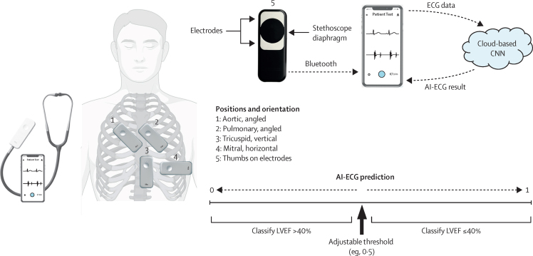
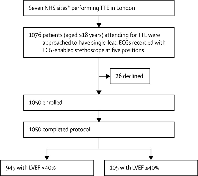
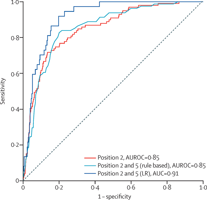
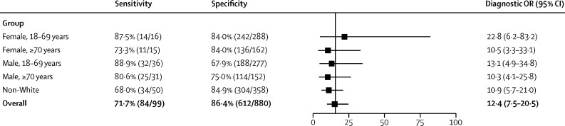
BACKGROUND: Most patients who have heart failure with a reduced ejection fraction, when left ventricular ejection fraction (LVEF) is 40% or lower, are diagnosed in hospital. This is despite previous presentations to primary care with symptoms. We aimed to test an artificial intelligence (AI) algorithm applied to a single-lead ECG, recorded during ECG-enabled stethoscope examination, to validate a potential point-of-care screening tool for LVEF of 40% or lower. METHODS: We conducted an observational, prospective, multicentre study of a convolutional neural network (known as AI-ECG) that was previously validated for the detection of reduced LVEF using 12-lead ECG as input. We used AI-ECG retrained to interpret single-lead ECG input alone. Patients (aged ≥18 years) attending for transthoracic echocardiogram in London (UK) were recruited. All participants had 15 s of supine, single-lead ECG recorded at the four standard anatomical positions for cardiac auscultation, plus one handheld position, using an ECG-enabled stethoscope. Transthoracic echocardiogram-derived percentage LVEF was used as ground truth. The primary outcome was performance of AI-ECG at classifying reduced LVEF (LVEF ≤40%), measured using metrics including the area under the receiver operating characteristic curve (AUROC), sensitivity, and specificity, with two-sided 95% CIs. The primary outcome was reported for each position individually and with an optimal combination of AI-ECG outputs (interval range 0-1) from two positions using a rule-based approach and several classification models. This study is registered with ClinicalTrials.gov, NCT04601415. FINDINGS: Between Feb 6 and May 27, 2021, we recruited 1050 patients (mean age 62 years [SD 17·4], 535 [51%] male, 432 [41%] non-White). 945 (90%) had an ejection fraction of at least 40%, and 105 (10%) had an ejection fraction of 40% or lower. Across all positions, ECGs were most frequently of adequate quality for AI-ECG interpretation at the pulmonary position (979 [93·3%] of 1050). Quality was lowest for the aortic position (846 [80·6%]). AI-ECG performed best at the pulmonary valve position (p=0·02), with an AUROC of 0·85 (95% CI 0·81-0·89), sensitivity of 84·8% (76·2-91·3), and specificity of 69·5% (66·4-72·6). Diagnostic odds ratios did not differ by age, sex, or non-White ethnicity. Taking the optimal combination of two positions (pulmonary and handheld positions), the rule-based approach resulted in an AUROC of 0·85 (0·81-0·89), sensitivity of 82·7% (72·7-90·2), and specificity of 79·9% (77·0-82·6). Using AI-ECG outputs from these two positions, a weighted logistic regression with l2 regularisation resulted in an AUROC of 0·91 (0·88-0·95), sensitivity of 91·9% (78·1-98·3), and specificity of 80·2% (75·5-84·3). INTERPRETATION: A deep learning system applied to single-lead ECGs acquired during a routine examination with an ECG-enabled stethoscope can detect LVEF of 40% or lower. These findings highlight the potential for inexpensive, non-invasive, workflow-adapted, point-of-care screening, for earlier diagnosis and prognostically beneficial treatment. FUNDING: NHS Accelerated Access Collaborative, NHSX, and the National Institute for Health Research.
Overview
The claims and statistics of this paper haven’t been checked yet.
Run the check — free, no account needed. It reads the open-access text and checks the sources it cites.
Integrity signals
Registry
Cites
Statistics
Figures
Verification
Claims the paper makes
The claims and statistics of this paper haven’t been checked yet.
Run the check — free, no account needed. It reads the open-access text and checks the sources it cites.
Claims other papers make about this one
Each is checked against this paper’s own text: does it say what the citing paper says it does?
The source backs part of the claim, not all of it — or backs it for a different population, condition or comparison.
In the citing paper
“Models trained on clinical ECGs have been applied to wearable device ECGs, but have traditionally shown significantly lower performance on wearable device ECGs than on held-out clinical ECG test sets14,15.”
From Detection of left ventricular systolic dysfunction from single-lead electrocardiography adapted for portable and wearable devices (2023) · cites this paper as [14]
Experimental — accuracy not yet measuredThe source backs part of the claim, not all of it — or backs it for a different population, condition or comparison.
In the citing paper
“Consequently, the performance of models for detecting LVSD from portable device ECGs may degrade in the real-world setting, with lower performance than observed in the original single-lead derivatives of the clinical development studies14,15.”
From Detection of left ventricular systolic dysfunction from single-lead electrocardiography adapted for portable and wearable devices (2023) · cites this paper as [14]
Experimental — accuracy not yet measuredThe cited source says what the claim attributes to it.
In the citing paper
“Separately, a prospective multicenter study showed that a deep-learning model applied to single-lead ECGs obtained during examination with an ECG-enabled stethoscope could identify patients with a left ventricular ejection fraction of 40% or lower [64].”
From Deep Learning Applied to 12-Lead ECGs for Detection of Structural, Metabolic and Systemic Disease (2026) · cites this paper as [64]
Self-check
Percentages, sums and reported differences re-computed from the paper’s own numbers.
The self-check needs the paper’s full text.
Citations
Cites
DeLong, E. R., DeLong, D. M., & Clarke-Pearson, D. L. (1988) · Biometrics · doi:10.2307/2531595 · Cited by 22,649
High-performance medicine: the convergence of human and artificial intelligence
Topol, E. J. (2019) · Nature Medicine · doi:10.1038/s41591-018-0300-7 · Cited by 10,450
Forecasting the Impact of Heart Failure in the United States
Heidenreich, P. A., Albert, N. M., Allen, L. A., Bluemke, D. A., Butler, J., Fonarow, G. C., Ikonomidis, J. S., Khavjou, O., Konstam, M. A., Maddox, T. M., et al. (2013) · Circulation Heart Failure · doi:10.1161/hhf.0b013e318291329a · Cited by 2,884
Global Public Health Burden of Heart Failure
Savarese, G., & Lund, L. H. (2017) · Cardiac failure review · doi:10.15420/cfr.2016:25:2 · Cited by 2,818
Angiotensin–Neprilysin Inhibition in Heart Failure with Preserved Ejection Fraction
Solomon, S. D., McMurray, J. J. V., Anand, I. S., Ge, J., Lam, C. S. P., Maggioni, A. P., Martinez, F., Packer, M., Pfeffer, M. A., Pieske, B., et al. (2019) · New England Journal of Medicine · doi:10.1056/nejmoa1908655 · Cited by 2,430
The Global Health and Economic Burden of Hospitalizations for Heart Failure
Ambrosy, A. P., Fonarow, G. C., Butler, J., Chioncel, O. D., Greene, S. J., Vaduganathan, M., Nodari, S., Lam, C. S. P., Sato, N., Shah, A. N., et al. (2014) · Journal of the American College of Cardiology · doi:10.1016/j.jacc.2013.11.053 · Cited by 2,110
Bozkurt, B., Coats, A. J. S., Tsutsui, H., Abdelhamid, C. M., Adamopoulos, S., Albert, N., Anker, S. D., Atherton, J., Böhm, M., Butler, J., et al. (2021) · European Journal of Heart Failure · doi:10.1002/ejhf.2115 · Cited by 1,665
Attia, Z. I., Noseworthy, P. A., Lopez-Jimenez, F., Asirvatham, S. J., Deshmukh, A. J., Gersh, B. J., Carter, R. E., Yao, X., Rabinstein, A. A., Erickson, B. J., et al. (2019) · The Lancet · doi:10.1016/s0140-6736(19)31721-0 · Cited by 1,613
Attia, Z. I., Kapa, S., Lopez-Jimenez, F., McKie, P. M., Ladewig, D. J., Satam, G., Pellikka, P. A., Enriquez-Sarano, M., Noseworthy, P. A., Munger, T. M., et al. (2019) · Nature Medicine · doi:10.1038/s41591-018-0240-2 · Cited by 1,389
Nagendran, M., Chen, Y., Lovejoy, C. A., Gordon, A. C., Komorowski, M., Harvey, H., Topol, E. J., Ioannidis, J. P. A., Collins, G. S., & Maruthappu, M. (2020) · BMJ · doi:10.1136/bmj.m689 · Cited by 1,152
Cited by
Sorted by citations
Armoundas, A. A., Narayan, S. M., Arnett, D. K., Spector-Bagdady, K., Bennett, D. A., Celi, L. A., Friedman, P. A., Gollob, M. H., Hall, J. L., Kwitek, A. E., et al. (2024) · Circulation · doi:10.1161/cir.0000000000001201 · Cited by 347
Artificial intelligence in clinical medicine: catalyzing a sustainable global healthcare paradigm
Krishnan, G., Singh, S., Pathania, M., Gosavi, S., Abhishek, S., Parchani, A., & Dhar, M. (2023) · Frontiers in Artificial Intelligence · doi:10.3389/frai.2023.1227091 · Cited by 340
Transforming Cardiovascular Care With Artificial Intelligence: From Discovery to Practice
Khera, R., Oikonomou, E. K., Nadkarni, G. N., Morley, J., Wiens, J., Butte, A. J., & Topol, E. J. (2024) · Journal of the American College of Cardiology · doi:10.1016/j.jacc.2024.05.003 · Cited by 324
The Integration of Artificial Intelligence into Clinical Practice
Karalis, V. D. (2024) · Applied Biosciences · doi:10.3390/applbiosci3010002 · Cited by 255
Huang, J.-D., Wang, J., Ramsey, E., Leavey, G., Chico, T., & Condell, J. V. (2022) · Sensors · doi:10.3390/s22208002 · Cited by 203
Deep Learning Electrocardiographic Analysis for Detection of Left-Sided Valvular Heart Disease
Elias, P., Poterucha, T. J., Rajaram, V., Moller, L. M., Rodriguez, V., Bhave, S., Hahn, R. T., Tison, G., Abreau, S. A., Barrios, J., et al. (2022) · Journal of the American College of Cardiology · doi:10.1016/j.jacc.2022.05.029 · Cited by 153
Artificial Intelligence for Cardiovascular Care—Part 1: Advances
Elias, P. A., Jain, S. S., Poterucha, T. J., Randazzo, M. J., Jimenez, F. L., Khera, R., Perez, M. V., Ouyang, D., Pirruccello, J. P., Salerno, M., et al. (2024) · Journal of the American College of Cardiology · doi:10.1016/j.jacc.2024.03.400 · Cited by 141
Yim, D., Khuntia, J., Parameswaran, V., & Meyers, A. D. (2024) · JMIR Medical Informatics · doi:10.2196/52073 · Cited by 140
Khunte, A., Sangha, V., Oikonomou, E. K., Dhingra, L. S., Aminorroaya, A., Mortazavi, B. J., Coppi, A., Brandt, C. A., Krumholz, H. M., & Khera, R. (2023) · npj Digital Medicine · doi:10.1038/s41746-023-00869-w · Cited by 95
Sau, A., Pastika, L., Sieliwończyk, E., Patlatzoglou, K., Ribeiro, A. H., McGurk, K. A., Zeidaabadi, B., Zhang, H., Macierzanka, K., Mandic, D. P., et al. (2024) · The Lancet Digital Health · doi:10.1016/s2589-7500(24)00172-9 · Cited by 103
Self-citationWu, Z., & Guo, C. (2025) · BioMedical Engineering OnLine · doi:10.1186/s12938-025-01349-w · Cited by 116
AI-Reinforced Wearable Sensors and Intelligent Point-of-Care Tests
Yammouri, G., & Lahcen, A. A. (2024) · Journal of Personalized Medicine · doi:10.3390/jpm14111088 · Cited by 92
Artificial Intelligence–Enabled Prediction of Heart Failure Risk From Single-Lead Electrocardiograms
Dhingra, L. S., Aminorroaya, A., Pedroso, A. F., Khunte, A., Sangha, V., McIntyre, D., Chow, C. K., Asselbergs, F. W., Brant, L. C. C., Barreto, S. M., et al. (2025) · JAMA Cardiology · doi:10.1001/jamacardio.2025.0492 · Cited by 65
Le, K. H., Pham, H., Nguyen, T., Nguyen, T. A., Thanh, T., & Do, C. (2023) · Biomedical Signal Processing and Control · doi:10.1016/j.bspc.2023.104963 · Cited by 74
Dhingra, L. S., Aminorroaya, A., Sangha, V., Pedroso, A. F., Asselbergs, F. W., Brant, L. C. C., Barreto, S. M., Ribeiro, A. L. P., Krumholz, H. M., Oikonomou, E. K., et al. (2025) · European Heart Journal · doi:10.1093/eurheartj/ehae914 · Cited by 79
Adedinsewo, D. A., Morales-Lara, A. C., Afolabi, B. B., Kushimo, O. A., Mbakwem, A. C., Ibiyemi, K. F., Ogunmodede, J. A., Raji, H. O., Ringim, S. H., Habib, A. A., et al. (2024) · Nature Medicine · doi:10.1038/s41591-024-03243-9 · Cited by 63
Ding, C., Yao, T., Wu, C., & Ni, J. (2024) · Biosensors and Bioelectronics · doi:10.1016/j.bios.2024.117073 · Cited by 78
Recent advancements and applications of deep learning in heart failure: Α systematic review
Πετμεζάς, Γ., Papageorgiou, V. Ε., Vassilikos, V. P., Pagourelias, E. D., Tsaklidis, G. M., Katsaggelos, A. K., & Maglaveras, N. (2024) · Computers in Biology and Medicine · doi:10.1016/j.compbiomed.2024.108557 · Cited by 53
Promises and Perils of Consumer Mobile Technologies in Cardiovascular Care
Varma, N., Han, J. K., PASSMAN, R. S., Rosman, L., Ghanbari, H., Noseworthy, P. A., Silva, J. N. A., Deshmukh, A. J., Sanders, P., Hindricks, G., et al. (2024) · Journal of the American College of Cardiology · doi:10.1016/j.jacc.2023.11.024 · Cited by 52
Stamate, E., Piraianu, A. I., Ciobotaru, O. R., Crassas, R., Duca, O.-M., Fulga, A., Grigore, I., Vintila, V., Fulga, I., & Ciobotaru, O. C. (2024) · Diagnostics · doi:10.3390/diagnostics14111103 · Cited by 49
Docherty, K. F., Lam, C. S. P., Rakisheva, A., Coats, A. J. S., Greenhalgh, T., Metra, M., Petrie, M. C., & Rosano, G. M. C. (2023) · European Journal of Heart Failure · doi:10.1002/ejhf.2946 · Cited by 58
AI and Primary Care: Scoping Review
Katonai, G., Árvai, N., & Meskó, B. (2025) · Journal of Medical Internet Research · doi:10.2196/65950 · Cited by 56
Marsden, H., Kemos, P., Venzi, M., Noy, M., Maheswaran, S., Francis, N., Hyde, C., Mullarkey, D., Kalsi, D., & Thomas, L. (2024) · Frontiers in Medicine · doi:10.3389/fmed.2024.1302363 · Cited by 39
Yang, Q., Bee, Y. M., Lim, C. C., Sabanayagam, C., Yim-Lui Cheung, C., Wong, T. Y., Ting, D. S. W., Lim, L.-L., Li, H., He, M., et al. (2025) · eClinicalMedicine · doi:10.1016/j.eclinm.2025.103089 · Cited by 38
Adopting artificial intelligence in cardiovascular medicine: a scoping review
Makimoto, H., & Kohro, T. (2023) · Hypertension Research · doi:10.1038/s41440-023-01469-7 · Cited by 38
Artificial intelligence in cardiology: The past, present and future
Gupta, M. D., Kunal, S., Girish, M. P., Gupta, A., & Yadav, R. K. (2022) · Indian Heart Journal · doi:10.1016/j.ihj.2022.07.004 · Cited by 41
Applications of machine and deep learning to thyroid cytology and histopathology: a review
Slabaugh, G., Beltrán, L., Rizvi, H., Deloukas, P., & Marouli, E. (2023) · Frontiers in Oncology · doi:10.3389/fonc.2023.958310 · Cited by 38
Artificial intelligence-enhanced patient evaluation: bridging art and science
Oikonomou, E. K., & Khera, R. (2024) · European Heart Journal · doi:10.1093/eurheartj/ehae415 · Cited by 34
Availability of Evidence for Predictive Machine Learning Algorithms in Primary Care
Rakers, M. M., van Buchem, M. M., Kucenko, S., de Hond, A., Kant, I. M. J., van Smeden, M., Moons, K. G. M., Leeuwenberg, A., Chavannes, N. H., Villalobos-Quesada, M. J., et al. (2024) · JAMA Network Open · doi:10.1001/jamanetworkopen.2024.32990 · Cited by 39
Artificial Intelligence and ECG: A New Frontier in Cardiac Diagnostics and Prevention
Bartusik‐Aebisher, D., Rogóż, K., & Aebisher, D. (2025) · Biomedicines · doi:10.3390/biomedicines13071685 · Cited by 42
Langlais, É. L., Thériault-Lauzier, P., Marquis‐Gravel, G., Kulbay, M., So, D. Y. F., Tanguay, J., Ly, H. Q., Gallo, R., Lesage, F., & Avram, R. (2022) · Journal of Cardiovascular Translational Research · doi:10.1007/s12265-022-10260-x · Cited by 35
Digital tools in heart failure: addressing unmet needs
Myhre, P. L., Tromp, J., Ouwerkerk, W., Ting, D. S. W., Docherty, K. F., Gibson, C. M., & Lam, C. S. P. (2024) · The Lancet Digital Health · doi:10.1016/s2589-7500(24)00158-4 · Cited by 31
Randomized Trial of Remote Assessment of Patients After an Acute Coronary Syndrome
Alshahrani, N. S., Hartley, A., Howard, J. P., Hajhosseiny, R., Khawaja, S. A., Seligman, H., Akbari, T., Alharbi, B., Bassett, P., Al-Lamee, R. K., et al. (2024) · Journal of the American College of Cardiology · doi:10.1016/j.jacc.2024.03.398 · Cited by 35
Self-citationKelshiker, M. A., Bächtiger, P., Petri, C. F., Nakhare, S., Mansell, J., Chhatwal, K., Alrumayh, A., Zaman, J., Shah, M., Young, H., et al. (2026) · The Lancet · doi:10.1016/s0140-6736(25)02156-7 · Cited by 34
Self-citationOikonomou, E. K., & Khera, R. (2024) · Hellenic Journal of Cardiology · doi:10.1016/j.hjc.2024.07.003 · Cited by 26
Park, H., Kwon, O., Shim, J., Kim, D., Park, J., Kim, Y.-G., Yu, H. T., Kim, T., Uhm, J., Choi, J., et al. (2024) · npj Digital Medicine · doi:10.1038/s41746-024-01234-1 · Cited by 25
Prospective Human Validation of Artificial Intelligence Interventions in Cardiology
Moosavi, A., Huang, S., Vahabi, M., Motamedivafa, B., Tian, N., Mahmood, R., Liu, P. X., & Sun, C. L. F. (2024) · JACC Advances · doi:10.1016/j.jacadv.2024.101202 · Cited by 29
Artificial Intelligence and Its Role in Diagnosing Heart Failure: A Narrative Review
Medhi, D., Kamidi, S. R., Sree, K. P. M., Shaikh, S., Rasheed, S., Murichathil, A. H. T., & Nazir, Z. (2024) · Cureus · doi:10.7759/cureus.59661 · Cited by 27
Electrocardiogram Devices for Home Use: Technological and Clinical Scoping Review
Zepeda-Echavarria, A., van de Leur, R. R., van Sleuwen, M., Hassink, R. J., Wildbergh, T. X., Doevendans, P. A., Jaspers, J., & van Es, R. (2023) · JMIR Cardio · doi:10.2196/44003 · Cited by 24
Artificial intelligence–based screening for cardiomyopathy in an obstetric population: A pilot study
Adedinsewo, D. A., Morales-Lara, A. C., Hardway, H., Johnson, P. W., Young, K. A., Garzon-Siatoya, W. T., Tobah, Y. B., Rose, C. H., Burnette, D., Seccombe, K., et al. (2024) · Cardiovascular Digital Health Journal · doi:10.1016/j.cvdhj.2024.03.005 · Cited by 22
Real-world evaluation of the Stemoscope electronic tele-auscultation system
Fan, M., Wang, Q., Liu, J., Su, L., Wang, B., Xu, H., Li, Q., & Zhang, Z. (2022) · BioMedical Engineering OnLine · doi:10.1186/s12938-022-01032-4 · Cited by 21
Luo, H., Weerts, J., Bekkers, A., Achten, A., Lievens, S., Smeets, K., van Empel, V. P. M., Delhaas, T., & Prinzen, F. W. (2022) · European Heart Journal - Digital Health · doi:10.1093/ehjdh/ztac073 · Cited by 19
Bächtiger, P., Kelshiker, M. A., Petri, C. F., Gandhi, M., Shah, M., Kamalati, T., Khan, S. A., Hooper, G., Stephens, J., Alrumayh, A., et al. (2023) · BMJ Health & Care Informatics · doi:10.1136/bmjhci-2022-100718 · Cited by 18
Self-citationAutomated Detection of Reduced Ejection Fraction Using an ECG-Enabled Digital Stethoscope
Guo, L. T., Pressman, G. S., Kieu, S. N., Marrus, S. B., Mathew, G., Prince, J., Lastowski, E., McDonough, R. V., Currie, C., Tiwari, U., et al. (2025) · JACC Advances · doi:10.1016/j.jacadv.2025.101619 · Cited by 23
Lim, J., Lee, H. S., Han, G. I., Kang, S., Jang, J.-H., Jo, Y., Son, J. M., Lee, M. S., Kwon, J., & Lee, S. (2025) · European Heart Journal - Digital Health · doi:10.1093/ehjdh/ztaf025 · Cited by 16
The emerging role of artificial intelligence enabled electrocardiograms in healthcare
Sau, A., & Ng, F. S. (2023) · BMJ Medicine · doi:10.1136/bmjmed-2022-000193 · Cited by 19
Angelaki, E., Barmparis, G. D., KOCHIADAKIS, G. E., Maragkoudakis, S., Savva, E., Kampanieris, E., Kassotakis, S., Kalomoirakis, P., Vardas, P. E., Tsironis, G. P., et al. (2022) · Journal of Hypertension · doi:10.1097/hjh.0000000000003286 · Cited by 13
Artificial Intelligence Applied to Cardiomyopathies: Is It Time for Clinical Application?
Kim, K., Kwon, J. M., Pereira, T., Attia, Z. I., & Pereira, N. L. (2022) · Current Cardiology Reports · doi:10.1007/s11886-022-01776-4 · Cited by 14
Bota, P. J., Thambiraj, G., Bollepalli, S. C., & Armoundas, A. A. (2024) · Current Cardiology Reports · doi:10.1007/s11886-024-02146-y · Cited by 13
Niazai, A., Jamil, H., Hameed, M., Sheikh, S., & Nisar, M. R. (2025) · BMC Cardiovascular Disorders · doi:10.1186/s12872-025-05327-x · Cited by 26
Shiraga, T., Makimoto, H., Kohlmann, B., Magnisali, C.-E., Imai, Y., Itani, Y., Makimoto, A., Schölzel, F., Bejinariu, A., Kelm, M., et al. (2023) · Sensors · doi:10.3390/s23249834 · Cited by 16
Croon, P. M., Boonstra, M., Allaart, C. P., Arends, B. K. O., Dhingra, L. S., Huang, Y.-C., Mast, T. P., Khera, R., Kuo, C.-F., Kwon, J., et al. (2026) · JACC Advances · doi:10.1016/j.jacadv.2025.102572 · Cited by 15
Diagnostic Strategies Using AI and ML in Cardiovascular Diseases: Challenges and Future Perspectives
Rana, N., Sharma, K., & Sharma, A. (2025) · Algorithms for intelligent systems · doi:10.1007/978-981-96-1285-7_7 · Cited by 11
Simple models vs. deep learning in detecting low ejection fraction from the electrocardiogram
Hughes, J. W., Somani, S. S., Elias, P. A., Tooley, J., Rogers, A. J., Poterucha, T. J., Haggerty, C. M., Salerno, M., Ouyang, D., Ashley, E. A., et al. (2024) · European Heart Journal - Digital Health · doi:10.1093/ehjdh/ztae034 · Cited by 9
Ali, W., Alsabban, W. H., Shahbaz, M., Al-Laith, A. M. A., & Almogadwy, B. S. (2025) · PeerJ Computer Science · doi:10.7717/peerj-cs.2506 · Cited by 9
Michieli, L. D., Knott, J. D., Attia, Z. I., Ola, O. O., Mehta, R. A., Akula, A., Hodge, D. O., Gulati, R., Friedman, P. A., Jaffe, A. S., et al. (2022) · European Heart Journal Acute Cardiovascular Care · doi:10.1093/ehjacc/zuac156 · Cited by 9
Dhingra, L. S., Aminorroaya, A., Sangha, V., Pedroso, A. F., Asselbergs, F. W., Brant, L. C. C., Barreto, S. M., Ribeiro, A. L. P., Krumholz, H. M., Oikonomou, E. K., et al. (2024) · medRxiv · doi:10.1101/2024.04.02.24305232 · Cited by 9
AutoNet-Generated Deep Layer-Wise Convex Networks for ECG Classification
Shen, Y., Lü, L., Zhu, T., Wang, X., Clifton, L., Chen, Z., Clarke, R., & Clifton, D. A. (2024) · IEEE Transactions on Pattern Analysis and Machine Intelligence · doi:10.1109/tpami.2024.3378843 · Cited by 10
Angelaki, E., Barmparis, G. D., Fragkiadakis, K., Maragkoudakis, S., Zacharis, E. A., Plevritaki, A., Kampanieris, E., Kalomoirakis, P., Kassotakis, S., KOCHIADAKIS, G. E., et al. (2024) · Journal of Human Hypertension · doi:10.1038/s41371-024-00969-4 · Cited by 9
Lin, J.-Y., Wang, S., Xuan, W.-P., Chen, D., Liu, F., Chen, J.-K., Xia, S., Dong, S.-R., & Luo, J.-K. (2025) · Comput. Biol. Medicine · doi:10.1016/j.compbiomed.2025.110202 · Cited by 10
Alahdab, F., Saad, M. B., Ahmed, A. I., Al-Tashi, Q., Aminu, M., Han, Y., Moody, J. B., Murthy, V. L., Wu, J., & Al-Mallah, M. H. (2024) · Cell Reports Medicine · doi:10.1016/j.xcrm.2024.101746 · Cited by 10
Basem, J. I., Mani, R., Sun, S., Gilotra, K., Dianati‐Maleki, N., & Dashti, R. (2025) · Frontiers in Cardiovascular Medicine · doi:10.3389/fcvm.2025.1525966 · Cited by 8
Artificial intelligence analysis of the single-lead ECG predicts long-term clinical outcomes
Alrumayh, A., Bächtiger, P., Sau, A., Mansell, J., Almonte, M. T., Chhatwal, K., Ng, F. S., Kelshiker, M. A., & Peters, N. S. (2025) · European Heart Journal - Digital Health · doi:10.1093/ehjdh/ztaf057 · Cited by 7
Self-citationArtificial intelligence in emergency medicine: a narrative review
Rego, A., Arango-Ibañez, J. P., Taylor, R. A., Smith, M. E., Jones, D. D., Pelletier, J., Colletti, J. E., Gottlieb, M. A., & Long, B. (2026) · The American Journal of Emergency Medicine · doi:10.1016/j.ajem.2026.01.028 · Cited by 7
Ichihara, Y. K., Kohsaka, S., Kisanuki, M., Sandhu, A. T., & Kawana, M. (2023) · Journal of Cardiology · doi:10.1016/j.jjcc.2023.07.019 · Cited by 7
Diagnostic and Prognostic Electrocardiogram-Based Models for Rapid Clinical Applications
Islam, M. S., Kalmady, S. V., Hindle, A., Sandhu, R. K., Sun, W., Sepehrvand, N., Greiner, R., & Kaul, P. (2024) · Canadian Journal of Cardiology · doi:10.1016/j.cjca.2024.07.003 · Cited by 9
Huecker, M. R., Schutzman, C., French, J., El‐Kersh, K., Ghafghazi, S., Desai, R., Frick, D., & Thomas, J. J. (2024) · JMIR Cardio · doi:10.2196/57111 · Cited by 6
Kelshiker, M. A., Bächtiger, P., Mansell, J., Kramer, D. B., Nakhare, S., Almonte, M. T., Alrumayh, A., Petri, C. F., Peters, A., Costelloe, C., et al. (2025) · BMJ Open · doi:10.1136/bmjopen-2024-098030 · Cited by 7
Self-citationMangold, K. E., Carter, R. E., Siontis, K. C., Noseworthy, P. A., López-Jiménez, F. A., Asirvatham, S. J., Friedman, P. A., & Attia, Z. I. (2024) · European Heart Journal - Digital Health · doi:10.1093/ehjdh/ztae024 · Cited by 6
Point-of-care heart failure platform: where are we now and where are we going to?
Berezin, A. Е., & Berezin, A. A. (2022) · Expert Review of Cardiovascular Therapy · doi:10.1080/14779072.2022.2080657 · Cited by 6
Artificial intelligence: Revolutionizing pediatric emergency care - A narrative review
Saha, A., Shukla, A., & Bhaskar, V. (2025) · International Journal of Critical Illness and Injury Science · doi:10.4103/ijciis.ijciis_24_25 · Cited by 5
Perlman, G., Leroux, J., Mendoza, C. O. P., & Sharma, A. (2026) · Heart Failure Reviews · doi:10.1007/s10741-026-10596-5 · Cited by 5
Fassina, L., Muzio, F. P. L., Berboth, L., Otvos, J., Faragli, A., & Alogna, A. (2024) · Journal of Cardiovascular Translational Research · doi:10.1007/s12265-024-10546-2 · Cited by 5
Xie, M., Zhu, S., Liu, G., Wu, Y., Zhou, W., Yu, D., Wan, J., Xing, S., Wang, S., Gan, L., et al. (2023) · Journal of Cardiovascular Translational Research · doi:10.1007/s12265-023-10395-5 · Cited by 5
Howard, J. P., Vasudevan, N., Sarkar, S., Landman, S. R., Koehler, J. L., & Keene, D. (2024) · European Heart Journal - Digital Health · doi:10.1093/ehjdh/ztae035 · Cited by 5
Self-citationQi, Y., Li, G., Yang, J., Li, H., Qi, Y., Qu, M., Ning, H., & Wang, Y. (2025) · Artificial Intelligence in Medicine · doi:10.1016/j.artmed.2024.103065 · Cited by 5
Park, H., Kwon, O., Shim, J., Kim, D., Park, J., Kim, Y.-G., Yu, H. T., Kim, T., Uhm, J., Choi, J., et al. (2025) · European Heart Journal - Digital Health · doi:10.1093/ehjdh/ztaf054 · Cited by 8
Improvement initiatives in the diagnostic process of heart failure: a scoping review
Aguiar, D., Manzanares, R. G., Raya‐Cruz, M., Romero-Vigara, J. C., Mosteiro, C. S., Díaz, A. J. G., Pastor, V. I. G., de Miguel, A. U., & Ródenas‐Alesina, E. (2026) · Frontiers in Cardiovascular Medicine · doi:10.3389/fcvm.2025.1681976 · Cited by 4
Artificial Intelligence-Enabled Electrocardiography in Practice: A State-of-the-Art Review
Lee, H. S., Croon, P. M., Lee, M. S., Poterucha, T. J., Lin, C. H., Son, J. M., Jeon, K. H., Tarabanis, C., Lee, S., Kim, K.-H., et al. (2026) · Korean Circulation Journal · doi:10.4070/kcj.2025.0486 · Cited by 4
Rhee, T., Kang, S., Lee, M. S., Han, G. I., Yoo, A.-H., Jang, J.-H., Jo, Y., Son, J. M., Kwon, J., Choi, S., et al. (2026) · JACC Advances · doi:10.1016/j.jacadv.2026.102660 · Cited by 7
Sanyaolu, S. E. (2025) · Premier Journal of Cardiology · doi:10.70389/pjc.100006 · Cited by 4
Palazzuoli, A., Pellicori, P., & Cleland, J. G. F. (2025) · European Journal of Heart Failure · doi:10.1002/ejhf.3716 · Cited by 4
Artificial Intelligence–Enabled ECG Screening for LVSD in LBBB
Lee, H. S., Lee, S., Kang, S., Han, G. I., Yoo, A.-H., Jang, J.-H., Jo, Y., Son, J. M., Lee, M. S., Kwon, J., et al. (2025) · JACC Advances · doi:10.1016/j.jacadv.2025.102089 · Cited by 4
Automated HFrEF Diagnosis Using an Optimized TimeSformer Model in Echocardiography
Petmezas, G., Papageorgiou, V. E., Vassilikos, V., Pagourelias, E., Tachmatzidis, D., Tsaklidis, G., Katsaggelos, A. K., & Maglaveras, N. (2025) · Journal of Imaging Informatics in Medicine · doi:10.1007/s10278-025-01758-2 · Cited by 3
Computer-aided characterization of the arrhythmogenic substrate after myocardial infarction
Kloosterman, M., Smits, K. C., Stoks, J., Boonstra, M., Meijborg, V. M. F., Bhagirath, P., ter Bekke, R. M. A., Karel, J. M. H., Götte, M., Loh, P. K. K., et al. (2026) · EP Europace · doi:10.1093/europace/euag003 · Cited by 3
Hierarchical fusion of electrocardiogram and phonocardiogram data improves heart failure detection
Ma, F., Zhang, H., Fang, S., Wang, Q., Lin, F., Chao, L., Wang, Z., Li, Q., & Zhang, P. (2026) · Patterns · doi:10.1016/j.patter.2025.101448 · Cited by 3
McDonald, A., Gales, M., Rana, B. S., Shun-Shin, M. J., Lukban, B. F., Adrego, R., Papachristidis, A., Hajee, F., Shapiro, L. M., Wilson, J., et al. (2026) · npj Cardiovascular Health · doi:10.1038/s44325-026-00103-y · Cited by 3
Self-citationDimopoulou, M., Peterson, H., Stensöta, O., Karlsteen, M., Ljungvall, I., Rydén, J., & Skiöldebrand, E. (2024) · The Veterinary Journal · doi:10.1016/j.tvjl.2024.106261 · Cited by 3
Alrubayyi, A., Fahimi, R., HASSAN, M. S. M. A. E.-M. O. Y., Madan, J. J., & Uthman, O. A. (2025) · International Journal of Clinical Practice · doi:10.1155/ijcp/4742851 · Cited by 3
Towards an artificial intelligence-augmented, ECG-enabled physical exam
Shen, C. P., & Muse, E. D. (2022) · The Lancet Digital Health · doi:10.1016/s2589-7500(21)00281-8 · Cited by 3
Conti, D. M., Backer, V., Kirsten, B., Bjermer, L. H., Chaker, A. M., Dramburg, S., Gaga, M., Gappa, M., Gevaert, P., Hamelmann, E. H., et al. (2024) · Pediatric Allergy and Immunology · doi:10.1111/pai.14183 · Cited by 3
Revolutionizing Cardiac Care: Artificial Intelligence Applications in Heart Failure Management
Fareed, A., Vaid, R., Moradeyo, A. A., Sohail, A., Sarwar, A., & Khalid, A. (2025) · Cardiology in Review · doi:10.1097/crd.0000000000000851 · Cited by 3
Bian, J., Chee, K., Liu, C., Sun, H., Zhang, S., Chen, P., Renchen, D., & Ting, H.-N. (2025) · Scientific Reports · doi:10.1038/s41598-025-99039-z · Cited by 5
Morales-Lara, A. C., Garzon-Siatoya, W. T., Campos, B. A. F., & Adedinsewo, D. A. (2023) · JACC Advances · doi:10.1016/j.jacadv.2023.100272 · Cited by 4
Khan, S. J., Qayyum, K., Qadeer, A., Khalid, M., Anthony, S., khan, W., Ghulam, M., Jamil, Z., & Anthony, N. (2025) · Cureus · doi:10.7759/cureus.78683 · Cited by 3
Pande, A., Kumar, A., & Anjankar, A. P. (2025) · Multidisciplinary Reviews · doi:10.31893/multirev.2025218 · Cited by 3
Tran, H. H.-V., Thu, A., Fuertes, A., Twayana, A. R., Mahadevaiah, A., Mehta, K. A., James, M., Basta, M., Weissman, S. I., Frishman, W. H., et al. (2025) · Cardiology in Review · doi:10.1097/crd.0000000000000975 · Cited by 4
Brar, S. S., Yathindra, M. R., Arango, J. S. A., Gutierrez, E. A., Aldoohan, F., Chahal, P. S., Youssef, A., Narra, M. S., Tatineni, M. B., Saldaña, J. M. A., et al. (2025) · Current Cardiology Reviews · doi:10.2174/011573403x369978250818060357 · Cited by 2
Tarique, M., Ali, U., & Bang, D. (2025) · Journal of Sensor Science and Technology · doi:10.46670/jsst.2025.34.5.498 · Cited by 2
Bhandari, A., Riaz, I., Hariram, S., Vallejos, C. R. I., Gupta, S., Sharma, K., Palaparthi, E. C., Kyaw, P. H., Seelam, C. M. R., Tiwary, S., et al. (2025) · Cureus · doi:10.7759/cureus.94920 · Cited by 4
Metadata
- Authors
- Bachtiger, P., Petri, C. F., Scott, F. E., Ri Park, S., Kelshiker, M. A., Sahemey, H. K., Dumea, B., Alquero, R., Padam, P. S., Hatrick, I. R., Ali, A., Ribeiro, M., Cheung, W.-S., Bual, N., Rana, B., Shun-Shin, M., Kramer, D. B., Fragoyannis, A., Keene, D., Plymen, C. M., & Peters, N. S.
- Affiliations
- Imperial College London (GB)
- Imperial College Healthcare NHS Trust (GB)
- Beth Israel Deaconess Medical Center (US)
- Corresponding author
- Peters, Nicholas S
- ORCID
- Venue
- The Lancet Digital Health
- Publisher
- Elsevier BV
- Published
- 2022-02
- Volume
- 4
- Issue
- 2
- Pages
- e117-e125
- ISSN
- 2589-7500 (ISSN-L)
- PMC
- PMC8789562
- OpenAlex
- W4206756110
- Cited by
- 167
- References
- 31
- Citations by year
- 2026: 40 · 2025: 48 · 2024: 46 · 2023: 18 · 2022: 14
- FWCI
- 21.94
- Type
- journal article
- Language
- en
- Open access
- gold
- doi.org
- Licence
- Locations
- The Lancet Digital Health (publishedVersion, cc-by)
- PubMed Central (publishedVersion, cc-by)
- PubMed (publishedVersion)
- DOAJ (DOAJ: Directory of Open Access Journals) (submittedVersion, cc-by-sa)
- Spiral (Imperial College London) (submittedVersion, cc-by)
- Funders
- National Institute for Health Research
- Topics
- Health Sciences › Medicine › Cardiology and Cardiovascular Medicine › ECG Monitoring and Analysis
- Health Sciences › Health Professions › Health Information Management › Artificial Intelligence in Healthcare
- Health Sciences › Medicine › Health Informatics › Artificial Intelligence in Healthcare and Education
- Keywords
- heart failure with reduced ejection fraction, point-of-care screening, artificial intelligence, single-lead ECG, convolutional neural networks, left ventricular ejection fraction, transthoracic echocardiography, cardiac auscultation, primary care, early diagnosis, model generalization, sex differences
- MeSH terms
- Aged, Artificial Intelligence, Electrocardiography, Female, United Kingdom, Heart Failure, Humans, Male, Middle Aged, Physical Examination, Prospective Studies, Neural Networks, Computer, Stethoscopes, Point-of-Care Systems
About these checks
Registry, retraction, Cites and statistics results are computed, not judged, with one exception: a printed reference that no index lists is read once by a model before it counts as a citation error. Claims are extracted by a model and kept only when their quote appears verbatim in the text; each cited source is then checked, with a model’s help, for whether it says what the claim relies on. Those verdicts can be wrong — read the claim’s quote and the cited source before relying on one.
Statistics and arithmetic are recomputed, not judged.
Confidence bands are shown only for fields calibrated on our annotated test set. None is calibrated yet, so no band is shown.
What counts as a claim ›How we measure confidence ›How we check this ›
Are you an author, or do you see a problem with this page? Request a review
