

The DOI matches its Crossref record; 68 of the 68 references it cites were found in a registry or an index.
Screen for DNA‐damage‐responsive histone modifications identifies H3K9Ac and H3K56Ac in human cells
Open accessAbstract
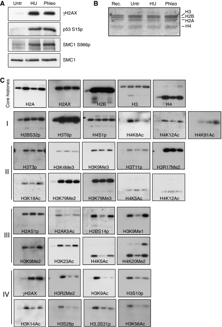
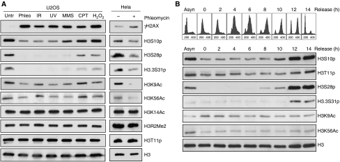
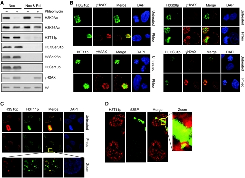
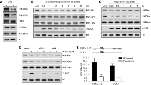
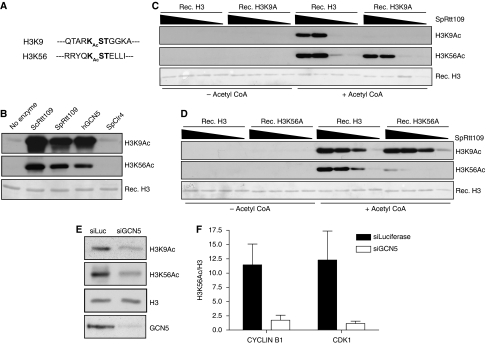
Recognition and repair of damaged DNA occurs within the context of chromatin. The key protein components of chromatin are histones, whose post-translational modifications control diverse chromatin functions. Here, we report our findings from a large-scale screen for DNA-damage-responsive histone modifications in human cells. We have identified specific phosphorylations and acetylations on histone H3 that decrease in response to DNA damage. Significantly, we find that DNA-damage-induced changes in H3S10p, H3S28p and H3.3S31p are a consequence of cell-cycle re-positioning rather than DNA damage per se. In contrast, H3K9Ac and H3K56Ac, a mark previously uncharacterized in human cells, are rapidly and reversibly reduced in response to DNA damage. Finally, we show that the histone acetyl-transferase GCN5/KAT2A acetylates H3K56 in vitro and in vivo. Collectively, our data indicate that though most histone modifications do not change appreciably after genotoxic stress, H3K9Ac and H3K56Ac are reduced in response to DNA damage in human cells.
Overview
The claims and statistics of this paper haven’t been checked yet.
Run the check — free, no account needed. It reads the open-access text and checks the sources it cites.
Integrity signals
Registry
Cites
Statistics
Figures
Verification
Claims the paper makes
The claims and statistics of this paper haven’t been checked yet.
Run the check — free, no account needed. It reads the open-access text and checks the sources it cites.
Claims other papers make about this one
Each is checked against this paper’s own text: does it say what the citing paper says it does?
In the citing paper
“we recently found that H3K9 and H3K56 acetylations (H3K9Ac and H3K56Ac) are negatively regulated by DNA damage20.”
From Human HDAC1 and HDAC2 function in the DNA-damage response to promote DNA nonhomologous end-joining (2010) · cites this paper as [20]
In the citing paper
“the observed decrease in H3K56Ac levels were not due to effects of DNA damage on cell-cycle distributions, as this histone mark is not cell cycle regulated20,35”
From Human HDAC1 and HDAC2 function in the DNA-damage response to promote DNA nonhomologous end-joining (2010) · cites this paper as [20]
Self-check
Percentages, sums and reported differences re-computed from the paper’s own numbers.
The self-check needs the paper’s full text.
Citations
Cites
Chromatin modifications and their function
Kouzarides, T. (2007) · Cell · doi:10.1016/j.cell.2007.02.005 · Cited by 10,883
DNA double-stranded breaks induce histone H2AX phosphorylation on serine 139
Rogakou, E. P., Pilch, D. R., Orr, A. H., Ivanova, V. S., & Bonner, W. M. (1998) · Journal of Biological Chemistry · doi:10.1074/jbc.273.10.5858 · Cited by 5,438
ATM and ATR Substrate Analysis Reveals Extensive Protein Networks Responsive to DNA Damage
Matsuoka, S., Ballif, B. A., Smogorzewska, A., McDonald, E. R., Hurov, K. E., Luo, J., Bakalarski, C. E., Zhao, Z., Solimini, N., Lerenthal, Y., et al. (2007) · Science · doi:10.1126/science.1140321 · Cited by 3,075
Checkpoints: Controls That Ensure the Order of Cell Cycle Events
Hartwell, L. H., & Weinert, T. A. (1989) · Science · doi:10.1126/science.2683079 · Cited by 3,053
The complex language of chromatin regulation during transcription
Berger, S. L. (2007) · Nature · doi:10.1038/nature05915 · Cited by 2,768
Combinatorial patterns of histone acetylations and methylations in the human genome
Wang, Z., Zang, C., Rosenfeld, J. A., Schones, D. E., Barski, A., Cuddapah, S., Cui, K., Roh, T.-Y., Peng, W., Zhang, M. Q., et al. (2008) · Nature Genetics · doi:10.1038/ng.154 · Cited by 2,331
The DNA Damage Response: Ten Years After
Harper, J. W., & Elledge, S. J. (2007) · Molecular Cell · doi:10.1016/j.molcel.2007.11.015 · Cited by 1,705
Functions of Site-Specific Histone Acetylation and Deacetylation
Shahbazian, M. D., & Grunstein, M. (2007) · Annual Review of Biochemistry · doi:10.1146/annurev.biochem.76.052705.162114 · Cited by 1,635
Hickson, I., Zhao, Y., Richardson, C. J., Green, S. J., Martin, N. M. B., Orr, A. I., Reaper, P. M., Jackson, S. P., Curtin, N. J., & Smith, G. C. M. (2004) · Cancer Research · doi:10.1158/0008-5472.can-04-2727 · Cited by 1,220
Self-citationPartitioning and Plasticity of Repressive Histone Methylation States in Mammalian Chromatin
Peters, A. H. F. M., Kubicek, S., Mechtler, K., O’Sullivan, R. J., Derijck, A. A. H. A., Perez-Burgos, L., Kohlmaier, A., Opravil, S., Tachibana, M., Shinkai, Y., et al. (2003) · Molecular Cell · doi:10.1016/s1097-2765(03)00477-5 · Cited by 1,208
Cited by
Sorted by citations
Items 301–351 of 351
hMOF and associated proteins in transcription and cancer
Classen, S. (2013) · Arrow@dit (Dublin Institute of Technology) · doi:10.13025/16867
da Rosa, J. L. (2012) · The Journal of the American Medical Association (JAMA) Network (American Medical Association) · doi:10.13028/w35r-7869
Histone deacetylase inhibition-mediated cytotoxicity of oligodendrocytes.
Dincman, T. A. (2011) · ThinkIR: The University of Louisville's Institutional Repository (University of Louisville) · doi:10.18297/etd/351
Regulation of Chromatin by FACT and NuA3
Chruscicki, A. (2014) · cIRcle (University of British Columbia) · doi:10.14288/1.0073776
The interaction between Rad9 and Tousled-like kinase 1 in the cell cycle and the DNA damage response
Kelly, R. M. (2013) · QSpace (Queen's University Library)
Proteomic Analysis of Core Histones and Their Variants in Tetrahymena Thermophila
Ashraf, K. (2015) · YorkSpace (York University)
Chromatin and Polycomb: Biology and bioinformatics
Kudrin, R., Mironov, A. A., & Stavrovskaya, E. D. (2017) · Molecular Biology · doi:10.1134/s0026893316060121
Meng, F., Qian, M., Peng, B., Wang, X., Peng, L., Zheng, K., Liu, Z., Tang, X., Zhang, S., Sun, S., et al. (2020) · bioRxiv (Cold Spring Harbor Laboratory) · doi:10.1101/2020.02.01.930172
Histone Variant H2A.Z's Effects on Nicotine Locomotor Sensitization and Reward
Steininger, C. F. D. (2019) · TSpace (University of Toronto)
Chlamydia trachomatis. Co-factor or factor in cancer of the cervix?
Núñez-Troconis, J. (2021) · Investigación Clínica · doi:10.22209/ic.v62n3a06
Dissecting the DNA damage-induced transcription stress response
Geijer, M. E. (2021) · RePub (Erasmus University Rotterdam)
Role of Histone Metabolism and Chromatin Structure in DNA Repair
Kari, V. (2014) · eDiss (Georg-August-Universität Göttingen) · doi:10.53846/goediss-4552
Modulating histone acetylationwith inhibitors and activators
Selvi, B. R., Mohankrishna, D. V., & Kundu, T. K. (2012) · Cambridge University Press eBooks · doi:10.1017/cbo9780511777271.032
Polyphenolic Acetates as Potential Therapeutics and Adjuvant in Radiotherapy of Cancer
Venkateswaran, K., Verma, A., & Dwarakanath, B. S. R. (2022) · Handbook of Oxidative Stress in Cancer: Therapeutic Aspects · doi:10.1007/978-981-16-1247-3_38-1
Plant Monoterpenes Camphor, Eucalyptol, Thujone, and DNA Repair
Nikolić, B., Mitić-Ćulafić, D., Vuković‐Gačić, B., & Knežević‐Vukčević, J. (2017) · Springer eBooks · doi:10.1007/978-3-319-31143-2_106-1
Polyphenolic Acetates as Potential Therapeutics and Adjuvant in Radiotherapy of Cancer
Venkateswaran, K., Verma, A., & Dwarakanath, B. S. R. (2022) · Handbook of Oxidative Stress in Cancer: Therapeutic Aspects · doi:10.1007/978-981-16-5422-0_38
The fork restart factor PHF6 interacts with RRM2 and binds to H3K56ac marked nascent DNAPreprint
Depestel, L., Bekaert, S.-L., Sanders, E., Damme, C. V., Eggermont, A., Loontiens, S., Tilleman, L., Nieuwerburgh, F. V., Delhaye, L., Vlierberghe, P. V., et al. (2023) · bioRxiv (Cold Spring Harbor Laboratory) · doi:10.1101/2023.03.08.531704
Mechanism of Histone Arginine Methylation Dynamic Change on Cellular StressPreprint
Ren, X., Li, W., Li, W., Yang, S., Yang, Z., Tong, Y., Chen, L., Liu, M., Zhang, B., & Yu, W. (2023) · SSRN Electronic Journal · doi:10.2139/ssrn.4619306
Yuan, S., Huang, T., Bao, Z., Wang, S.-Y., Wu, X., Liu, J., Liu, H., & Chen, Z. (2022) · Figshare · doi:10.6084/m9.figshare.21009549
Yuan, S., Huang, T., Bao, Z., Wang, S.-Y., Wu, X., Liu, J., Liu, H., & Chen, Z. (2022) · Figshare · doi:10.6084/m9.figshare.21009552
CoREST Complex Stabilizes MYC Protein to Promote Cancer Cell Genome StabilityPreprint
Khan, A. A., Kim, S., Sidrat, T., Byun, J., Aptekmann, A. A., Dalerba, P. D., Zakrzewski, J. L., Shin, J., & Ryu, B. (2024) · bioRxiv (Cold Spring Harbor Laboratory) · doi:10.1101/2024.12.23.630174
Pan, B., Liu, C., Su, J., & Xia, C. (2024) · Figshare · doi:10.6084/m9.figshare.25963130
Pan, B., Liu, C., Su, J., & Xia, C. (2024) · Figshare · doi:10.6084/m9.figshare.26728960
Kovaříková, A. S., Stixová, L., Kovařík, A., & Bártová, E. (2024) · Figshare · doi:10.6084/m9.figshare.26597312
Kovaříková, A. S., Stixová, L., Kovařík, A., & Bártová, E. (2024) · Figshare · doi:10.6084/m9.figshare.26597309
Pan, B., Liu, C., Su, J., & Xia, C. (2024) · Figshare · doi:10.6084/m9.figshare.25963256
Yuan, S., Huang, T., Bao, Z., Wang, S., Wu, X., Liu, J., Liu, H., & Chen, Z.-J. (2022) · Figshare · doi:10.6084/m9.figshare.21009546
Pan, B., Liu, C., Su, J., & Xia, C. (2024) · Figshare · doi:10.6084/m9.figshare.26728966
Pan, B., Liu, C., Su, J., & Xia, C. (2024) · Figshare · doi:10.6084/m9.figshare.26728969
Yuan, S., Huang, T., Bao, Z., Wang, S., Wu, X., Liu, J., Liu, H., & Chen, Z.-J. (2022) · Figshare · doi:10.6084/m9.figshare.21009555
Pan, B., Liu, C., Su, J., & Xia, C. (2024) · Figshare · doi:10.6084/m9.figshare.26728963
Role of the Asf1-Rad53 interaction in genomic stability in S.cerevisiae
Jiao, Y. (2011) · theses.fr (ABES) · doi:10.70675/6682a2b6z80c8z49e3z8704zd182d4f122f8
Development of new approaches to study the role of chromatin in dna damage response
Shoaib, M. (2011) · theses.fr (ABES) · doi:10.70675/e19260c9z46f4z4ce8za025z3d373bec66a5
Laboucarié, T. (2016) · theses.fr (ABES) · doi:10.70675/5c52847ez969az4bc3z9144zdedf14c308df
Stabilité du génome et rôle des INGs dans la réponse aux dommages de l'ADN
Mouche, A. (2017) · theses.fr (ABES) · doi:10.70675/2eac02ceze1a0z4c25z9effza47f3d916ad9
Double strand break repair within constitutive heterochromatin
Tsouroula, A. (2017) · theses.fr (ABES) · doi:10.70675/6345eaa6z96c3z47bdz8b53zab2527ff8785
Méthylation de H3K27 : régulation et rôle dans le cartilage articulaire
Allas, L. (2019) · theses.fr (ABES) · doi:10.70675/0b801552z0546z4765za411z263c0baf83ef
Etude des éléments cis-régulateurs dans les pathologies associées au gène CFTR
Collobert, M. (2021) · theses.fr (ABES) · doi:10.70675/88d0128fzfaa1z423czbbe8z64062873c0a2
Liu, L., Zhu, R., Yin, J., Zhang, F., Lu, G., Fang, L., Okyere, J., Opoku, M., Lv, X., Lin, F., et al. (2026) · FEBS Letters · doi:10.1002/1873-3468.70410
Newly synthesized histones: passive or active players in the regulation of epigenetic inheritance?
Martin, C. J., & Parthun, M. R. (2026) · Nucleic Acids Research · doi:10.1093/nar/gkag819
Li, H., Maben, C., Young, L., Pal, D., Davern, S., & McCord, R. P. (2026) · Frontiers in Cell and Developmental Biology · doi:10.3389/fcell.2026.1691057
Epigenetic alterations induced by ionizing radiation: pathways to cancer and prognostic strategies
Behera, A., Ghosh, K., Pandey, R., Sharma, A., Rath, G. K., Adhikary, A., & Sharma, A. K. (2026) · International Journal of Radiation Biology · doi:10.1080/09553002.2026.2708816
The structural analysis of histone H3 lysine 56 acetylation and related histone chaperone complexes
Hammond, C. M. (2013) · Discovery Research Portal (University of Dundee)
Understanding the Role of Lysine-Specific Demethylase 1 in Embryonic Gene Regulation
Foster, C. T. (2012) · OpenGrey (Institut de l'Information Scientifique et Technique)
Metadata
- Authors
- Tjeertes, J. V., Miller, K. M., & Jackson, S. P.
- Affiliations
- University of Cambridge (GB)
- Corresponding author
- Miller, Kyle M
- Jackson, Stephen P
- Venue
- The EMBO Journal
- Published
- 2009-04-30
- Volume
- 28
- Issue
- 13
- Pages
- 1878-1889
- ISSN
- 0261-4189 (ISSN-L)
- 1460-2075
- PMC
- PMC2684025
- OpenAlex
- W2046691617
- Cited by
- 347
- References
- 68
- Citations by year
- 2026: 7 · 2025: 10 · 2024: 21 · 2023: 15 · 2022: 16 · 2021: 18 · 2020: 14 · 2019: 17 · 2018: 13 · 2017: 21 · 2016: 21 · 2015: 22 · 2014: 20 · 2013: 31 · 2012: 31
- FWCI
- 13.54
- Type
- journal article
- Language
- en
- Open access
- hybrid
- doi.org
- Licence
- Locations
- The EMBO Journal (publishedVersion, cc-by-nc-nd)
- PubMed Central (publishedVersion, cc-by-nc-nd)
- PubMed (publishedVersion)
- Funders
- Wellcome Trust (086861/Z/08/Z, 086861)
- Cancer Research UK (A5290)
- Directorate for Biological Sciences
- Biotechnology and Biological Sciences Research Council
- Topics
- Life Sciences › Biochemistry, Genetics and Molecular Biology › Molecular Biology › Genomics and Chromatin Dynamics
- Life Sciences › Biochemistry, Genetics and Molecular Biology › Molecular Biology › DNA Repair Mechanisms
- Life Sciences › Biochemistry, Genetics and Molecular Biology › Molecular Biology › Epigenetics and DNA Methylation
- Keywords
- DNA damage response, histone modifications, H3K9 acetylation, H3K56 acetylation, histone acetylation, histone phosphorylation, histone acetyltransferase, chromatin, genotoxic stress
- MeSH terms
- p300-CBP-Associated Factor, Acetylation, Cell Cycle, DNA Damage, DNA Repair, HeLa Cells, Histones, Humans, Phosphorylation, Protein Processing, Post-Translational, Histone Code, Cell Line, Tumor, Histone Acetyltransferases
About these checks
Registry, retraction, Cites and statistics results are computed, not judged, with one exception: a printed reference that no index lists is read once by a model before it counts as a citation error. Claims are extracted by a model and kept only when their quote appears verbatim in the text; each cited source is then checked, with a model’s help, for whether it says what the claim relies on. Those verdicts can be wrong — read the claim’s quote and the cited source before relying on one.
Statistics and arithmetic are recomputed, not judged.
Confidence bands are shown only for fields calibrated on our annotated test set. None is calibrated yet, so no band is shown.
What counts as a claim ›How we measure confidence ›How we check this ›
Are you an author, or do you see a problem with this page? Request a review
