

The DOI matches its Crossref record; 33 of the 34 references it cites were found in a registry or an index.
Characterization of the receptor-binding domain (RBD) of 2019 novel coronavirus: implication for development of RBD protein as a viral attachment inhibitor and vaccine
Open accessAbstract
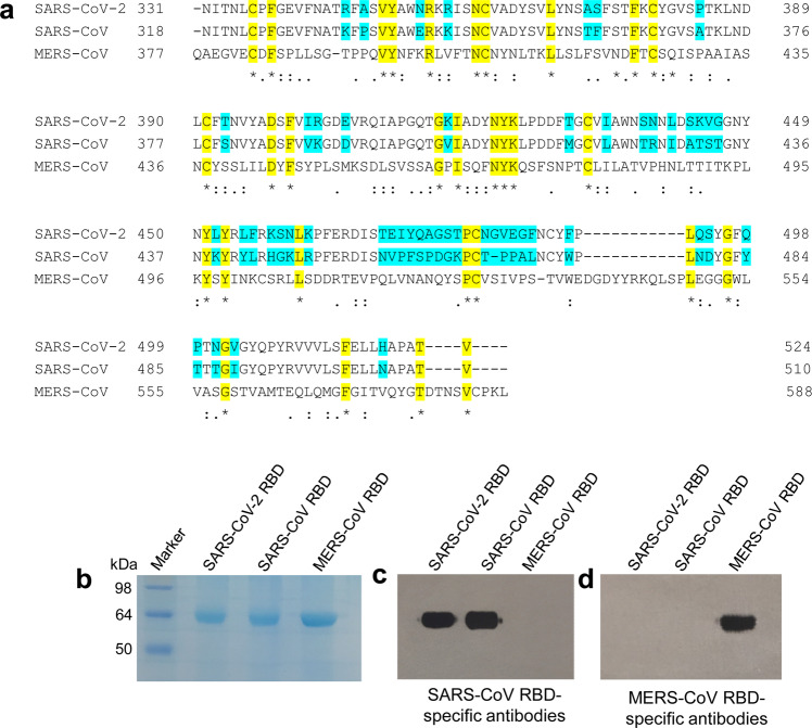
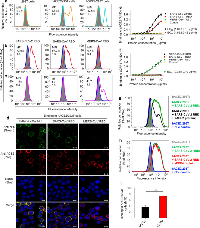
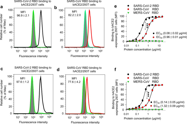
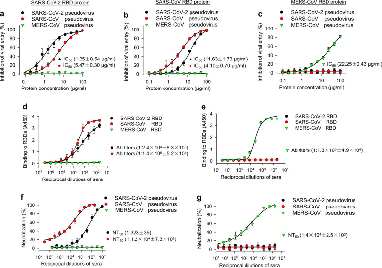
The outbreak of Coronavirus Disease 2019 (COVID-19) has posed a serious threat to global public health, calling for the development of safe and effective prophylactics and therapeutics against infection of its causative agent, severe acute respiratory syndrome coronavirus 2 (SARS-CoV-2), also known as 2019 novel coronavirus (2019-nCoV). The CoV spike (S) protein plays the most important roles in viral attachment, fusion and entry, and serves as a target for development of antibodies, entry inhibitors and vaccines. Here, we identified the receptor-binding domain (RBD) in SARS-CoV-2 S protein and found that the RBD protein bound strongly to human and bat angiotensin-converting enzyme 2 (ACE2) receptors. SARS-CoV-2 RBD exhibited significantly higher binding affinity to ACE2 receptor than SARS-CoV RBD and could block the binding and, hence, attachment of SARS-CoV-2 RBD and SARS-CoV RBD to ACE2-expressing cells, thus inhibiting their infection to host cells. SARS-CoV RBD-specific antibodies could cross-react with SARS-CoV-2 RBD protein, and SARS-CoV RBD-induced antisera could cross-neutralize SARS-CoV-2, suggesting the potential to develop SARS-CoV RBD-based vaccines for prevention of SARS-CoV-2 and SARS-CoV infection.
Overview
The claims and statistics of this paper haven’t been checked yet.
Run the check — free, no account needed. It reads the open-access text and checks the sources it cites.
Integrity signals
Registry
Cites
Statistics
Figures
Verification
Claims the paper makes
The claims and statistics of this paper haven’t been checked yet.
Run the check — free, no account needed. It reads the open-access text and checks the sources it cites.
Claims other papers make about this one
Each is checked against this paper’s own text: does it say what the citing paper says it does?
The source backs part of the claim, not all of it — or backs it for a different population, condition or comparison.
In the citing paper
“Some recent studies also demonstrated that immunization with the recombinant RBD of SARS-CoV-2 induced high titers of neutralizing antibodies in the absence of antibody-dependent enhancement (ADE) of infection (Quinlan et al., 2020; Tai et al., 2020).”
From A thermostable mRNA vaccine against COVID-19 (2020) · cites this paper as Tai et al., 2020
Experimental — accuracy not yet measured
Self-check
Percentages, sums and reported differences re-computed from the paper’s own numbers.
The self-check needs the paper’s full text.
Citations
Cites
A novel coronavirus from patients with pneumonia in China, 2019
Zhu, N., Zhang, D., Wang, W., Li, X., Yang, B., Song, J., Zhao, X., Huang, B., Shi, W., Lu, R., et al. (2020) · New England Journal of Medicine · doi:10.1056/nejmoa2001017 · Cited by 28,244
A pneumonia outbreak associated with a new coronavirus of probable bat origin
Zhou, P., Yang, X.-L., Wang, X.-G., Hu, B., Zhang, L., Zhang, W., Si, H.-R., Zhu, Y., Li, B., Huang, C.-L., et al. (2020) · Nature · doi:10.1038/s41586-020-2012-7 · Cited by 21,969 · cited 3×
“Since SARS-CoV-2 is more phylogenetically related to SARS-CoV than MERS-CoV, 22 we further detected the cross-reactivity of SARS-CoV RBD-specific antibodies with SARS-CoV-2 RBD and crossneutralizing activity of SARS-CoV RBD-specific antibodies against pseudotyped SARS-CoV-2.”
BackgroundResultInfluentialGorbalenya, A. E., Baker, S. C., Baric, R. S., de Groot, R. J., Drosten, C., Gulyaeva, A. A., Haagmans, B. L., Lauber, C., Leontovich, A. M., Neuman, B. W., et al. (2020) · Nature Microbiology · doi:10.1038/s41564-020-0695-z · Cited by 8,745
Angiotensin-converting enzyme 2 is a functional receptor for the SARS coronavirus
Li, W., Moore, M. J., Vasilieva, N., Sui, J., Wong, S. K., Berne, M. A., Somasundaran, M., Sullivan, J. L., Luzuriaga, K., Greenough, T. C., et al. (2003) · Nature · doi:10.1038/nature02145 · Cited by 5,923
Isolation of a Novel Coronavirus from a Man with Pneumonia in Saudi Arabia
Zaki, A. M., van Boheemen, S., Bestebroer, T. M., Osterhaus, A. D. M. E., & Fouchier, R. A. M. (2012) · New England Journal of Medicine · doi:10.1056/nejmoa1211721 · Cited by 5,677
Chou, T.-C. (2006) · Pharmacological Reviews · doi:10.1124/pr.58.3.10 · Cited by 5,163
Bats Are Natural Reservoirs of SARS-Like Coronaviruses
Li, W., Shi, Z., Yu, M., Ren, W., Smith, C. S., Epstein, J. H., Wang, H., Crameri, G. S., Hú, Z., Zhang, H., et al. (2005) · Science · doi:10.1126/science.1118391 · Cited by 2,571
Dipeptidyl peptidase 4 is a functional receptor for the emerging human coronavirus-EMC
Raj, V. S., Mou, H., Smits, S. L., Dekkers, D. H. W., Müller, M. A., Dijkman, R., Muth, D., Demmers, J. A. A., Zaki, A., Fouchier, R. A. M., et al. (2013) · Nature · doi:10.1038/nature12005 · Cited by 2,061
Structure of SARS Coronavirus Spike Receptor-Binding Domain Complexed with Receptor
Li, F., Li, W., Farzan, M., & Harrison, S. C. (2005) · Science · doi:10.1126/science.1116480 · Cited by 2,035
The spike protein of SARS-CoV — a target for vaccine and therapeutic development
Du, L., He, Y., Zhou, Y., Liu, S., Zheng, B.-J., & Jiang, S. (2009) · Nature Reviews Microbiology · doi:10.1038/nrmicro2090 · Cited by 1,678
Cited by
Sorted by citations
Items 301–400 of 1,001
Silver-Based Nanomaterials as Therapeutic Agents Against Coronaviruses: A Review
Das, C., Paul, S. S., Saha, A., Singh, T. P., Saha, A., Im, J., & Biswas, G. K. (2020) · International Journal of Nanomedicine · doi:10.2147/ijn.s280976 · Cited by 55
Bats, pangolins, minks and other animals - villains or victims of SARS-CoV-2?
do Vale, B., Lopes, A. P., Fontes, M. D. C., Silvestre, A. M., Cardoso, L., & Coelho, A. C. (2021) · Veterinary Research Communications · doi:10.1007/s11259-021-09787-2 · Cited by 55
1,2,3,4,6-Pentagalloyl Glucose, a RBD-ACE2 Binding Inhibitor to Prevent SARS-CoV-2 Infection
Chen, R. H., Yang, L., Hamdoun, S., Chung, S. K., Lam, C. W.-K., Zhang, K. X., Guo, X., Xia, C., Law, B. Y. K., & Wong, V. K. W. (2021) · Frontiers in Pharmacology · doi:10.3389/fphar.2021.634176 · Cited by 51
“A recent study characterized the cross-neutralizing activity of SARS-CoV2, SARS-CoV and MERS-RBD protein in hACE2/293 cells and the results showed that SARS-RBD exhibits competitive inhibition with SARS-CoV-2-RBD, but not with MERS-RBD, indicating that SARS-CoV-2 and SARS-CoV have similar infection mechanism (Tai et a…”
ResultEfficient production of recombinant SARS-CoV-2 spike protein using the baculovirus-silkworm system
Fujita, R., Hino, M., Ebihara, T., Nagasato, T., Masuda, A., Lee, J. M., Fujii, T., Mon, H., Kakino, K., Nagai, R., et al. (2020) · Biochemical and Biophysical Research Communications · doi:10.1016/j.bbrc.2020.06.020 · Cited by 51
A contemporary review on pathogenesis and immunity of COVID-19 infection
Mirzaei, R., Karampoor, S., Sholeh, M., Moradi, P., Ranjbar, R., & Ghasemi, F. (2020) · Molecular Biology Reports · doi:10.1007/s11033-020-05621-1 · Cited by 57
“SARS-CoV-2 binds to special cellular receptors via the S protein, that makes deformation of the S proteins and then intercedes the fusion among the viral and cell membranes, leading to discharging the nucleocapsid into the host cell [16, 17].”
BackgroundPathogenesis‐directed therapy of 2019 novel coronavirus disease
Stratton, C. W., Tang, Y., & Lu, H. (2020) · Journal of Medical Virology · doi:10.1002/jmv.26610 · Cited by 59
Development of Diagnostic Tests for Detection of SARS-CoV-2
Nguyen, N. N. T., McCarthy, C., Lantigua, D., & Camci‐Unal, G. (2020) · Diagnostics · doi:10.3390/diagnostics10110905 · Cited by 56
“The S proteins bind to a host receptor through the receptor-binding domain (RBD) in the S1 subunit and fuse the viral and host membranes through the S2 subunit [31].”
BackgroundMor, M., Werbner, M., Alter, J., Safra, M., Chomsky, E., Lee, J. C., Hada-Neeman, S., Polonsky, K., Nowell, C. J., Clark, A. E., et al. (2021) · PLoS Pathogens · doi:10.1371/journal.ppat.1009165 · Cited by 52
“Within the Spike protein, the receptor binding domain (RBD) is the minimal subunit that mediates viral entry [4,5] and was reported to be the main target for neutralizing antibodies elicited during infection [7,17–24].”
BackgroundKonda, M., Dodda, B., Konala, V. M., Naramala, S., & Adapa, S. (2020) · Cureus · doi:10.7759/cureus.8932 · Cited by 52
“The S2 polypeptide of S protein that makes up fusion peptide fuses viral and host membranes [25-27].”
BackgroundPatel, A. D., Rajendran, M., Shah, A. P., Patel, H., Pakala, S. B., & Karyala, P. (2021) · Journal of Biomolecular Structure and Dynamics · doi:10.1080/07391102.2020.1868338 · Cited by 53
“The X-ray crystal structure of spike surface glycoprotein of SARS-CoV (PDB entry: 6CRV, resolution ¼ 3.2 Å) and co-crystallized structure of SARS-CoV-2 RBD with human ACE2 Protein (PDB entry: 6M0J, resolution ¼ 2.45Å) was retrieved from Protein Data Bank (https:/www.rscb.org/pdb).”
BackgroundInfluentialLiu, M., Zheng, B. J., Zhang, Y., & Li, J. (2020) · Chronic Diseases and Translational Medicine · doi:10.1016/j.cdtm.2020.05.003 · Cited by 53
“ACE2 was recognized as a target gene for hepatocyte nuclear factor 1b (HNF-1b), as binding sites for HNF-1b were identified in the promoter regions of ACE2.”
InfluentialQuiles, J. L., Rivas‐García, L., Varela‐López, A., Llopis, J., Battino, M. A., & Sánchez‐González, C. (2020) · Environmental Research · doi:10.1016/j.envres.2020.110053 · Cited by 53
Khan, N., Chen, X., & Geiger, J. D. (2020) · Frontiers in Pharmacology · doi:10.3389/fphar.2020.595888 · Cited by 51
“SARS-CoV-2 spike is a trimer with three receptor-binding domains (RBDs) of S1 heads on top of a trimeric S2 stalk (Gui et al., 2017; Shang et al., 2020a; Walls et al., 2020).”
BackgroundCOVID-19 patients may become predisposed to pulmonary arterial hypertension
Suzuki, Y., Nikolaienko, S. I., Shults, N. V., & Gychka, S. G. (2021) · Medical Hypotheses · doi:10.1016/j.mehy.2021.110483 · Cited by 52
“SARS-CoV-2 uses angiotensin converting enzyme 2 (ACE2) as a receptor to enter the host cells [3,4], and some infected individuals develop severe pneumonia and acute respiratory distress syndrome (ARDS) [5].”
BackgroundNeutrobodyPlex—monitoring SARS-CoV-2 neutralizing immune responses using nanobodies
Wagner, T. R., Kaiser, P. D., Gramlich, M., Ostertag, E., Ruétalo, N., Junker, D., Häring, J., Traenkle, B., Becker, M., Dulovic, A., et al. (2021) · EMBO Reports · doi:10.15252/embr.202052325 · Cited by 50
Molecular biology of the SARs‐CoV‐2 spike protein: A review of current knowledge
Zhu, C., He, G., Yin, Q., Zeng, L., Ye, X., Shi, Y., & Xu, W. (2021) · Journal of Medical Virology · doi:10.1002/jmv.27132 · Cited by 61
“S1 S1 Mediate receptor recognition and viral attachment to initiate host cell entry Walls et al.27 NTD Contribute to determine host range Sironi et al.28 RBD Recognize and strongly bind to hACE2 receptors Tai et al.29 and Lan et al.30 RBM Bind and interact with hACE2 Lan et al.30 S2 S2 Mediate host‐virus membrane fusi…”
BackgroundKar, P., Kumar, V., Vellingiri, B., Sen, A., Jaishee, N., Anandraj, A., Malhotra, H., Bhattacharyya, S., Mukhopadhyay, S., Kinoshita, M., et al. (2020) · Journal of Biomolecular Structure and Dynamics · doi:10.1080/07391102.2020.1860133 · Cited by 57
“…proteins encoded by the viral genome including the spike (S) glycoprotein, the main protease enzyme (Mpro) and the RNA-dependent RNA polymerase (RdRp) have attracted interest as promising druggable and vaccine targets given their pivotal roles in viral replication and infection (Tai et al., 2020).”
BackgroundSARS-CoV-2 and endothelial cell interaction in COVID-19: molecular perspectives
Giordo, R., Paliogiannis, P., Mangoni, A. A., & Pintus, G. (2021) · Vascular Biology · doi:10.1530/vb-20-0017 · Cited by 54
Immunogenetic Predictors of Severe COVID-19
Malkova, A. M., Kudlay, D. A., Kudryavtsev, I. V., Starshinova, A. A., Yаblonskiy, P. K., & Shoenfeld, Y. (2021) · Vaccines · doi:10.3390/vaccines9030211 · Cited by 53
“In particular, of the 33 amino acids in this region (amino acids 460-492) in the SARS-CoV S protein, which contains critical residues and binds to ACE-2 52, less than half of them (15/33) are conservative in SARS-CoV-2 [52].”
BackgroundTetz, G. V., & Tetz, V. (2022) · Microorganisms · doi:10.3390/microorganisms10020280 · Cited by 54
“Like other β-CoVs, the genome of the novel SARS-CoV-2 virus encodes structural proteins required for the efficient formation of infectious virions; these include the spike (S), envelope (E), membrane (M) and nucleocapsid (N) proteins [7].”
BackgroundRapid characterization of spike variants via mammalian cell surface display
Javanmardi, K., Chou, C., Terrace, C. I., Annapareddy, A., Kaoud, T. S., Guo, Q., Lutgens, J., Zorkic, H., Horton, A. P., Gardner, E. C., et al. (2021) · Molecular Cell · doi:10.1016/j.molcel.2021.11.024 · Cited by 54
Malaiyan, J., Suresh, A., Mohan, K., & Radhakrishnan, G. G. (2020) · Journal of Medical Virology · doi:10.1002/jmv.26261 · Cited by 54
“RBD of S protein in SARS‐CoV‐2 binds strongly to human, pangolin, and bat angiotensin‐converting enzyme 2 (ACE2) receptors.(19,33,34) Studies have confirmed that S protein in the SARS‐CoV‐2 uses the ACE2, found in the lower respiratory tract of humans,(1,9) and other certain species (pangolin, civet, swine, cow, buffa…”
BackgroundThe Physiological Mechanisms of the Sex-Based Difference in Outcomes of COVID19 Infection
Wray, S., & Arrowsmith, S. (2021) · Frontiers in Physiology · doi:10.3389/fphys.2021.627260 · Cited by 50
COVID-19: Molecular and Cellular Response
Alipoor, S. D., Mortaz, E., Jamaati, H., Tabarsi, P., Bayram, H., Varahram, M., & Adcock, I. M. (2021) · Frontiers in Cellular and Infection Microbiology · doi:10.3389/fcimb.2021.563085 · Cited by 53
Genetic emergence of B.1.617.2 in COVID-19
Kirola, L. (2021) · New Microbes and New Infections · doi:10.1016/j.nmni.2021.100929 · Cited by 51
“Many studies have exhibited an amino acid change inside 438–506 position in the Spike protein (also called the receptorbinding domain) could be considerably associated with an increase in virus transmission, infection, or evade immunity [12].”
Hu, S., Wang, J., Zhang, Y., Bai, H., Wang, C., Wang, N., & He, L. (2021) · Journal of Medical Virology · doi:10.1002/jmv.26874 · Cited by 51
“As with SARS‐CoV, the receptor binding domain (RBD) of the 2019‐nCoV S protein binds to the angiotensin‐ converting enzyme 2 (ACE2) cell receptor.7–9 However, the RBD of 2019‐nCoV has a higher binding affinity for ACE2 than that of SARS‐CoV.6 RBDs of S proteins of SARS‐CoV or 2019‐nCoV can both be used as vaccines to…”
MethodInfluentialPranata, R., Huang, I., & Raharjo, S. B. (2020) · Indian Pacing and Electrophysiology Journal · doi:10.1016/j.ipej.2020.08.001 · Cited by 50
“After activation of the viral surface spike (S) protein of SARS-CoV-2 by the transmembrane protease serine 2 (TMPRSS2), the virus binds to angiotensin-converting enzyme 2 (ACE2) [22,23], which has high affinity to the human receptor [24,25].”
Zha, L., Chang, X., Zhao, H., Mohsen, M. O., Hong, L., Zhou, Y., Chen, H., Liu, X., Zhang, J., Li, D., et al. (2021) · Vaccines · doi:10.3390/vaccines9040395 · Cited by 49
“of neutralizing antibodies, blocking the interaction of the virus with its receptor [27].”
BackgroundPrediction of Drug-Induced Liver Toxicity Using SVM and Optimal Descriptor Sets
Jaganathan, K., Tayara, H., & Chong, K. T. (2021) · International Journal of Molecular Sciences · doi:10.3390/ijms22158073 · Cited by 55
Paper-based sensors for rapid important biomarkers detection
Kuswandi, B., Hidayat, M. A., & Noviana, E. (2022) · Biosensors and Bioelectronics X · doi:10.1016/j.biosx.2022.100246 · Cited by 50
Barh, D., Tiwari, S., Rodrigues Gomes, L. G., Ramalho Pinto, C. H., Andrade, B. S., Ahmad, S., Aljabali, A. A. A., Alzahrani, K. J., Banjer, H. J., Hassan, S. S., et al. (2023) · Inflammation · doi:10.1007/s10753-022-01734-w · Cited by 50
Mbatha, L. S., Akinyelu, J., Chukwuma, C. I., Mokoena, M. P., & Kudanga, T. (2023) · Viruses · doi:10.3390/v15030741 · Cited by 54
Chourasia, R., Padhi, S., Phukon, L. C., Abedin, M. M., Singh, S. P., & Rai, A. K. (2020) · Frontiers in Molecular Biosciences · doi:10.3389/fmolb.2020.601753 · Cited by 49
“RBM is supported by two alpha helices, two beta sheets, and connecting loops (Tai et al., 2020).”
BackgroundPossible host-adaptation of SARS-CoV-2 due to improved ACE2 receptor binding in mink
Welkers, M. R. A., Han, A. X., Reusken, C. B. E. M., & Eggink, D. (2020) · Virus Evolution · doi:10.1093/ve/veaa094 · Cited by 47
“Antibodies targeting the RBD of the spike protein offer protection by inhibiting the attachment of SARS-CoV-2 to target cells (Tai et al. 2020).”
AI-guided discovery of the invariant host response to viral pandemics
Sahoo, D., Katkar, G. D., Khandelwal, S., Behroozikhah, M., Claire, A., Castillo, V., Tindle, C., Fuller, M., Taheri, S., Rogers, T. F., et al. (2021) · EBioMedicine · doi:10.1016/j.ebiom.2021.103390 · Cited by 51
“Three relatively widely accepted facts shaped our approach using Angiotensin-converting enzyme 2 (ACE2) as ‘seed’ gene in our computational studies: (i) ACE2 is the most well-known portal for SARS-CoV-2 entry into the host cell [27,28]; its expression in cell lines correlates with the expression of innate immune genes…”
Vashi, Y., Jagrit, V., & Kumar, S. (2020) · Infection Genetics and Evolution · doi:10.1016/j.meegid.2020.104382 · Cited by 48
“However, some suggest that SARS-CoV-2 binds ACE2 with higher affinity than SARS-CoV (Tai et al., 2020; Wang et al., 2020; Wrapp et al., 2020).”
Luo, Z., Ang, M. J. Y., Chan, S. Y., Yi, Z., Goh, Y. Y., Yan, S., Tao, J., Liu, K., Li, X., Zhang, H., et al. (2020) · Research · doi:10.34133/2020/6925296 · Cited by 51
Kwofie, S. K., Broni, E., Asiedu, S. O., Kwarko, G. B., Dankwa, B., Enninful, K. S., Tiburu, E. K., & Wilson, M. D. (2021) · Molecules · doi:10.3390/molecules26020406 · Cited by 51
“The inhibition of SARS-CoV-2 spike proteins is a critical antiviral strategy, since it serves as the major barrier to block the infection [15,77].”
BackgroundInfluentialImpact of Expressing Cells on Glycosylation and Glycan of the SARS-CoV-2 Spike Glycoprotein
Wang, Y., Wu, Z., Hu, W., Hao, P., & Yang, S. (2021) · ACS Omega · doi:10.1021/acsomega.1c01785 · Cited by 50
“58 Given that SARS-CoV-2 can be infected and transmitted through many media (lungs, oral, eyes, intestine, etc.), consideration should be given to selecting suitable host cell lines for diagnostic applications and the development of inhibitors, antibodies, or vaccines.”
Sari, A. K., Hartati, Y. W., Gaffar, S., Anshori, I., Hidayat, D., & Wiraswati, H. L. (2022) · Journal of Electrochemical Science and Engineering · doi:10.5599/jese.1206 · Cited by 49
Innate Immune Cells and Hypertension: Neutrophils and Neutrophil Extracellular Traps (NETs)
McCarthy, C. G., Saha, P., Golonka, R. M., Wenceslau, C. F., Joe, B., & Vijay–Kumar, M. (2021) · Comprehensive physiology · doi:10.1002/cphy.c200020 · Cited by 48
Naidoo, D., Roy, A., Kar, P., Mutanda, T., & Anandraj, A. (2020) · Journal of Biomolecular Structure and Dynamics · doi:10.1080/07391102.2020.1794972 · Cited by 52
“…spike (S) protein is an important structural protein that facilitates viral attachment, fusion and subsequent entry into the human host via its interaction with the human (host) cellular receptor angiotensin converting enzyme II (ACE 2) (Abdelli et al., 2020; Sinha et al., 2020; Tai et al., 2020).”
BackgroundGalimberti, S., Petrini, M., Baratè, C., Ricci, F., Balducci, S., Grassi, S., Guerrini, F., Ciabatti, E., Mechelli, S., Paolo, A. D., et al. (2020) · Frontiers in Oncology · doi:10.3389/fonc.2020.01428 · Cited by 48
Goh, Y. S., Chavatte, J., Jieling, A. L., Lee, B. T. K., Hor, P. X., Amrun, S. N., Lee, C. Y., Chee, R. S., Wang, B., Lee, C. Y., et al. (2020) · Cell Reports Medicine · doi:10.1016/j.xcrm.2021.100193 · Cited by 48
Ameratunga, R., Longhurst, H. J., Steele, R. H., Lehnert, K., Leung, E. Y., Brooks, A. E. S., & Woon, S. (2021) · The Journal of Allergy and Clinical Immunology In Practice · doi:10.1016/j.jaip.2021.06.019 · Cited by 49
Morsy, M., Alakeel, K. A., Ahmed, A. E., Abbas, A. M., Omara, A. I., Abdelsalam, N. R., & Emaish, H. H. (2022) · Saudi Journal of Biological Sciences · doi:10.1016/j.sjbs.2022.02.046 · Cited by 48
“mer adobe brick, and these results agree with (Tai et al., 2020; Hany et al., 2021).”
ResultPharmacotherapy in COVID-19 patients: a review of ACE2-raising drugs and their clinical safety
Akhtar, S., Benter, I. F., Danjuma, M. I.-M., Doi, S. A. R., Hasan, S. S., & Habib, A. M. (2020) · Journal of drug targeting · doi:10.1080/1061186x.2020.1797754 · Cited by 49
Jörrißen, P., Schütz, P., Weiand, M., Vollenberg, R., Schrempf, I. M., Ochs, K., Frömmel, C., Tepasse, P., Schmidt, H., & Zibert, A. (2021) · Frontiers in Immunology · doi:10.3389/fimmu.2021.679841 · Cited by 50
“The RBD domain of the S protein has been proposed to be of high importance for induction of immunity against COVID-19 (31).”
BackgroundRole for Anti-Cytokine Therapies in Severe Coronavirus Disease 2019
Buckley, L. F., Wohlford, G. F., Ting, C., Alahmed, A. A., Tassell, B. W. V., Abbate, A., Devlin, J. W., & Libby, P. (2020) · Critical Care Explorations · doi:10.1097/cce.0000000000000178 · Cited by 48
“The receptor-binding domain of the SARS-CoV-2 spike protein possesses greater affinity for ACE2 than that of SARS-CoV-1, the causative agent of the 2002–2003 SARS pandemic (9, 10).”
BackgroundDitte, K., Le, T. A. N., Ditzer, O., Bojórquez, D. I. S., Chae, S., Bachmann, M. P., Baraban, L., & Lissel, F. (2021) · ACS Biomaterials Science & Engineering · doi:10.1021/acsbiomaterials.1c00727 · Cited by 49
Long COVID, neuropsychiatric disorders, psychotropics, present and future
Tang, S. W., Leonard, B. E., & Helmeste, D. M. (2022) · Acta Neuropsychiatrica · doi:10.1017/neu.2022.6 · Cited by 45
Coronavirus Disease 2019 and the Cerebrovascular‐Cardiovascular Systems: What Do We Know So Far?
Larson, A. S., Savastano, L. E., Kadirvel, R., Kallmes, D. F., Hassan, A. E., & Brinjikji, W. (2020) · Journal of the American Heart Association · doi:10.1161/jaha.120.016793 · Cited by 46
“Coronavirus Disease 2019 and the Cerebrovascular-Cardiovascular Systems: What Do We Know So Far? Journal of the American Heart Association, 9(13), e016793.”
Llanes, A., Restrepo, C. M., Zuleima Caballero, E., Rajeev, S., Kennedy, M. A., & Lleonart, R. (2020) · International Journal of Molecular Sciences · doi:10.3390/ijms21124546 · Cited by 52
“Sarbecovirus comprises the severe acute respiratory syndrome (SARS)-related coronaviruses such as SARS-CoV and SARS-CoV-2 (2019-nCoV), respectively responsible for the © 2020 by the author(s).”
BackgroundInfluentialLi, H., Zhao, C., Zhang, Y., Yuan, F., Zhang, Q., Shi, X., Zhang, L., Qin, C., & Zheng, A. (2020) · Emerging Microbes & Infections · doi:10.1080/22221751.2020.1830715 · Cited by 44
“The receptor binding domain (RBD) of the spike protein is proved to inhibit the entry of SARS-CoV2 by binding to the ACE2 receptor on the plasma membrane [20].”
BackgroundDetection of Infectious Viruses Using CRISPR-Cas12-Based Assay
Talwar, C. S., Park, K., Ahn, W., Kim, Y.-S., Kwon, O. S., Yong, D., Kang, T., & Woo, E. (2021) · Biosensors · doi:10.3390/bios11090301 · Cited by 47
“surface and the receptor-binding domain (RBD) in the spike protein of the virus coded by the S gene [27].”
BackgroundMabrouk, M. T., Chiem, K., Rujas, E., Huang, W., Jahagirdar, D., Quinn, B., Nair, M. S., Nissly, R. H., Cavener, V. S., Boyle, N. R., et al. (2021) · Science Advances · doi:10.1126/sciadv.abj1476 · Cited by 47
Radbel, J. M., Jagpal, S. K., Roy, J., Brooks, A., Tischfield, J. A., Sheldon, M., Bixby, C. J., Witt, D., Gennaro, M. L., Horton, D. B., et al. (2020) · Journal of Molecular Diagnostics · doi:10.1016/j.jmoldx.2020.04.209 · Cited by 47
“Testing can focus on any or all of the four SARS-Cov-2 structural proteins, including the spike, envelope, membrane, and nucleocapsid proteins, or on any of their protein domains.(13) The fact that results for all three viral genes tested were strongly correlated across samples from multiple subjects supports the robu…”
Islam, M. A. (2023) · Infectious Medicine · doi:10.1016/j.imj.2023.08.005 · Cited by 46
“Nsp2 is an enzyme-like protease that splits the SARS-CoV-2 polyprotein into functional nsps [26] .”
BackgroundProtein subunit vaccines: Promising frontiers against COVID-19
Chavda, V. P., Ghali, E. N. H. K., Balar, P. C., Chauhan, S. C., Tiwari, N., Shukla, S., Athalye, M., Patravale, V. B., Apostolopoulos, V., & Yallapu, M. M. (2024) · Journal of Controlled Release · doi:10.1016/j.jconrel.2024.01.017 · Cited by 48
SARS‐CoV‐2 and the possible connection to ERs, ACE2, and RAGE: Focus on susceptibility factors
Stilhano, R. S., Costa, A. J., Nishino, M. S., Shams, S., Bartolomeo, C. S., Breithaupt‐Faloppa, A. C., Silva, E. A., Ramirez, A. L., Prado, C. M., & Ureshino, R. P. (2020) · The FASEB Journal · doi:10.1096/fj.202001394rr · Cited by 48
SARS-COV-2, infection, transmission, transcription, translation, proteins, and treatment: A review
Emrani, J., Ahmed, M., Jeffers-Francis, L. K., Teleha, J. C., Mowa, C. N., Newman, R. H., & Thomas, M. D. (2021) · International Journal of Biological Macromolecules · doi:10.1016/j.ijbiomac.2021.10.172 · Cited by 49
“Interestingly, the relatively small number of differences between the two S-proteins has a dramatic influence on parameters such as affinity between the S-protein and its receptor (ACE2) [170,171].”
Mechanisms contributing to adverse outcomes of COVID-19 in obesity
Sudhakar, M., Beaula, W. S., Meiyazhagan, G., & Parvathi, V. D. (2022) · Molecular and Cellular Biochemistry · doi:10.1007/s11010-022-04356-w · Cited by 50
Dampalla, C. S., Rathnayake, A. D., Kankanamalage, A. C. G., Kim, Y., Perera, K. D., Nguyen, H. N., Miller, M. J., Madden, T. K., Picard, H. R., Thurman, H. A., et al. (2022) · Journal of Medicinal Chemistry · doi:10.1021/acs.jmedchem.2c00224 · Cited by 47
Wang, X., Xia, S., Wang, Q., Xu, W., Li, W., Lu, L., & Jiang, S. (2020) · International Journal of Molecular Sciences · doi:10.3390/ijms21113843 · Cited by 46
“At the boundary of the S1/S2 subunits, a study found that SARS-CoV-2 S protein contains a furin cleavage site that is cleaved during biosynthesis, which is different from SARS-CoV and SARSr-CoVs [17] but is similar to MERS-CoV [33].”
BackgroundInfluentialSARS-CoV-2: An Update on Potential Antivirals in Light of SARS-CoV Antiviral Drug Discoveries
Elshabrawy, H. A. (2020) · Vaccines · doi:10.3390/vaccines8020335 · Cited by 46
Computational Design of 25-mer Peptide Binders of SARS-CoV‑2
Sitthiyotha, T., & Chunsrivirot, S. (2020) · The Journal of Physical Chemistry B · doi:10.1021/acs.jpcb.0c07890 · Cited by 41
Genomic analysis and comparative multiple sequences of SARS-CoV2
Chang, T.-J., Yang, D., Wang, M., Liang, K.-H., Tsai, P., Chiou, S.-H., Lin, T., & Wang, C.-T. (2020) · Journal of the Chinese Medical Association · doi:10.1097/jcma.0000000000000335 · Cited by 44
Phosphodiesterase Inhibitors: Could They Be Beneficial for the Treatment of COVID-19?
Giorgi, M., Cardarelli, S., Ragusa, F., Saliola, M., Biagioni, S., Poiana, G., Naro, F., & Massimi, M. (2020) · International Journal of Molecular Sciences · doi:10.3390/ijms21155338 · Cited by 43
“The mechanisms of the entry of the virus into the cell have already been reported in great details [2], while the immediate consequence of the infection is the activation of the innate immunity reaction, decisive for the outcome of the infection, with negligible or very severe effects [3].”
BackgroundWhole-genome sequencing of SARS-CoV-2 reveals the detection of G614 variant in Pakistan
Umair, M., Ikram, A., Salman, M., Khurshid, A., Alam, M., Badar, N., Suleman, R., Tahir, F., Sharif, S., Montgomery, J. M., et al. (2021) · PLoS ONE · doi:10.1371/journal.pone.0248371 · Cited by 42
“The receptor binding domain (RBD) in the S protein interacts with ACE2 receptor that eventually leads to the fusion of virus to host cell membranes [3].”
BackgroundPotential Genes Associated with COVID-19 and Comorbidity
Feng, S., Song, F.-Q., Guo, W., Tan, J., Zhang, X., Qiao, F., Guo, J., Zhang, L., & Jia, X. (2022) · International Journal of Medical Sciences · doi:10.7150/ijms.67815 · Cited by 44
“The RBD region of the S1 subunit can be used as a vital target for developing drugs and vaccines [10].”
BackgroundOverview of Targets and Potential Drugs of SARS-CoV-2 According to the Viral Replication
Zhang, Y., & Tang, L. V. (2020) · Journal of Proteome Research · doi:10.1021/acs.jproteome.0c00526 · Cited by 50
Probiotic-Based Vaccines May Provide Effective Protection against COVID-19 Acute Respiratory Disease
Taghinezhad‐S, S., Mohseni, A. H., Bermúdez‐Humarán, L. G., Casolaro, V., Cortes‐Perez, N. G., Keyvani, H., & Simal‐Gándara, J. (2021) · Vaccines · doi:10.3390/vaccines9050466 · Cited by 46
“The S protein is responsible for attachment to the host cell following recognition of the human angiotensinconverting enzyme 2 (hACE2) receptor, which plays a pivotal role in provoking the immune response during the progression of disease and is targeted by host neutralizing antibodies (Figure 1) [7,8].”
BackgroundInfluentialRice, A., Verma, M. S., Shin, A., Zakin, L., Sieling, P. A., Tanaka, S., Balint, J. P., Dinkins, K., Adisetiyo, H., Morimoto, B., et al. (2021) · Scientific Reports · doi:10.1038/s41598-021-94364-5 · Cited by 43
Ghareeb, D. A., Saleh, S. R., Seadawy, M. G., Nofal, M. S., Abdulmalek, S. A., Hassan, S. F., Khedr, S. M., Wahab, M. A., Sobhy, A. A., Abdelhamid, A. S. A., et al. (2021) · Journal of Pharmaceutical Investigation · doi:10.1007/s40005-021-00544-w · Cited by 44
HBD-2 binds SARS-CoV-2 RBD and blocks viral entry: Strategy to combat COVID-19
Zhang, L., Ghosh, S. K., Basavarajappa, S. C., Chen, Y., Shrestha, P., Penfield, J., Brewer, A., Ramakrishnan, P., Buck, M., & Weinberg, A. (2022) · iScience · doi:10.1016/j.isci.2022.103856 · Cited by 46
“He et al., 2020), using the same computational method, who calculated a free energy for the RBD:ACE2 interaction of 50.4 kcal/mol.”
InfluentialAn updated review of SARS‐CoV ‐2 detection methods in the context of a novel coronavirus pandemic
Zhang, Y., Huang, Z., Zhu, J., Li, C., Fang, Z., Chen, K., & Zhang, Y. (2022) · Bioengineering & Translational Medicine · doi:10.1002/btm2.10356 · Cited by 45
Role of Dipeptidyl Peptidase-4 (DPP4) on COVID-19 Physiopathology
Sebastián-Martín, A., Sánchez, B. G., Mora-Rodríguez, J. M., Bort, A., & Díaz‐Laviada, I. (2022) · Biomedicines · doi:10.3390/biomedicines10082026 · Cited by 48
“However, an experimental demonstration of the interaction between SARS-CoV-2 and DPP4 has not yet been reported, and in vitro studies could not demonstrate that SARS-CoV-2 RBD binds to DPP4-expressing cells [71].”
BackgroundSohag, A. A. M., Hannan, M. A., Rahman, S., Hossain, M., Hasan, M., Khan, M. K., Khatun, A., Dash, R., & Uddin, M. J. (2020) · Drug Development Research · doi:10.1002/ddr.21709 · Cited by 43
“Recently, Wan et al. (2020) and Tai et al. (2020) sequenced and characterized RBD of SARS-CoV-2, including its receptor-binding motif (RBM) that directly associates with ACE2.”
BackgroundDollman, N. L., Griffin, J. H., & Downard, K. M. (2020) · ACS Infectious Diseases · doi:10.1021/acsinfecdis.0c00664 · Cited by 42
Natural product remedies for COVID-19: A focus on safety
Omokhua, A. G., & Staden, J. V. (2021) · South African Journal of Botany · doi:10.1016/j.sajb.2021.03.012 · Cited by 43
Yayehrad, A. T., Siraj, E. A., Wondie, G. B., Alemie, A. A., Derseh, M. T., & Ambaye, A. S. (2021) · International Journal of Nanomedicine · doi:10.2147/ijn.s327334 · Cited by 43
Monoclonal antibody therapies in the management of SARS-CoV-2 infection
Rey, E. M., Choi, D., Kim, S., Yoon, S., & Săndulescu, O. (2022) · Expert Opinion on Investigational Drugs · doi:10.1080/13543784.2022.2030310 · Cited by 46
Majdič, G. (2020) · Frontiers in Cellular and Infection Microbiology · doi:10.3389/fcimb.2020.00327 · Cited by 42
Hyperthrombotic Milieu in COVID-19 Patients
Kamel, M. H., Yin, W., Zavaro, C., Francis, J. M., & Chitalia, V. C. (2020) · Cells · doi:10.3390/cells9112392 · Cited by 42
“accessory m olecules in this m ode [11,12].”
BackgroundJomhori, M., Mosaddeghi, H., & Farzin, H. (2021) · Computers in Biology and Medicine · doi:10.1016/j.compbiomed.2021.104692 · Cited by 42
Schrörs, B., Riesgo-Ferreiro, P., Sorn, P., Gudimella, R., Bukur, T., Rösler, T., Löwer, M., & Şahin, U. (2021) · PLoS ONE · doi:10.1371/journal.pone.0249254 · Cited by 41
Evolution of the SARS-CoV-2 genome and emergence of variants of concern
Safari, I., & Elahi, E. (2021) · Archives of Virology · doi:10.1007/s00705-021-05295-5 · Cited by 41
“The SARSCoV-2 spike protein has two functional subunits, S1 and S2, which play crucial roles, respectively, in the attachment of the virus to the host-cell receptor (angiotensinconverting enzyme 2; ACE2) through its receptor-binding domain (RBD) and mediation of fusion of viral and host membranes, leading to entry of…”
BackgroundNejat, R., Torshizi, M. F., & Najafi, D. J. (2023) · Vaccines · doi:10.3390/vaccines11020204 · Cited by 44
Mousavi, S. M., Kalashgrani, M. Y., Gholami, A., Omidifar, N., Binazadeh, M., & Chiang, W. (2023) · Biosensors · doi:10.3390/bios13080786 · Cited by 43
Intoxication With Endogenous Angiotensin II: A COVID-19 Hypothesis
Sfera, A., Osorio, C., Jafri, N., Diaz, E. L., & Maldonado, J. E. C. (2020) · Frontiers in Immunology · doi:10.3389/fimmu.2020.01472 · Cited by 42
Deciphering SARS-CoV-2 Virologic and Immunologic Features
Lebeau, G., Vagner, D., Frumence, É., Ah-Pine, F., Guillot, X., Nobécourt, E., Raffray, L., & Gasque, P. (2020) · International Journal of Molecular Sciences · doi:10.3390/ijms21165932 · Cited by 54
“Among the viral proteins that we presented, some are potential targets for novel drug development or drug repurposing, with already established lead compounds, notably due to computational methods [18,23,24].”
Behera, S. K., Mahapatra, N., Tripathy, C. S., & Pati, S. (2021) · The Indian Journal of Medical Research · doi:10.4103/ijmr.ijmr_1132_20 · Cited by 42
Merkuleva, I. A., Щербаков, Д. Н., Borgoyakova, M. B., Васильевич, Ш. Д., Рудометов, А. П., Карпенко, Л. И., Belenkaya, S. V., Исаева, А. А., Nesmeyanova, V. S., Kazachinskaia, E. I., et al. (2022) · Vaccines · doi:10.3390/vaccines10010096 · Cited by 41
Angiotensin-converting enzyme 2 (ACE2) is upregulated in Alzheimer’s disease brainPreprint
Ding, Q., Shults, N. V., Harris, B. T., & Suzuki, Y. (2020) · bioRxiv (Cold Spring Harbor Laboratory) · doi:10.1101/2020.10.08.331157 · Cited by 42
Analysis and comparison of genetic variants and mutations of the novel coronavirus SARS-CoV-2
Almubaid, Z., & Al-Mubaid, H. (2021) · Gene Reports · doi:10.1016/j.genrep.2021.101064 · Cited by 41
Xiao, S., Alshahrani, M., Gupta, G., Tao, P., & Verkhivker, G. M. (2023) · Journal of Chemical Information and Modeling · doi:10.1021/acs.jcim.3c00778 · Cited by 37
Diacerein: A potential multi-target therapeutic drug for COVID-19
de Oliveira, P. G., Termini, L., Durigon, E. L., Lepique, A. P., Sposito, A. C., & Boccardo, E. (2020) · Medical Hypotheses · doi:10.1016/j.mehy.2020.109920 · Cited by 41
Naeem, S. M., Mabrouk, M. S., Marzouk, S. Y., & Eldosoky, M. A. A. (2020) · Briefings in Bioinformatics · doi:10.1093/bib/bbaa170 · Cited by 41
“[1] studied the characteristics of the COVID-19 receptor-binding domain (RBD) by implication for the development of RBD protein as a viral attachment inhibitor and vaccine.”
BackgroundBinding of SARS‐CoV‐2 to Cell Receptors: A Tale of Molecular Evolution
Gómez, S. A., Rojas‐Valencia, N., Gómez, S., Egidi, F., Cappelli, C., & Restrepo, A. A. (2020) · ChemBioChem · doi:10.1002/cbic.202000618 · Cited by 40
Targeting the viral‐entry facilitators of SARS‐CoV‐2 as a therapeutic strategy in COVID‐19
Muralidar, S., Gopal, G., & Ambi, S. V. (2021) · Journal of Medical Virology · doi:10.1002/jmv.27019 · Cited by 42
Metadata
- Authors
- Tai, W., He, L., Zhang, X., Pu, J., Voronin, D., Jiang, S., Zhou, Y., & Du, L.
- Affiliations
- New York Blood Center (US)
- Beijing Institute of Microbiology and Epidemiology (CN)
- Shanghai Medical College of Fudan University (CN)
- Corresponding author
- Jiang, Shibo
- Zhou, Yusen
- Du, Lanying
- Venue
- Cellular & Molecular Immunology
- Published
- 2020-03-19
- Volume
- 17
- Issue
- 6
- Pages
- 613-620
- ISSN
- 1672-7681 (ISSN-L)
- 2042-0226
- PMC
- PMC7091888
- OpenAlex
- W3011537081
- Cited by
- 1,654
- References
- 34
- Citations by year
- 2026: 28 · 2025: 68 · 2024: 120 · 2023: 184 · 2022: 296 · 2021: 482 · 2020: 475
- FWCI
- 47.20
- Type
- journal article
- Language
- en
- Open access
- hybrid
- doi.org
- Locations
- Cellular and Molecular Immunology (publishedVersion, cc-by)
- PubMed Central (publishedVersion, cc-by)
- PubMed (publishedVersion)
- Informationszentrum Lebenswissenschaften (ZB MED) (submittedVersion, cc-by)
- Funders
- U.S. Department of Health & Human Services | National Institutes of Health (R01AI137472, R01AI139092)
- Topics
- Health Sciences › Medicine › Infectious Diseases › SARS-CoV-2 and COVID-19 Research
- Life Sciences › Agricultural and Biological Sciences › Animal Science and Zoology › Animal Virus Infections Studies
- Life Sciences › Biochemistry, Genetics and Molecular Biology › Molecular Biology › vaccines and immunoinformatics approaches
- Keywords
- SARS-CoV-2, receptor binding domain, spike protein, ACE2 receptor, viral attachment inhibition, entry inhibitors, vaccine development, cross-reactive antibodies, SARS-CoV, COVID-19 pandemic, neutralizing antibodies, receptor binding affinity
- MeSH terms
- Betacoronavirus, COVID-19, SARS-CoV-2, COVID-19 Vaccines, Amino Acid Sequence, Animals, Antibodies, Viral, Binding Sites, Chiroptera, Cross Reactions, Humans, Pneumonia, Viral, Protein Binding, Viral Vaccines, Sequence Alignment, Coronavirus Infections, Receptor, Angiotensin, Type 2, Severe acute respiratory syndrome-related coronavirus, Mice, Virus Internalization, HEK293 Cells, Pandemics, Spike Glycoprotein, Coronavirus
About these checks
Registry, retraction, Cites and statistics results are computed, not judged, with one exception: a printed reference that no index lists is read once by a model before it counts as a citation error. Claims are extracted by a model and kept only when their quote appears verbatim in the text; each cited source is then checked, with a model’s help, for whether it says what the claim relies on. Those verdicts can be wrong — read the claim’s quote and the cited source before relying on one.
Statistics and arithmetic are recomputed, not judged.
Confidence bands are shown only for fields calibrated on our annotated test set. None is calibrated yet, so no band is shown.
What counts as a claim ›How we measure confidence ›How we check this ›
Are you an author, or do you see a problem with this page? Request a review
