

The DOI matches its Crossref record; 10 of the 11 references it cites were found in a registry or an index.
Functional exhaustion of antiviral lymphocytes in COVID-19 patients
Open accessAbstract
In December 2019, a novel coronavirus was first reported in Wuhan, China. 1 It was named by the World Health Organization as severe acute respiratory syndrome coronavirus 2 (SARS-CoV-2) and is responsible for coronavirus disease 2019 (COVID-19). Up to 28 February 2020, 79,394 cases have been confirmed according to China’s National Health Commission. Outside China, the virus has spread rapidly to over 36 countries and territories.
Overview
The claims and statistics of this paper haven’t been checked yet.
Run the check — free, no account needed. It reads the open-access text and checks the sources it cites.
Integrity signals
Registry
Cites
Statistics
Figures
Verification
Claims the paper makes
The claims and statistics of this paper haven’t been checked yet.
Run the check — free, no account needed. It reads the open-access text and checks the sources it cites.
Claims other papers make about this one
Each is checked against this paper’s own text: does it say what the citing paper says it does?
The cited source says what the claim attributes to it.
In the citing paper
“In ex vivo studies, NK cells from patients with COVID-19 displayed impaired cytotoxicity (64, 65)”
From SARS-CoV-2 variants of concern partially escape humoral but not T cell responses in COVID-19 convalescent donors and vaccine recipients (2021) · cites this paper as [64]
In the citing paper
“NK cells were shown to be decreased in patients with COVID-19 in association with disease severity (64).”
From SARS-CoV-2 variants of concern partially escape humoral but not T cell responses in COVID-19 convalescent donors and vaccine recipients (2021) · cites this paper as [64]
Self-check
Percentages, sums and reported differences re-computed from the paper’s own numbers.
The self-check needs the paper’s full text.
Citations
Cites
A novel coronavirus from patients with pneumonia in China, 2019
Zhu, N., Zhang, D., Wang, W., Li, X., Yang, B., Song, J., Zhao, X., Huang, B., Shi, W., Lu, R., et al. (2020) · New England Journal of Medicine · doi:10.1056/nejmoa2001017 · Cited by 28,244
A pneumonia outbreak associated with a new coronavirus of probable bat origin
Zhou, P., Yang, X.-L., Wang, X.-G., Hu, B., Zhang, L., Zhang, W., Si, H.-R., Zhu, Y., Li, B., Huang, C.-L., et al. (2020) · Nature · doi:10.1038/s41586-020-2012-7 · Cited by 21,969
Chen, N., Zhou, M., Dong, X., Qu, J., Gong, F., Han, Y., Qiu, Y., Wang, J., Liu, Y., Wei, Y., et al. (2020) · The Lancet · doi:10.1016/s0140-6736(20)30211-7 · Cited by 21,415
Restoring function in exhausted CD8 T cells during chronic viral infection
Barber, D. L., Wherry, E. J., Masopust, D., Zhu, B., Allison, J. P., Sharpe, A. H., Freeman, G. J., & Ahmed, R. (2005) · Nature · doi:10.1038/nature04444 · Cited by 4,057
André, P., Denis, C., Soulas, C., Bourbon, C., Lopez, J., Arnoux, T., Bléry, M., Bonnafous, C., Gauthier, L., Morel, A., et al. (2018) · Cell · doi:10.1016/j.cell.2018.10.014 · Cited by 1,257
Zhang, Q., Bi, J., Zheng, X., Chen, Y., Wang, H., Wu, W., Wang, Z., Wu, Q., Peng, H. C., Wei, H., et al. (2018) · Nature Immunology · doi:10.1038/s41590-018-0132-0 · Cited by 1,155
Self-citationLi, F., Wei, H., Wei, H., Gao, Y., Xu, L., Yin, W., Sun, R., & Tian, Z. (2013) · Gastroenterology · doi:10.1053/j.gastro.2012.10.039 · Cited by 164
Self-citationNKG2A is a NK cell exhaustion checkpoint for HCV persistence
Zhang, C., Wang, X., Li, S.-R., Twelkmeyer, T., Wang, W., Zhang, S.-Y., Wang, S., Chen, J., Jin, X., Wu, Y., et al. (2019) · Nature Communications · doi:10.1038/s41467-019-09212-y · Cited by 148
NKG2A, a New Kid on the Immune Checkpoint Block
Haanen, J. B. A. G., & Cerundolo, V. (2018) · Cell · doi:10.1016/j.cell.2018.11.048 · Cited by 104
Barathan, M., Mohamed, R., Yong, Y. K., Kannan, M., Vadivelu, J., Saeidi, A., Larsson, M., & Shankar, E. M. (2018) · Cells · doi:10.3390/cells7100165 · Cited by 37
Cited by
Sorted by citations
Items 101–200 of 1,000
Tang, H., Gao, Y., Li, Z., Miao, Y., Huang, Z., Liu, X., Xie, L., Li, H., Wen, W., Zheng, Y., et al. (2020) · Clinical and Translational Medicine · doi:10.1002/ctm2.200 · Cited by 157
Self-citationMa, X., Guan, C., Chen, R., Wang, Y., Feng, S., Wang, R., Qu, G., Zhao, S., Wang, F., Wang, X., et al. (2020) · Cellular and Molecular Immunology · doi:10.1038/s41423-020-00604-5 · Cited by 160
Comparison of rhesus and cynomolgus macaques as an infection model for COVID-19
Salguero, F. J., White, A., Slack, G. S., Fotheringham, S. A., Bewley, K. R., Gooch, K. E., Longet, S., Humphries, H. E., Watson, R. J., Hunter, L. E., et al. (2021) · Nature Communications · doi:10.1038/s41467-021-21389-9 · Cited by 163
Gut-lung axis and dysbiosis in COVID-19
Aktaş, B., & Aslım, B. (2020) · TURKISH JOURNAL OF BIOLOGY · doi:10.3906/biy-2005-102 · Cited by 170
“Increased proinflammatory cytokines and lymphocytopenia are associated with severe SARS-CoV-2 infection (Huang et al., 2020; Zheng et al., 2020).”
BackgroundLi, M., Guo, W., Dong, Y., Wang, X., Dai, D., Liu, X., Wu, Y., Li, M., Zhang, W., Zhou, H., et al. (2020) · Frontiers in Immunology · doi:10.3389/fimmu.2020.580237 · Cited by 157
T-Helper Cell Subset Response Is a Determining Factor in COVID-19 Progression
Gil‐Etayo, F. J., Suàrez-Fernández, P., Cabrera-Marante, Ó., Arroyo‐Sánchez, D., Garcinuño, S., Naranjo, L., Pleguezuelo, D. E., Allende, L. M., Mancebo, E., Lalueza, A., et al. (2021) · Frontiers in Cellular and Infection Microbiology · doi:10.3389/fcimb.2021.624483 · Cited by 157
Complete blood count alterations in COVID-19 patients
Palladino, M. (2021) · Biochemia Medica · doi:10.11613/bm.2021.030501 · Cited by 162
“The results remained consistent despite the differences in the definition of disease severity across the studies (27-29).”
Xie, J., Hungerford, D., Chen, H., Abrams, S. T., Li, S., Wang, G., Wang, Y., Kang, H., Bonnett, L. J., Zheng, R., et al. (2020) · SSRN Electronic Journal · doi:10.1101/2020.03.28.20045997 · Cited by 162
“of symptoms to hospital admission was no different in the developmental 10 [7-14] and validation 10 [7-13] cohorts.”
BackgroundSelf-citationImmune responses during COVID-19 infection
Melenotte, C., Silvin, A., Goubet, A., Lahmar, I., Dubuisson, A., Zumla, A. I., Raoult, D., Merad-Boudia, M., Gachot, B., Henon, C., et al. (2020) · OncoImmunology · doi:10.1080/2162402x.2020.1807836 · Cited by 167
Pathogenesis of Respiratory Viral and Fungal Coinfections
Salazar, F., Bignell, E., Brown, G. D., Cook, P. C., & Warris, A. (2021) · Clinical Microbiology Reviews · doi:10.1128/cmr.00094-21 · Cited by 164
Flattening the COVID-19 Curve With Natural Killer Cell Based Immunotherapies
Market, M., Angka, L., Martel, A. B., Bastin, D. J., Olanubi, O., Tennakoon, G., Boucher, D. M., Ng, J., Ardolino, M., & Auer, R. C. (2020) · Frontiers in Immunology · doi:10.3389/fimmu.2020.01512 · Cited by 158
“NKG2A expression is increased in circulating (127) and BALF NK cells from COVID19 patients, in contrast to NKG2C, an activating receptor closely related to NKG2A, which remains unchanged (129).”
BackgroundResultInfluentialSustained cellular immune dysregulation in individuals recovering from SARS-CoV-2 infection
Files, J. K., Boppana, S., Perez, M. D., Sarkar, S., Lowman, K. E., Qin, K., Sterrett, S., Carlin, E., Bansal, A., Sabbaj, S., et al. (2020) · Journal of Clinical Investigation · doi:10.1172/jci140491 · Cited by 158
Immunotherapies for COVID-19: lessons learned from sepsis
Remy, K. E., Brakenridge, S. C., François, B., Daix, T., Deutschman, C. S., Monneret, G., Jeannet, R., Laterre, P., Hotchkiss, R. S., & Moldawer, L. L. (2020) · The Lancet Respiratory Medicine · doi:10.1016/s2213-2600(20)30217-4 · Cited by 153
Filgueira, T. O., Castoldi, Â., Santos, L. E. R., de Amorim, G. J., Fernandes, M. S. D. S., Anastácio, W. D. L. D. N., Campos, E. Z., Santos, T. M., & Souto, F. O. (2021) · Frontiers in Immunology · doi:10.3389/fimmu.2021.587146 · Cited by 163
“Upon reaching the bloodstream, it takes seven days for the viral load to increase due to the reduction of systemic immune cells, mainly NK and CD8 T cells (51).”
BackgroundResultInfluentialChakraborty, C., Sharma, A. R., Bhattacharya, M., & Lee, S. (2022) · Frontiers in Immunology · doi:10.3389/fimmu.2022.801522 · Cited by 154
Metabolic programs tailor T cell immunity in viral infection, cancer, and aging
Møller, S. H., Hsueh, P., Yu, Y.-R., Zhang, L., & Ho, P. (2022) · Cell Metabolism · doi:10.1016/j.cmet.2022.02.003 · Cited by 154
Herpes zoster following inactivated COVID-19 vaccine: a coexistence or coincidence?
Bostan, E., & Yalici‐Armagan, B. (2021) · Journal of Cosmetic Dermatology · doi:10.1111/jocd.14035 · Cited by 148
Covid-19: Perspectives on Innate Immune Evasion
Taefehshokr, N., Taefehshokr, S., Hemmat, N., & Heit, B. (2020) · Frontiers in Immunology · doi:10.3389/fimmu.2020.580641 · Cited by 152
“Natural killer (NK) cells are essential to the control of viral infections, and functional impairment of NK cells correlate with persistence of SARS-CoV-2 (115, 116).”
BackgroundLymphopenia an important immunological abnormality in patients with COVID‐19: Possible mechanisms
Jafarzadeh, A., Jafarzadeh, S., Nozari, P., Mokhtari, P., & Nemati, M. (2020) · Scandinavian Journal of Immunology · doi:10.1111/sji.12967 · Cited by 155
Mesenchymal stem cell therapy for acute respiratory distress syndrome: from basic to clinics
Qin, H., & Zhao, A. (2020) · Protein & Cell · doi:10.1007/s13238-020-00738-2 · Cited by 147
“The apoptosis of lymphocytes induced by SARS-CoV-2 infection leads to lymphocytopenia in critically ill patients with COVID-19 pneumonia (Chen et al., 2020b; Zheng et al., 2020).”
BackgroundDysbiosis, malnutrition and enhanced gut-lung axis contribute to age-related respiratory diseases
Saint‐Criq, V., Lugo‐Villarino, G., & Thomas, M. (2020) · Ageing Research Reviews · doi:10.1016/j.arr.2020.101235 · Cited by 152
“Beyond alteration in abundance, it appears that the function of CD8 T cells in COVID-19 patients becomes exhausted (Zhang et al., 2019; Zheng et al., 2020).”
The Role of Cytokines and Chemokines in Severe Acute Respiratory Syndrome Coronavirus 2 Infections
Hsu, R., Yu, W.-C., Peng, G.-R., Ye, C.-H., Hu, S., Chong, P. C. T., Yap, K. Y., Lee, J. Y. C., Lin, W., & Yu, S. (2022) · Frontiers in Immunology · doi:10.3389/fimmu.2022.832394 · Cited by 148
The key role of Warburg effect in SARS-CoV-2 replication and associated inflammatory response
Icard, P., Lincet, H., Wu, Z., Coquerel, A., Forgez, P., Alifano, M., & Fournel, L. (2020) · Biochimie · doi:10.1016/j.biochi.2020.11.010 · Cited by 141
“In favor, an increased serum lactate dehydrogenase (LDH) level reflects the severity of COVID-19 [74], as well as lymphopenia which means a decreased number of NK cells and cytotoxic T lymphocytes [75,76].”
Pelaia, C., Tinello, C., Vatrella, A., Sarro, G. D., & Pelaia, G. (2020) · Therapeutic Advances in Respiratory Disease · doi:10.1177/1753466620933508 · Cited by 136
Azithromycin: The First Broad-spectrum Therapeutic
Firth, A., & Prathapan, P. (2020) · European Journal of Medicinal Chemistry · doi:10.1016/j.ejmech.2020.112739 · Cited by 147
“symptoms which result from deficient IFN production [55].”
Reappearance of effector T cells is associated with recovery from COVID-19
Odak, I., Barros‐Martins, J., Bošnjak, B., Stahl, K., David, S., Wiesner, O., Busch, M., Hoeper, M. M., Pink, I., Welte, T., et al. (2020) · EBioMedicine · doi:10.1016/j.ebiom.2020.102885 · Cited by 139
“It is thus reasonable to assume that these cells contribute to keeping the disease from progressing, as suggested before [8].”
InfluentialSARS-CoV-2 and coagulation disorders in different organs
Vinayagam, S., & Kamaraj, S. (2020) · Life Sciences · doi:10.1016/j.lfs.2020.118431 · Cited by 135
“The mechanism of SARS-CoV-2 is similar to the mechanism of SARS which includes the infection directly; lymphocyte destruction [26] and cytokine-induced destruction of the lymphocyte [27].”
Vietzen, H., Zoufaly, A., Traugott, M. T., Aberle, J. H., Aberle, S. W., & Puchhammer‐Stöckl, E. (2021) · Genetics in Medicine · doi:10.1038/s41436-020-01077-7 · Cited by 134
Impaired natural killer cell counts and cytolytic activity in patients with severe COVID-19
Osman, M. S., Faridi, R. M., Sligl, W., Shabani‐Rad, M., Dharmani‐Khan, P., Parker, A., Kalra, A., Tripathi, M. B., Storek, J., Tervaert, J. W. C., et al. (2020) · Blood Advances · doi:10.1182/bloodadvances.2020002650 · Cited by 131
“17,18 18-20 Furthermore, other RNA viruses causing pulmonary infections such as influenza A have also been reported to reduce NK-cell activity and levels.”
Influenza Vaccination and COVID-19 Mortality in the USA: An Ecological Study
Zanettini, C., Omar, M., Dinalankara, W., Imada, E. L., Colantuoni, E., Parmigiani, G., & Marchionni, L. (2021) · Vaccines · doi:10.3390/vaccines9050427 · Cited by 132
Shah, S., Danda, D., Kavadichanda, C. G., Das, S., Adarsh, M. B., & Negi, V. S. (2020) · Rheumatology International · doi:10.1007/s00296-020-04639-9 · Cited by 130
Exposure to SARS-CoV-2 generates T-cell memory in the absence of a detectable viral infection
Wang, Z., Yang, X., Zhong, J., Zhou, Y., Tang, Z., Zhou, H., He, J., Mei, X., Tang, Y., Lin, B., et al. (2021) · Nature Communications · doi:10.1038/s41467-021-22036-z · Cited by 131
T cell immunobiology and cytokine storm of COVID‐19
Luo, X., Zhu, Y., Mao, J., & Du, R. (2020) · Scandinavian Journal of Immunology · doi:10.1111/sji.12989 · Cited by 127
“Further analysis indicates a distinct reduction of T-cell counts, especially CD8 + T cells during COVID-19 (12, 13, 15-19).”
BackgroundResultInfluentialSaikh, K. U. (2021) · Immunologic Research · doi:10.1007/s12026-021-09188-2 · Cited by 131
Immune Parameters and COVID-19 Infection – Associations With Clinical Severity and Disease Prognosis
Jeseňák, M., Brndiarová, M., Urbančíková, I., Rennerová, Z., Vojtková, J., Bobčáková, A., Ostró, R., & Bánovčin, P. (2020) · Frontiers in Cellular and Infection Microbiology · doi:10.3389/fcimb.2020.00364 · Cited by 126
COVID-19 Hyperinflammation: What about Neutrophils?
Didangelos, A. (2020) · mSphere · doi:10.1128/msphere.00367-20 · Cited by 125
“In current COVID-19 literature, an increased peripheral neutrophil-to-lymphocyte ratio is observed in severe cases and is likely associated with unfavorable prognosis (13).”
BackgroundCastelli, V., Cimini, A., & Ferri, C. (2020) · Frontiers in Immunology · doi:10.3389/fimmu.2020.02132 · Cited by 129
Pišlar, A. H., Mitrović, A., Sabotič, J., Fonović, U. P., Nanut, M. P., Jakoš, T., Senjor, E., & Kos, J. (2020) · PLoS Pathogens · doi:10.1371/journal.ppat.1009013 · Cited by 124
The role of Interleukin 6 inhibitors in therapy of severe COVID-19
Nasonov, E. L., & MIu, S. (2020) · Biomedicine & Pharmacotherapy · doi:10.1016/j.biopha.2020.110698 · Cited by 126
Does Serum Vitamin D Level Affect COVID-19 Infection and Its Severity?-A Case-Control Study
Ye, K., Tang, F., Liao, X., Shaw, B. A., Deng, M., Huang, G., Qin, Z., Peng, X., Xiao, H., Chen, C., et al. (2020) · Journal of the American College of Nutrition · doi:10.1080/07315724.2020.1826005 · Cited by 123
“Although the exact biological mechanism is unknown, several studies had proposed that an excessive autoimmune response, or dysregulation of one’s immune response, may contribute to COVID-19, these study also suggested that the host’s innate immune response is a key determinant of COVID-19 severity (3, 31, 32).”
BackgroundSharing CD4+ T Cell Loss: When COVID-19 and HIV Collide on Immune System
Peng, X., Ouyang, J., Isnard, S., Lin, J. C.-Y., Fombuena, B., Zhu, B., & Routy, J. (2020) · Frontiers in Immunology · doi:10.3389/fimmu.2020.596631 · Cited by 126
“Studies have demonstrated that decreases in polyfunctionality (multiple cytokine secretion) and cytotoxicity of T cells were correlated with disease progression (21, 49).”
BackgroundGangaev, A., Ketelaars, S. L. C., Isaeva, O. I., Patiwael, S., Dopler, A., Hoefakker, K., De Biasi, S., Gibellini, L., Mussini, C., Guaraldi, G., et al. (2021) · Nature Communications · doi:10.1038/s41467-021-22811-y · Cited by 111
Favorable outcomes of COVID-19 in recipients of hematopoietic cell transplantation
Shah, G. L., DeWolf, S., Lee, Y. J., Tamari, R., Dahi, P. B., Lavery, J. A., Ruiz, J. D., Devlin, S. M., Cho, C., Peled, J. U., et al. (2020) · Journal of Clinical Investigation · doi:10.1172/jci141777 · Cited by 121
“Early data suggest an exhausted phenotype in CD8 T cells in patients with SARS-CoV-2 infection (10, 11), which may reflect an active viral infection but may also be part of the picture of why some patients with SARS-CoV-2 infection are unable to mount an adequate antiviral response (8).”
BackgroundResultMoutchia, J., Pokharel, P., Kerri, A., McGaw, K., Uchai, S., Nji, M., & Goodman, M. G. (2020) · PLoS ONE · doi:10.1371/journal.pone.0239802 · Cited by 119
“[37] Hospital(s): The First Affiliated Hospital of Anhui Medical University and Fuyang second people’s Hospital Number of non-severe COVID-19 cases: 55 Medium Number of severe/critical COVID-19 cases: 13 Age: 47.”
BackgroundThe potential of JAK/STAT pathway inhibition by ruxolitinib in the treatment of COVID-19
Bağca, B. G., & Avcı, Ç. B. (2020) · Cytokine & Growth Factor Reviews · doi:10.1016/j.cytogfr.2020.06.013 · Cited by 119
Pathology of TB/COVID-19 Co-Infection: The phantom menace
Mousquer, G. T., Peres, A., & Fiegenbaum, M. (2020) · Tuberculosis · doi:10.1016/j.tube.2020.102020 · Cited by 120
“At first, the TB/COVID-19 co-infection should delay or jeopardize the response against SARS-CoV-2, while successive inflammatory stimuli over time would result in a generalized exhaustion of T cells [44,45].”
Brough, H. A., Kalayci, O., Sediva, A., Untersmayr, E., Munblit, D., Rodriguez del Rio, P., Vazquez‐Ortiz, M., Arasi, S., Alvaro‐Lozano, M., Tsabouri, S., et al. (2020) · Pediatric Allergy and Immunology · doi:10.1111/pai.13262 · Cited by 111
Animal Models for COVID-19: Hamsters, Mouse, Ferret, Mink, Tree Shrew, and Non-human Primates
Shou, S., Liu, M., Yang, Y., Kang, N., Song, Y., Tan, D., Liu, N., Wang, F., Liu, J., & Xie, Y. (2021) · Frontiers in Microbiology · doi:10.3389/fmicb.2021.626553 · Cited by 120
Bošnjak, B., Stein, S. C., Willenzon, S., Cordes, A. K., Puppe, W., Bernhardt, G., Ravens, I., Ritter, C., Schultze-Florey, C. R., Gödecke, N., et al. (2020) · Cellular and Molecular Immunology · doi:10.1038/s41423-020-00573-9 · Cited by 115
Mutations of SARS-CoV-2 spike protein: Implications on immune evasion and vaccine-induced immunity
Mengist, H. M., Kombe, A. J. K., Mekonnen, D. S., Abebaw, A., Getachew, M., & Jin, T. (2021) · Seminars in Immunology · doi:10.1016/j.smim.2021.101533 · Cited by 115
Single-cell immunology of SARS-CoV-2 infection
Tian, Y., Carpp, L. N., Miller, H. E. R., Zager, M. A., Newell, E. W., & Gottardo, R. (2021) · Nature Biotechnology · doi:10.1038/s41587-021-01131-y · Cited by 114
Kumar, A., Parihar, A., Panda, U., & Parihar, D. S. (2022) · ACS Applied Bio Materials · doi:10.1021/acsabm.1c01320 · Cited by 116
SARS-CoV-2 Spike 1 Protein Controls Natural Killer Cell Activation via the HLA-E/NKG2A Pathway
Bortolotti, D., Gentili, V., Rizzo, S., Rotola, A., & Rizzo, R. (2020) · Cells · doi:10.3390/cells9091975 · Cited by 127
“Recent studies have documented a modification in the NK cell number and phenotype [5,8,30].”
BackgroundAl-Hatamleh, M. A. I., Hatmal, M. M., Sattar, K., Ahmad, S., Mustafa, M. Z., Bittencourt, M. D. C., & Mohamud, R. (2020) · Molecules · doi:10.3390/molecules25215017 · Cited by 120
“Recently published work demonstrated that SARS-CoV-2 also causes excessive inflammatory responses [76], where it infects T lymphocytes through the S protein-mediated membrane fusion [77] and leads to lymphocytopenia, associated with the mortality rate of COVID-19 [78,79].”
BackgroundSacchi, A., Grassi, G., Bordoni, V., Lorenzini, P., Cimini, E., Casetti, R., Tartaglia, E., Marchioni, L., Petrosillo, N., Palmieri, F., et al. (2020) · Cell Death and Disease · doi:10.1038/s41419-020-03125-1 · Cited by 115
Herpes zoster emergence following mRNA COVID‐19 vaccine
Eid, E., Abdullah, L., Kurban, M., & Abbas, O. (2021) · Journal of Medical Virology · doi:10.1002/jmv.27036 · Cited by 112
“The rationale for the increased susceptibility to herpes zoster reactivation in COVID‐19 patients most likely centers on the tendency of COVID‐19 to produce an immunosuppressive state secondary to the functional impairment and a concomitant quantitative decrease in T lymphocytes, particularly CD4+ T cells, CD8+ T cell…”
Handling of allergen immunotherapy in the COVID‐19 pandemic: An ARIA‐EAACI statement
Klimek, L., Jutel, M., Akdiş, C. A., Bousquet, J., Akdiş, M., Bachert, C., Agache, I. O., Ansotegui, I. J., Bedbrook, A., Bosnic-Anticevich, S. Z., et al. (2020) · Allergy · doi:10.1111/all.14336 · Cited by 111
“The total number of NK and CD8+ T cells was markedly decreased in patients with SARS-CoV-2 infection (38).”
BackgroundSelf-citationPathogenesis, Clinical Manifestations and Complications of COVID-19
Kordzadeh-Kermani, E., Khalili, H., & Karimzadeh, I. (2020) · Future Microbiology · doi:10.2217/fmb-2020-0110 · Cited by 121
“In patients with severe disease, a significant reduction in lymphocyte count is observed [24,25,32].”
BackgroundT cell immunity to SARS-CoV-2 following natural infection and vaccination
DiPiazza, A. T., Graham, B. S., & Ruckwardt, T. J. (2020) · Biochemical and Biophysical Research Communications · doi:10.1016/j.bbrc.2020.10.060 · Cited by 109
“in inhibitory marker expression returned to baseline in one study [29] and anamnestic responses of memory cells from convalescent samples in another report showed antigen-specific cell proliferation and IFN-g and TNF cytokine production following restimulation in vitro [31].”
Immune dysregulation and system pathology in COVID-19
Jamal, M., Bangash, H. I., Habiba, M., Lei, Y., Xie, T., Sun, J., Wei, Z., Hong, Z., Shao, L., & Zhang, Q. (2021) · Virulence · doi:10.1080/21505594.2021.1898790 · Cited by 121
“exhaustion marker expression, such as NKG2A on T cells and NK cells have been reported in fully recovered or recovering patients [43,44].”
BackgroundGoldman, J. D., Robinson, P. C., Uldrick, T. S., & Ljungman, P. T. (2021) · Journal for ImmunoTherapy of Cancer · doi:10.1136/jitc-2021-002630 · Cited by 112
Zhang, Y., Coats, A. J. S., Zheng, Z., Adamo, M., Ambrosio, G., Anker, S. D., Butler, J., Xu, D., Mao, J., Khan, M. S., et al. (2020) · European Journal of Heart Failure · doi:10.1002/ejhf.1915 · Cited by 112
Can existing live vaccines prevent COVID-19?
Chumakov, K., Benn, C. S., Aaby, P., Kottilil, S., & Gallo, R. C. (2020) · Science · doi:10.1126/science.abc4262 · Cited by 110
Rheumatological complications of Covid 19
Zacharias, H., Dubey, S., Koduri, G., & D’Cruz, D. P. (2021) · Autoimmunity Reviews · doi:10.1016/j.autrev.2021.102883 · Cited by 118
“vere COVID-19, and is strongly associated with increased mortality rates [61,62].”
NK cells: A double edge sword against SARS-CoV-2
Masselli, E., Vaccarezza, M., Carubbi, C., Pozzi, G., Presta, V., Mirandola, P., & Vitale, M. (2020) · Advances in Biological Regulation · doi:10.1016/j.jbior.2020.100737 · Cited by 110
Haematological manifestations of COVID‐19: From cytopenia to coagulopathy
Agbuduwe, C., & Basu, S. (2020) · European Journal Of Haematology · doi:10.1111/ejh.13491 · Cited by 112
Cañas, C. A. (2020) · Medical Hypotheses · doi:10.1016/j.mehy.2020.110345 · Cited by 116
The deregulated immune reaction and cytokines release storm (CRS) in COVID-19 disease
Pasrija, R., & Naime, M. (2020) · International Immunopharmacology · doi:10.1016/j.intimp.2020.107225 · Cited by 112
“Another report document that, soon after activation, CD8+ cells start expressing repressing receptors like PD1, TIM3, LAG3, CTLA4, NKG2A and CD39, resulting in Tc cell exhaustion [63,64].”
The role of vitamin D in the age of COVID‐19: A systematic review and meta‐analysis
Ghasemian, R., Shamshirian, A., Heydari, K., Malekan, M., Alizadeh‐Navaei, R., Ebrahimzadeh, M. A., Warkiani, M. E., Jafarpour, H., Bazaz, S. R., Shahmirzadi, A. R., et al. (2021) · International Journal of Clinical Practice · doi:10.1111/ijcp.14675 · Cited by 114
Abnormal immunity of non-survivors with COVID-19: predictors for mortality
Zhao, Y., Nie, H., Hu, K., Wu, X., Zhang, Y., Wang, M. E., Wang, T., Zheng, Z., Li, X., & Zeng, S. (2020) · Infectious Diseases of Poverty · doi:10.1186/s40249-020-00723-1 · Cited by 107
Rosa, B. A., Ahmed, M., Singh, D. K., Choreño-Parra, J. A., Cole, J., Jiménez-Álvarez, L. A., Rodríguez-Reyna, T. S., Singh, B., Gonzalez, O., Carrion, R., et al. (2021) · Communications Biology · doi:10.1038/s42003-021-01829-4 · Cited by 106
Contribution of STAT3 to the pathogenesis of COVID-19
Jafarzadeh, A., Nemati, M., & Jafarzadeh, S. (2021) · Microbial Pathogenesis · doi:10.1016/j.micpath.2021.104836 · Cited by 110
Bordoni, V., Sacchi, A., Cimini, E., Notari, S., Grassi, G., Tartaglia, E., Casetti, R., Giancola, M. L., Bevilacqua, N., Maeurer, M. J., et al. (2020) · Clinical Infectious Diseases · doi:10.1093/cid/ciaa577 · Cited by 105
“granzyme content, suggesting an impaired cytotoxic immune response during SARS-CoV-2 infection [6].”
BackgroundThe pathophysiology of SARS-CoV-2: A suggested model and therapeutic approach
Morris, G., Bortolasci, C. C., Puri, B. K., OLIVE, L. S., Marx, W., O'Neil, A. E., Athan, E., Carvalho, A. F., Maes, M. H. J., Walder, K. R., et al. (2020) · Life Sciences · doi:10.1016/j.lfs.2020.118166 · Cited by 107
The pandemic exposes human nature: 10 evolutionary insights
Seitz, B. M., Aktipis, A., Buss, D. M., Alcock, J., Bloom, P. A., Gelfand, M. J., Harris, S., Lieberman, D., Natterson-Horowitz, B., Pinker, S. A., et al. (2020) · Proceedings of the National Academy of Sciences · doi:10.1073/pnas.2009787117 · Cited by 101
Age-related mitochondrial dysfunction as a key factor in COVID-19 disease
Fernández‐Ayala, D. J. M., Navas, P., & López‐Lluch, G. (2020) · Experimental Gerontology · doi:10.1016/j.exger.2020.111147 · Cited by 110
Coronavirus Disease 2019 (COVID–19): A Short Review on Hematological Manifestations
Słomka, A., Kowalewski, M., & Żekanowska, E. (2020) · Pathogens · doi:10.3390/pathogens9060493 · Cited by 106
“These hypothetical considerations are now supported by the observations that the virus can infect T cells through receptor–dependent, S protein–mediated membrane fusion [57], which results in depletion of the cytotoxic capacity of lymphocytes [58].”
BackgroundCharacterization of humoral and SARS-CoV-2 specific T cell responses in people living with HIV
Alrubayyi, A., Gea‐Mallorquí, E., Touizer, E., Hameiri‐Bowen, D., Kopycinski, J. T., Charlton, B. G., Fisher-Pearson, N., Muir, L., Rosa, A., Roustan, C., et al. (2021) · Nature Communications · doi:10.1038/s41467-021-26137-7 · Cited by 107
Herpes zoster might be an indicator for latent COVID 19 infection
Elsaie, M. L., Youssef, E. A., & Nada, H. (2020) · Dermatologic Therapy · doi:10.1111/dth.13666 · Cited by 102
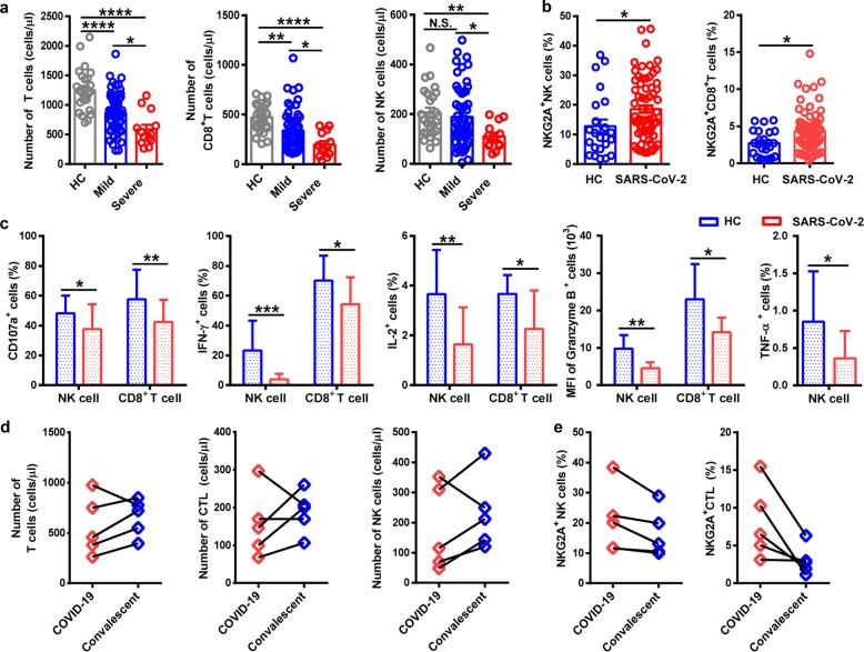
“Lower percentages of T cell functional markers is also observed with in the T Lymphocytes population as COVID-19 patients showed decreased percentages of CD107a+ CD8+, IFN-γ+CD8+, and IL-2+CD8+ T cells and MFI of granzyme B+CD8+ T cells when compared to normal controls (13).”
BackgroundNon‐steroidal anti‐inflammatory drugs, prostaglandins, and COVID‐19
Robb, C. T., Goepp, M., Rossi, A. G., & Yao, C. (2020) · British Journal of Pharmacology · doi:10.1111/bph.15206 · Cited by 104
“Like T cells, NK cells are also depleted in peripheral blood of severe but not mild COVID-19 patients (Zheng et al., 2020; Wen et al., 2020; Wilk et al., 2020).”
BackgroundInfluentialImmune Response and COVID-19: A mirror image of Sepsis
López‐Collazo, E., Ortiz, J. A., Martín‐Quirós, A., & Aguirre, L. A. (2020) · International Journal of Biological Sciences · doi:10.7150/ijbs.48400 · Cited by 103
“NK cell levels have been found to be reduced and to present an anergic status in patients with COVID-19 [90].”
BackgroundLongitudinal transcriptome analyses show robust T cell immunity during recovery from COVID-19
Zheng, H., Xu, M., Yang, C.-X., Tian, R.-R., Zhang, M., Li, J. J., Wang, X.-C., Ding, Z.-L., Li, G.-M., Li, X., et al. (2020) · Signal Transduction and Targeted Therapy · doi:10.1038/s41392-020-00457-4 · Cited by 103
“A large number of studies have shown that SARS-CoV-2 infection can cause a more pronounced lymphocytopenia in patients with moderate and severe COVID-19, as reflected by the decreased level of T cells, including Th1, Treg, and CD8 T cells.(1,39,40) In addition, neutrophils increased and monocytes decreased in COVID-19…”
ResultKreutmair, S., Unger, S., Núñez, N. G., Ingelfinger, F., Alberti, C., Feo, D. D., Krishnarajah, S., Kauffmann, M., Friebel, E., Babaei, S., et al. (2021) · Immunity · doi:10.1016/j.immuni.2021.05.002 · Cited by 109
“Multiple studies have shown differences in the proportions of immune cell populations in the peripheral blood of COVID-19 patients compared with healthy individuals, in particular a marked lymphopenia that is accompanied by changes to the lymphocyte activation and exhaustion phenotypes, some of which are partly associ…”
Age-severity matched cytokine profiling reveals specific signatures in Covid-19 patients
Angioni, R., Sánchez‐Rodríguez, R., Munari, F., Bertoldi, N., Arcidiacono, D., Cavinato, S., Marturano, D., Zaramella, A., Realdon, S., Cattelan, A. M., et al. (2020) · Cell Death and Disease · doi:10.1038/s41419-020-03151-z · Cited by 101
The immunology of SARS-CoV-2 infections and vaccines
Grigoryan, L., & Pulendran, B. (2020) · Seminars in Immunology · doi:10.1016/j.smim.2020.101422 · Cited by 102
Zhang, Y.-Y., Li, B.-R., & Ning, B. (2020) · Frontiers in Immunology · doi:10.3389/fimmu.2020.02033 · Cited by 103
Calming the Storm: Natural Immunosuppressants as Adjuvants to Target the Cytokine Storm in COVID-19
Peter, A. E., Sandeep, B. V., Rao, B. G., & Kalpana, V. L. (2021) · Frontiers in Pharmacology · doi:10.3389/fphar.2020.583777 · Cited by 106
Parallels in Sepsis and COVID-19 Conditions: Implications for Managing Severe COVID-19
Olwal, C. O., Nganyewo, N. N., Tapela, K., Zune, A. L. D., Owoicho, O., Bediako, Y., & Duodu, S. (2021) · Frontiers in Immunology · doi:10.3389/fimmu.2021.602848 · Cited by 97
“Increased T lymphopenia, particularly CD4+ and CD8+ T cells have been shown to decrease markedly in COVID-19 patients, implying damage of lymphocytes by the SARS-CoV-2 virus (13, 20, 21).”
BackgroundObesity and its impact on COVID-19
de Leeuw, A. J. M., Luttikhuis, M. A. M. O., Wellen, A. C., Müller, C., & Calkhoven, C. F. (2021) · Journal of Molecular Medicine · doi:10.1007/s00109-021-02072-4 · Cited by 115
Early Differences in Cytokine Production by Severity of Coronavirus Disease 2019
Tjan, L. H., Furukawa, K., Nagano, T., Kiriu, T., Nishimura, M., Arii, J., Hino, Y., Iwata, S., Nishimura, Y., & Mori, Y. (2021) · The Journal of Infectious Diseases · doi:10.1093/infdis/jiab005 · Cited by 96
“Interestingly, several studies showed that the numbers of peripheral NK cells were significantly lower in patients with severe COVID-19 than in healthy individuals [2, 8] or patients with mild COVID-19 [2].”
BackgroundResultNKG2A and COVID-19: another brick in the wall
Antonioli, L., Fornai, M., Pellegrini, C., & Blandizzi, C. (2020) · Cellular and Molecular Immunology · doi:10.1038/s41423-020-0450-7 · Cited by 97
“expression was markedly reduced.(14) These findings allow to hypothesize that the functional exhaustion of cytotoxic lymphocytes associated with COVID-19 infection breaks down the antiviral immunity, and that the enhanced expression of NKG2A, as specifically observed in CD8 and NK cells, could contribute to the mainte…”
BackgroundResultMorris, G., Bortolasci, C. C., Puri, B. K., Marx, W., O'Neil, A. E., Athan, E., Walder, K. R., Berk, M., OLIVE, L. S., Carvalho, A. F., et al. (2021) · Cytokine · doi:10.1016/j.cyto.2021.155593 · Cited by 101
Gagiannis, D., Steinestel, J., Hackenbroch, C., Schreiner, B., Hannemann, M., Bloch, W., Umathum, V. G., Gebauer, N., Rother, C., Stahl, M., et al. (2020) · Frontiers in Immunology · doi:10.3389/fimmu.2020.587517 · Cited by 101
“Coronavirus disease 2019 (COVID-19), caused by severe acute respiratory syndrome coronavirus 2 (SARS-CoV-2), has caused or contributed to hundreds of thousands of deaths and led to almost complete shutdown of social and economic life in many countries (1).”
BackgroundXu, R., Zhou, Y., Cai, L., Wang, L., Han, J.-B., Yang, X., Chen, J., Chen, J., Ma, C., & Shen, L.-L. (2020) · British Journal of Dermatology · doi:10.1111/bjd.19484 · Cited by 95
Notz, Q., Schmalzing, M., Wedekink, F., Schlesinger, T., Gernert, M., Herrmann, J., Sorger, L., Weismann, D., Schmid, B., Sitter, M., et al. (2020) · Frontiers in Immunology · doi:10.3389/fimmu.2020.581338 · Cited by 98
“Exhaustion of NK cells and CD8+ T cells has been previously described in COVID-19 and was associated with reduced CD107a degranulation and granzyme B production (34).”
BackgroundNazerian, Y., Ghasemi, M., Yassaghi, Y., Nazerian, A., & Hashemi, S. M. (2022) · International Immunopharmacology · doi:10.1016/j.intimp.2022.109428 · Cited by 96
“significantly higher in COVID-19 patients, especially in severe conditions [38-40].”
BackgroundCOVID‐19: Current knowledge in clinical features, immunological responses, and vaccine development
Singh, R., Kang, A., Luo, X., Jeyanathan, M., Gillgrass, A. E., Afkhami, S., & Xing, Z. (2021) · The FASEB Journal · doi:10.1096/fj.202002662r · Cited by 101
Helal, M. A., Shouman, S., Abdelwaly, A., Elmehrath, A. O., Essawy, M., Sayed, S. M., Saleh, A. H., & El‐Badri, N. (2020) · Journal of Biomolecular Structure and Dynamics · doi:10.1080/07391102.2020.1822208 · Cited by 96
“, 2020), especially in the CD8þ, CD4þ, and CD3þ T lymphocyte populations (Zeng, 2020; Zheng et al., 2020).”
BackgroundImmunobiology and immunotherapy of COVID‐19: A clinically updated overview
Esmaeilzadeh, A., & Elahi, R. (2020) · Journal of Cellular Physiology · doi:10.1002/jcp.30076 · Cited by 94
“The upregulated expression of NKG2A reduces the production of proinflammatory cytokine by NK cells and T lymphocytes (Zheng et al., 2020).”
BackgroundThe Effects of COVID-19 on the Placenta During Pregnancy
Sadeghirad, H., Röhl, J., Stylianou, N., Allenby, M. C., Bazaz, S. R., Warkiani, M. E., Souza-Fonseca-Guimarães, F., Clifton, V. L., & Kulasinghe, A. (2021) · Frontiers in Immunology · doi:10.3389/fimmu.2021.743022 · Cited by 94
Metadata
- Authors
- Zheng, M., Gao, Y., Wang, G., Song, G., Liu, S., Sun, D., Xu, Y., & Tian, Z.
- Affiliations
- First Affiliated Hospital of Anhui Medical University (CN)
- Anhui Medical University (CN)
- University of Science and Technology of China (CN)
- Corresponding author
- Zheng, Meijuan
- Xu, Yuanhong
- Tian, Zhigang
- Venue
- Cellular & Molecular Immunology
- Published
- 2020-03-19
- Volume
- 17
- Issue
- 5
- Pages
- 533-535
- ISSN
- 1672-7681 (ISSN-L)
- 2042-0226
- PMC
- PMC7091858
- OpenAlex
- W3011508332
- Cited by
- 1,768
- References
- 11
- Citations by year
- 2026: 26 · 2025: 63 · 2024: 119 · 2023: 186 · 2022: 329 · 2021: 514 · 2020: 531
- Type
- journal article
- Language
- en
- Open access
- hybrid
- doi.org
- Locations
- Cellular and Molecular Immunology (publishedVersion, cc-by)
- PubMed Central (publishedVersion, cc-by)
- PubMed (publishedVersion)
- Funders
- National Natural Science Foundation of China (81788101, 81771685)
- Chinese Academy of Sciences (XDB29030000, 81788101)
- Anhui Medical University (YJGG202002)
- Topics
- Health Sciences › Medicine › Infectious Diseases › SARS-CoV-2 and COVID-19 Research
- Health Sciences › Medicine › Infectious Diseases › COVID-19 Clinical Research Studies
- Life Sciences › Immunology and Microbiology › Immunology › T-cell and B-cell Immunology
- Keywords
- COVID-19 pandemic, SARS-CoV-2, T cell exhaustion, T cell dysfunction
- MeSH terms
- Betacoronavirus, COVID-19, SARS-CoV-2, Antiviral Agents, Hepatitis C, Humans, Killer Cells, Natural, Pneumonia, Viral, Coronavirus Infections, Pandemics
About these checks
Registry, retraction, Cites and statistics results are computed, not judged, with one exception: a printed reference that no index lists is read once by a model before it counts as a citation error. Claims are extracted by a model and kept only when their quote appears verbatim in the text; each cited source is then checked, with a model’s help, for whether it says what the claim relies on. Those verdicts can be wrong — read the claim’s quote and the cited source before relying on one.
Statistics and arithmetic are recomputed, not judged.
Confidence bands are shown only for fields calibrated on our annotated test set. None is calibrated yet, so no band is shown.
What counts as a claim ›How we measure confidence ›How we check this ›
Are you an author, or do you see a problem with this page? Request a review
