

The DOI matches its Crossref record; 10 of the 11 references it cites were found in a registry or an index.
Functional exhaustion of antiviral lymphocytes in COVID-19 patients
Open accessAbstract
In December 2019, a novel coronavirus was first reported in Wuhan, China. 1 It was named by the World Health Organization as severe acute respiratory syndrome coronavirus 2 (SARS-CoV-2) and is responsible for coronavirus disease 2019 (COVID-19). Up to 28 February 2020, 79,394 cases have been confirmed according to China’s National Health Commission. Outside China, the virus has spread rapidly to over 36 countries and territories.
Overview
The claims and statistics of this paper haven’t been checked yet.
Run the check — free, no account needed. It reads the open-access text and checks the sources it cites.
Integrity signals
Registry
Cites
Statistics
Figures
Verification
Claims the paper makes
The claims and statistics of this paper haven’t been checked yet.
Run the check — free, no account needed. It reads the open-access text and checks the sources it cites.
Claims other papers make about this one
Each is checked against this paper’s own text: does it say what the citing paper says it does?
The cited source says what the claim attributes to it.
In the citing paper
“In ex vivo studies, NK cells from patients with COVID-19 displayed impaired cytotoxicity (64, 65)”
From SARS-CoV-2 variants of concern partially escape humoral but not T cell responses in COVID-19 convalescent donors and vaccine recipients (2021) · cites this paper as [64]
In the citing paper
“NK cells were shown to be decreased in patients with COVID-19 in association with disease severity (64).”
From SARS-CoV-2 variants of concern partially escape humoral but not T cell responses in COVID-19 convalescent donors and vaccine recipients (2021) · cites this paper as [64]
Self-check
Percentages, sums and reported differences re-computed from the paper’s own numbers.
The self-check needs the paper’s full text.
Citations
Cites
A novel coronavirus from patients with pneumonia in China, 2019
Zhu, N., Zhang, D., Wang, W., Li, X., Yang, B., Song, J., Zhao, X., Huang, B., Shi, W., Lu, R., et al. (2020) · New England Journal of Medicine · doi:10.1056/nejmoa2001017 · Cited by 28,244
A pneumonia outbreak associated with a new coronavirus of probable bat origin
Zhou, P., Yang, X.-L., Wang, X.-G., Hu, B., Zhang, L., Zhang, W., Si, H.-R., Zhu, Y., Li, B., Huang, C.-L., et al. (2020) · Nature · doi:10.1038/s41586-020-2012-7 · Cited by 21,969
Chen, N., Zhou, M., Dong, X., Qu, J., Gong, F., Han, Y., Qiu, Y., Wang, J., Liu, Y., Wei, Y., et al. (2020) · The Lancet · doi:10.1016/s0140-6736(20)30211-7 · Cited by 21,415
Restoring function in exhausted CD8 T cells during chronic viral infection
Barber, D. L., Wherry, E. J., Masopust, D., Zhu, B., Allison, J. P., Sharpe, A. H., Freeman, G. J., & Ahmed, R. (2005) · Nature · doi:10.1038/nature04444 · Cited by 4,057
André, P., Denis, C., Soulas, C., Bourbon, C., Lopez, J., Arnoux, T., Bléry, M., Bonnafous, C., Gauthier, L., Morel, A., et al. (2018) · Cell · doi:10.1016/j.cell.2018.10.014 · Cited by 1,257
Zhang, Q., Bi, J., Zheng, X., Chen, Y., Wang, H., Wu, W., Wang, Z., Wu, Q., Peng, H. C., Wei, H., et al. (2018) · Nature Immunology · doi:10.1038/s41590-018-0132-0 · Cited by 1,155
Self-citationLi, F., Wei, H., Wei, H., Gao, Y., Xu, L., Yin, W., Sun, R., & Tian, Z. (2013) · Gastroenterology · doi:10.1053/j.gastro.2012.10.039 · Cited by 164
Self-citationNKG2A is a NK cell exhaustion checkpoint for HCV persistence
Zhang, C., Wang, X., Li, S.-R., Twelkmeyer, T., Wang, W., Zhang, S.-Y., Wang, S., Chen, J., Jin, X., Wu, Y., et al. (2019) · Nature Communications · doi:10.1038/s41467-019-09212-y · Cited by 148
NKG2A, a New Kid on the Immune Checkpoint Block
Haanen, J. B. A. G., & Cerundolo, V. (2018) · Cell · doi:10.1016/j.cell.2018.11.048 · Cited by 104
Barathan, M., Mohamed, R., Yong, Y. K., Kannan, M., Vadivelu, J., Saeidi, A., Larsson, M., & Shankar, E. M. (2018) · Cells · doi:10.3390/cells7100165 · Cited by 37
Cited by
Sorted by citations
Items 201–300 of 1,000
Development of multi-epitope peptide-based vaccines against SARS-CoV-2
Lim, H. X., Lim, J., Jazayeri, S. D., Poppema, S., & Poh, C. L. (2020) · Biomedical Journal · doi:10.1016/j.bj.2020.09.005 · Cited by 91
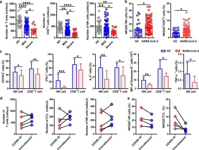
“CD8þ T-and NK-cells from COVID-19 patients had increased expression of the inhibitory receptor NKG2A with diminished productions of CD107a, IFN-g, IL-2 and granzyme B which indicated the functional exhaustion of NK-and CD8þ T-cells [29].”
The current understanding and potential therapeutic options to combat COVID-19
Pooladanda, V., Thatikonda, S., & Godugu, C. (2020) · Life Sciences · doi:10.1016/j.lfs.2020.117765 · Cited by 90
“reported that CD8+ Tcells and NK cells were significantly decreased, while NKG2A expression was dramatically increased in SARS-CoV-2 infected patients [204].”
Park, H. H., Park, W., Lee, Y. Y., Kim, H., Seo, H. S., Choi, D. W., Kwon, H., Na, D. H., Kim, T. P., Choy, Y. B., et al. (2020) · Advanced Science · doi:10.1002/advs.202001940 · Cited by 89
Aging, inflammaging and immunosenescence as risk factors of severe COVID-19
Tizazu, A. M., Mengist, H. M., & Demeke, G. (2022) · Immunity & Ageing · doi:10.1186/s12979-022-00309-5 · Cited by 96
“An increased expression of the inhibitory marker NKG2A results in the decreased level of IFNγ, IL-2, TNFα, CD107a and granzyme B in COVID-19 patients [131].”
BackgroundAdvances and Challenges in Sepsis Management: Modern Tools and Future Directions
Santacroce, E., D’Angerio, M., Ciobanu, A. L., Masini, L., Tartaro, D. L., Coloretti, I., Busani, S., Rubio, I., Meschiari, M., Franceschini, E., et al. (2024) · Cells · doi:10.3390/cells13050439 · Cited by 102
“However, prolonged hyperactivation leads to impaired NK cell function underlined by downregulation of cytotoxic genes [128] and overexpression of the inhibitory molecule NKG2A [129,130].”
BackgroundImmunology, immunopathogenesis and immunotherapeutics of COVID-19; an overview
Khosroshahi, L. M., Rokni, M., Mokhtari, T., & Noorbakhsh, F. (2021) · International Immunopharmacology · doi:10.1016/j.intimp.2020.107364 · Cited by 90
“results from NK cell trafficking to infected tissues or cell death in severe COVID-19 cases remains unknown [76,85,87].”
The role of type I interferon in the treatment of COVID‐19
Sodeifian, F., Nikfarjam, M., Kian, N., Mohamed, K., & Rezaei, N. (2021) · Journal of Medical Virology · doi:10.1002/jmv.27317 · Cited by 85
“Moreover, SARS‐CoV‐2 simultaneously upregulate expression of NKG2A receptor (a family of C‐type lectin receptors on the surface of NK cells), leading to the exhaustion of NK and CD8 T cells.(26,27) In contrast to reduced lymphocytes, the levels of neutrophils, leukocytes, and neutrophil–lymphocyte‐ratio are increased…”
BackgroundTherapeutic opportunities to manage COVID-19/SARS-CoV-2 infection: Present and future
Sethu, S., Shetty, R., Ghosh, A., Honavar, S. G., & Khamar, P. M. (2020) · Indian Journal of Ophthalmology · doi:10.4103/ijo.ijo_639_20 · Cited by 87
Brown, C., & Ravallion, M. D. (2020) · National Bureau of Economic Research · doi:10.3386/w27549 · Cited by 86
Yaqinuddin, A., & Kashir, J. (2020) · Medical Hypotheses · doi:10.1016/j.mehy.2020.109777 · Cited by 85
Keam, S., Megawati, D., Patel, S. K., Tiwari, R., Dhama, K., & Harapan, H. (2020) · Reviews in Medical Virology · doi:10.1002/rmv.2123 · Cited by 85
“A recent study has found a significant decrease of total T lymphocytes, CD8 T cells, and NK cells in patients with COVID-19 and this reduction was more prominent among those with severe disease compared with those with a mild one.(70) Moreover, NK cells become”
BackgroundResultInfluentialKai, H., Wang, C., Vagts, C. L., Raguveer, V., Finn, P. W., & Perkins, D. L. (2022) · PLoS ONE · doi:10.1371/journal.pone.0261242 · Cited by 84
Padilla, E. M., Yarce, O., Ortega, C., Fernández, S., Pascual, N., Gómez, C., Alvarez, M. A., Molina, I. J., Lama, R., & Santamaria, M. L. (2020) · The Journal of Allergy and Clinical Immunology In Practice · doi:10.1016/j.jaip.2020.06.046 · Cited by 84
“8 g/kg/24 h and aggressive antibiotic therapy), together with persistent fever and bilateral pneumonia, prompted us to consider the possibility that natural killer (NK)-cell and T-cell exhaustion might be an underlying cause for immunologic failure, as shown elsewhere.(5) We assessed this possibility in lung lymphoid…”
BackgroundAn Effective COVID-19 Vaccine Needs to Engage T Cells
Sauer, K., & Harris, T. (2020) · Frontiers in Immunology · doi:10.3389/fimmu.2020.581807 · Cited by 85
“Conversely, recovering patients had increased virus specific and general CD4 TFH cells, and their SARS-CoV-2 reactive T cells expressed reduced inhibitory markers and elevated effector molecules (25, 34, 44, 46, 47).”
BackgroundIncreased IL‐10‐producing regulatory T cells are characteristic of severe cases of COVID‐19
Neumann, J., Prezzemolo, T., Vanderbeke, L., Roca, C. P., Gerbaux, M., Janssens, S., Willemsen, M., Burton, O. T., Mol, P. V., Herck, Y. V., et al. (2020) · Clinical & Translational Immunology · doi:10.1002/cti2.1204 · Cited by 84
“Functional defects within T cells have been reported, with an increased number of non-functional CD4 T cells and impaired T-cell cytokine production.(2,3,4) This was confirmed in an independent study, but without significant changes between moderate and severe patients,(5) while another study reported no change.”
BackgroundImmunomodulatory Effects of Azithromycin Revisited: Potential Applications to COVID-19
Venditto, V. J., Haydar, D., Abdel‐Latif, A., Gensel, J. C., Anstead, M., Pitts, M. G., Creameans, J. W., Kopper, T. J., Peng, C., & Feola, D. J. (2021) · Frontiers in Immunology · doi:10.3389/fimmu.2021.574425 · Cited by 90
“Additionally, highlighting the complexity of these interactions, expression of genes and surface proteins associated with T cell and NK cell exhaustion has also been associated with severe disease (184, 221).”
BackgroundRabaan, A. A., Al-Ahmed, S. H., Garout, M. A., Al‐Qaaneh, A. M., Sule, A. A., Tirupathi, R., Mutair, A. S. A., Alhumaid, S., Hasan, A., Dhawan, M. K., et al. (2021) · Pathogens · doi:10.3390/pathogens10050565 · Cited by 84
“CD4+ and CD8+ T-cells are the primary T-cell subsets that are used as bioindicators and prognostic immune markers during acute viral infections.”
BackgroundInfluentialCOVID-19 disease and immune dysregulation
Davitt, E., Davitt, C., Mazer, M. B., Areti, S. S., Hotchkiss, R. S., & Remy, K. E. (2022) · Best Practice & Research Clinical Haematology · doi:10.1016/j.beha.2022.101401 · Cited by 84
“A proposed mechanism for IL-6/STAT3 signaling to induce the inactive phenotypes of NK cells and CD8 T cells in COVID-19 is by increasing expression of NKG2A, an inhibitory receptor [21,22].”
InfluentialCytokine storm syndrome in severe COVID-19
Ruscitti, P., Berardicurti, O., Iagnocco, A., & Giacomelli, R. (2020) · Autoimmunity Reviews · doi:10.1016/j.autrev.2020.102562 · Cited by 85
“Taking together all these observations, a defect in granule-mediated cytotoxicity could be postulated as a further mechanism facilitating the cytokine storm syndrome in patients with severe COVID-19, who are characterised by a decrease of NK cells [21,22].”
Drug repurposing against COVID-19: focus on anticancer agents
Ciliberto, G., Mancini, R., & Paggi, M. G. (2020) · Journal of Experimental & Clinical Cancer Research · doi:10.1186/s13046-020-01590-2 · Cited by 81
“Interestingly, NKG2A appears overexpressed in cytotoxic T lymphocytes and natural killer cells in SARS-CoV-2-infected patients [73, 74], where it may reestablish the host immune response and increase survival in patients with severe pneumonia.”
BackgroundPaliogiannis, P., Mangoni, A. A., Cangemi, M., Fois, A. G., Carru, C., & Zinellu, A. (2021) · Clinical and Experimental Medicine · doi:10.1007/s10238-021-00686-z · Cited by 82
Implications of NKG2A in immunity and immune-mediated diseases
Wang, X., Xiong, H., & Ning, Z. (2022) · Frontiers in Immunology · doi:10.3389/fimmu.2022.960852 · Cited by 82
“Interestingly, the NKG2A cytotoxic lymphocytes proportion was reduced among recovered patients (17).”
BackgroundPotential neurological effects of severe COVID-19 infection
Nuzzo, D., & Picone, P. (2020) · Neuroscience Research · doi:10.1016/j.neures.2020.06.009 · Cited by 81
Characterization of low-density granulocytes in COVID-19
Cabrera, L. E., Pekkarinen, P. T., Alander, M., Nowlan, K., Nguyen, N.-A. A., Jokiranta, S. T., Kuivanen, S. H., Patjas, A., Mero, S., Pakkanen, S. H., et al. (2021) · PLoS Pathogens · doi:10.1371/journal.ppat.1009721 · Cited by 83
“Interestingly, increased neutrophil recruitment is consistently observed in severe COVID-19 [1,3–7] and the neutrophil count positively correlates with disease severity, while lymphocyte numbers are depleted in patients with a poorer outcome [8].”
BackgroundNing, Q., Wu, D., Wang, X., Xi, D., Chen, T., Chen, G., Wang, H., Lü, H., Wang, M., Zhu, L., et al. (2022) · Signal Transduction and Targeted Therapy · doi:10.1038/s41392-022-00907-1 · Cited by 86
“Besides, multiple studies have shown decreased numbers and functionally exhaustion of natural killer (NK) cells during SARS-CoV-2 infection.(59) The diminished NK cell cytotoxicity and immune regulation result in a critical inflammatory phenotype in COVID-19.”
BackgroundVillena, J., & Kitazawa, H. (2020) · Frontiers in Physiology · doi:10.3389/fphys.2020.00699 · Cited by 83
Repurposing anticancer drugs for the management of COVID-19
Bairi, K. E., Trapani, D., Petrillo, A., Page, C. L., Zbakh, H., Daniele, B., Belbaraka, R., Curigliano, G., & Afqir, S. (2020) · European Journal of Cancer · doi:10.1016/j.ejca.2020.09.014 · Cited by 82
Profiling of immune dysfunction in COVID-19 patients allows early prediction of disease progression
Rendeiro, A. F., Casano, J., Vorkas, C. K., Singh, H. K., Morales, A. E., Desimone, R. A., Ellsworth, G. B., Soave, R., Kapadia, S. N., Saito, K., et al. (2020) · Life Science Alliance · doi:10.26508/lsa.202000955 · Cited by 83
“Indeed, similar to previous reports (De Biasi et al, 2020; Zheng et al, 2020a, 2020b), T cells displayed an overall exhausted phenotype, with overexpression of VISTA, TIM3, LAG3, TIGIT, and PD-1 co-inhibitory receptors in COVID-19 patient T cell populations.”
BackgroundResultInfluentialTocilizumab treatment for COVID-19 patients: a systematic review and meta-analysis
Wei, Q., Hua, L., Wei, R., Chen, N., He, F., Zou, D.-H., & Wei, J. (2021) · Infectious Diseases of Poverty · doi:10.1186/s40249-021-00857-w · Cited by 88
Loretelli, C., Abdelsalam, A., D’Addio, F., Nasr, M. B., Assi, E., Usuelli, V., Maestroni, A., Seelam, A. J., Ippolito, E., Maggio, S. D., et al. (2021) · JCI Insight · doi:10.1172/jci.insight.146701 · Cited by 81
Zuo, W., & Zhao, X. (2021) · Clinical Immunology · doi:10.1016/j.clim.2021.108727 · Cited by 79
Immunogenic and reactogenic efficacy of Covaxin and Covishield: a comparative review
Das, S., Kar, S. S., Samanta, S., Banerjee, J., Giri, B., & Dash, S. K. (2022) · Immunologic Research · doi:10.1007/s12026-022-09265-0 · Cited by 82
“It was quickly discovered that the overall number of NK cells in COVID-19 patients is significantly reduced [49].”
BackgroundMüller, L., & Benedetto, S. D. (2024) · International Journal of Molecular Sciences · doi:10.3390/ijms25020753 · Cited by 84
PATHOLOGICAL ANATOMY OF INFECTION CAUSED BY SARS-COV-2
Kogan, E., Коган, Е. А., Berezovsky, Y., Березовский, Ю. С., Protsenko, D. D., Проценко, Д. Д., Bagdasaryan, T. R., Багдасарян, Т. Р., Gretsov, E., Грецов, Е. М., et al. (2020) · Russian Journal of Forensic Medicine · doi:10.19048/2411-8729-2020-6-2-8-30 · Cited by 41
“Hematoxylin and eosin staining При иммуногистохимическом исследовании выявлено небольшое количество CD4+- и CD8+-лимфоцитов [65].”
Severe COVID-19: what have we learned with the immunopathogenesis?
Corrêa, B. B., Rodrigues, M. B., Cortez, A. F., Vieira, M., & Pinheiro, M. M. (2020) · Revista Brasileira de Reumatologia · doi:10.1186/s42358-020-00151-7 · Cited by 83
“This altered expression is normalized in convalescent patients [84].”
BackgroundInfluentialPresence of antibody-dependent cellular cytotoxicity (ADCC) against SARS-CoV-2 in COVID-19 plasma
Tso, F. Y., Lidenge, S. J., Poppe, L. K., Peña, P. B., Privatt, S. R., Bennett, S. J., Ngowi, J. R., Mwaiselage, J., Belshan, M., Siedlik, J. A., et al. (2021) · PLOS ONE · doi:10.1371/journal.pone.0247640 · Cited by 68
“Several studies have suggested dysfunction and decreased number of NK cells in patients with severe COVID-19 disease, which may undermine the role that NK cells and ADCC play in disease recovery [26, 27].”
BackgroundBaker, D., Amor, S., Kang, A. S., Schmierer, K., & Giovannoni, G. (2020) · Multiple Sclerosis and Related Disorders · doi:10.1016/j.msard.2020.102174 · Cited by 75
“This may reflect senescence and exhaustion of the anti-viral CD8 response (Zheng et al., 2020; Cossarizza et al., 2020).”
Vivarelli, S., Falzone, L., Grillo, C. M., Scandurra, G., Torino, F., & Libra, M. (2020) · Cancers · doi:10.3390/cancers12082237 · Cited by 75
“As described above, following a first hyper-inflammatory phase triggered by the CS, SARS-CoV-2 prolonged infection might induce T-cell hyperactivation and finally exhaustion, associated with concurrent patients’ lymphopenia, as well as sepsis [67].”
BackgroundInfluentialViral coinfections in COVID‐19
Aghbash, P. S., Eslami, N., Shirvaliloo, M., & Baghi, H. B. (2021) · Journal of Medical Virology · doi:10.1002/jmv.27102 · Cited by 82
Identification of druggable inhibitory immune checkpoints on Natural Killer cells in COVID-19
Demaria, O., Carvelli, J., Batista, L., Thibult, M., Morel, A., André, P., Morel, Y., Vely, F., & Vivier, É. (2020) · Cellular and Molecular Immunology · doi:10.1038/s41423-020-0493-9 · Cited by 75
“Thus, circulating NK cells in COVID-19 patients display an upregulation of the inhibitory receptor NKG2A that is associated with disease severity, consistent with recent results.(8) Bronchoalveolar lavage fluid (BALF) analysis showed a lack of CD16CD57 mature NK cells in the lungs of ARDS patients (Fig.”
Spectrum of Headaches Associated With SARS‐CoV‐2 Infection: Study of Healthcare Professionals
Porta-Etessam, J., Matías‐Guiu, J. A., González-García, N., Iglesias, P. G., Santos-Bueso, E., Arriola‐Villalobos, P., García‐Azorín, D., & Matías‐Guiu, J. (2020) · Headache The Journal of Head and Face Pain · doi:10.1111/head.13902 · Cited by 77
Cellular Immune Response to COVID-19 and Potential Immune Modulators
Zhou, X. K., & Ye, Q. (2021) · Frontiers in Immunology · doi:10.3389/fimmu.2021.646333 · Cited by 77
“The reduced abundance and exhausting function of circulating NK cells were noticed in severe COVID-19 patients (65).”
BackgroundSheerin, D., Abhimanyu, A., Peton, N., Vo, W., Allison, C. C., Wang, X., Johnson, W. E., & Coussens, A. K. (2022) · iScience · doi:10.1016/j.isci.2022.104464 · Cited by 78
Potential therapeutic effects of Resveratrol against SARS-CoV-2
Ramdani, L. H., & Bacharı, K. (2020) · Acta Virologica · doi:10.4149/av_2020_309 · Cited by 75
NF-κB signalling as a pharmacological target in COVID-19: potential roles for IKKβ inhibitors
Kandasamy, M. (2021) · Naunyn-Schmiedeberg s Archives of Pharmacology · doi:10.1007/s00210-020-02035-5 · Cited by 76
Severe COVID-19 and Sepsis: Immune Pathogenesis and Laboratory Markers
Zafer, M. M., El‐Mahallawy, H. A., & Ashour, H. M. (2021) · Microorganisms · doi:10.3390/microorganisms9010159 · Cited by 74
“That might explain why lymphocyte counts are markedly reduced in patients with severe COVID-19 [59].”
BackgroundCytokine Signature Induced by SARS-CoV-2 Spike Protein in a Mouse Model
Gu, T., Zhao, S., Jin, G., Song, M., Zhi, Y., Zhao, R., Ma, F., Zheng, Y., Wang, K., Liu, H., et al. (2021) · Frontiers in Immunology · doi:10.3389/fimmu.2020.621441 · Cited by 74
Tan, L., Kang, X., Ji, X., Li, G., Wang, Q. M., Li, Y., Wang, Q., & Miao, H. (2020) · Med · doi:10.1016/j.medj.2020.05.002 · Cited by 73
COVID-19: etiology, clinical picture, treatment
MIu, S., Колобухина, Л. В., Burgasova, O., Сергеевна, К. И., & Vv, M. (2020) · Russian Journal of Infection and Immunity · doi:10.15789/2220-7619-cec-1473 · Cited by 73
Márquez, E. J., Trowbridge, J. J., Kuchel, G. A., Banchereau, J., & Ucar, D. (2020) · Immunity & Ageing · doi:10.1186/s12979-020-00183-z · Cited by 73
“Lymphopenia (having reduced numbers of lymphocytes in the blood) has been reported in severe cases of COVID-19 in multiple studies [21, 23, 52, 53], including drastic declines in CD4+ and CD8+ T cells as well as NK and B cells.”
BackgroundGhayda, R. A., Lee, J., Lee, J. Y., Kim, D. K., Lee, K. H., Hong, S. H., Han, Y. J., Kim, J., Yang, J. W., Kronbichler, A., et al. (2020) · International Journal of Environmental Research and Public Health · doi:10.3390/ijerph17145026 · Cited by 72
“It has been well established that COVID-19 is associated with immune system dysregulation [35,36].”
Tea Bioactive Modulate Innate Immunity: In Perception to COVID-19 Pandemic
Chowdhury, P., & Barooah, A. K. (2020) · Frontiers in Immunology · doi:10.3389/fimmu.2020.590716 · Cited by 71
“Treatment with polyphenols in mice model with upper airway inflammation showed increase in NK cells degranulation (37).”
BackgroundInnate lymphoid cells and COVID-19 severity in SARS-CoV-2 infection
Silverstein, N. J., Wang, Y., Manickas-Hill, Z., Carbone, C., Dauphin, A., Boribong, B. P., Loiselle, M., Davis, J., Leonard, M. M., Kuri-Cervantes, L., et al. (2022) · eLife · doi:10.7554/elife.74681 · Cited by 73
Witkowski, J. M., Fülöp, T., & Bryl, E. (2022) · Mechanisms of Ageing and Development · doi:10.1016/j.mad.2022.111672 · Cited by 86
“of COVID-19 patients show reduced numbers and an exhausted phenotype (changed expression of NKG2A antigen), which may aggravate the severity of the disease by compromising viral clearance (Cunningham et al., 2021; Zheng et al., 2020).”
BackgroundCell-Mediated Immune Responses to COVID-19 Infection
Guihot, A., Litvinova, E., Autran, B., Debré, P., & Vieillard, V. (2020) · Frontiers in Immunology · doi:10.3389/fimmu.2020.01662 · Cited by 70
“It was rapidly determined that in COVID-19 patients, the total number of NK cells is markedly decreased (54), as previously reported for the SARS (55).”
BackgroundResultInfluentialMolecular Regulation of NK Cell Maturation
Bi, J., & Wang, X. (2020) · Frontiers in Immunology · doi:10.3389/fimmu.2020.01945 · Cited by 70
“proliferation, activity, and expression of activating receptors, as well as increased apoptosis and expression of inhibitory receptors (92, 93).”
BackgroundA scoping review of the pathophysiology of COVID-19
Marik, P. E., Iglesias, J. I., Varón, J., & Kory, P. D. (2021) · International Journal of Immunopathology and Pharmacology · doi:10.1177/20587384211048026 · Cited by 74
A Review on SARS-CoV-2 Virology, Pathophysiology, Animal Models, and Anti-Viral Interventions
Neerukonda, S. N., & Katneni, U. (2020) · Pathogens · doi:10.3390/pathogens9060426 · Cited by 70
“In addition, NK‐ and CD8+ T‐cell populations in COVID‐19 patients are functionally exhausted (NKG2A+), whereas convalescing patients recover functionality [25].”
BackgroundDiNicolantonio, J. J., & O’Keefe, J. H. (2021) · Europe PMC (PubMed Central) · Cited by 68
Bobčáková, A., Petriskova, J., Vyšehradský, R., Kocan, I., Kapustova, L., Barnova, M., Diamant, Z., & Jeseňák, M. (2021) · Frontiers in Cellular and Infection Microbiology · doi:10.3389/fcimb.2021.646688 · Cited by 68
Natural Killer Cells in SARS-CoV-2 Infection: Pathophysiology and Therapeutic Implications
Vito, C. D., Calcaterra, F., Coianiz, N., Terzoli, S., Voza, A., Mikulak, J., Bella, S. D., & Mavilio, D. (2022) · Frontiers in Immunology · doi:10.3389/fimmu.2022.888248 · Cited by 69
“Several studies observed a significant increase in circulating NK cells after the resolution of the infection (40, 41), while others showed normal NK cell counts in convalescent individuals (34, 42).”
BackgroundResultInfluentialInnate immune evasion by SARS‐CoV ‐2: Comparison with SARS‐CoV
Bouayad, A. (2020) · Reviews in Medical Virology · doi:10.1002/rmv.2135 · Cited by 70
“The upregulation of human inhibitory receptors is one more strategy that SARS-CoV-2 uses to modulate NK cell-mediated cytotoxicity.(93)”
BackgroundRoberts, J., Pritchard, A. L., Treweeke, A. T., Rossi, A. G., Brace, N., Cahill, P. A., MacRury, S. M., Wei, J., & Megson, I. L. (2021) · Frontiers in Cardiovascular Medicine · doi:10.3389/fcvm.2020.629933 · Cited by 68
He, S., Liu, W., Jiang, M., Huang, P., Xiang, Z., Deng, D., Chen, P., & Xie, L. (2021) · PLoS ONE · doi:10.1371/journal.pone.0249668 · Cited by 69
“Similarly, it is possible that SARS-CoV-2 would also affect dendritic cells, and therefore leading to the release of many cytokines and T cell apoptosis, eventually leading to immune system disfunction [24, 26].”
BackgroundClinical characteristics of Egyptian male patients with COVID‐19 acute respiratory distress syndrome
Doghish, A. S., Elkhatib, W. F., Hassan, E. A., Elkhateeb, A. F., Mahmoud, E. E., Ahmed, M. I., & Khalil, M. A. F. (2021) · PLoS ONE · doi:10.1371/journal.pone.0249346 · Cited by 68
“Lymphopenia is a prominent characteristic of critically ill patients with COVID-19, resulting from the direct destruction of lymphocytes, especially CD4 T and CD8 T cells [15], or cytokinemediated lymphocyte destruction [16].”
BackgroundThe intersection of COVID-19 and cancer: signaling pathways and treatment implications
Zong, Z., Wei, Y., Ren, J., Zhang, L., & Zhou, F. (2021) · Molecular Cancer · doi:10.1186/s12943-021-01363-1 · Cited by 74
Fernández-Francos, S., Eiró, N., González-Galiano, N., & Vizoso, F. J. (2021) · International Journal of Molecular Sciences · doi:10.3390/ijms22157850 · Cited by 70
“In addition, it is a remarkable recent finding suggesting that MSC may reduce the apoptosis of lymphocytes induced by SARS-CoV-2 infection, which leads to lymphocytopenia in critically ill patients with COVID-19 pneumonia [52].”
BackgroundHasan, M. Z., Islam, S., Matsumoto, K., & Kawai, T. (2021) · Scientific Reports · doi:10.1038/s41598-021-96110-3 · Cited by 69
Wilairatana, P., Masangkay, F. R., Kotepui, K. U., Milanez, G. D. J., & Kotepui, M. (2021) · PLoS neglected tropical diseases · doi:10.1371/journal.pntd.0009766 · Cited by 65
Mahmoudi, S., Rezaei, M. S., Mansouri, N., Marjani, M., & Mansouri, D. (2020) · Journal of Clinical Immunology · doi:10.1007/s10875-020-00824-4 · Cited by 66
Duration of post–COVID-19 symptoms is associated with sustained SARS-CoV-2–specific immune responses
Files, J. K., Sarkar, S., Fram, T., Boppana, S., Sterrett, S., Qin, K., Bansal, A., Long, D. M., Sabbaj, S., Kobie, J. J., et al. (2021) · JCI Insight · doi:10.1172/jci.insight.151544 · Cited by 69
Role of Artificial Intelligence in COVID-19 Detection
Gudigar, A., Raghavendra, U., Nayak, S., Ooi, C. P., Chan, W. Y., Gangavarapu, M. R., Dharmik, C., Samanth, J., Kadri, N. A., Hasikin, K. B., et al. (2021) · Sensors · doi:10.3390/s21238045 · Cited by 70
Liu, X., Cao, W., & Li, T. (2020) · Frontiers in Immunology · doi:10.3389/fimmu.2020.01660 · Cited by 68
“An accompanying functional exhaustion of CD8+ T cells is furthermore detected in severe COVID-19 patients (131).”
BackgroundResultBaricitinib: From Rheumatoid Arthritis to COVID‐19
Assadiasl, S., Fatahi, Y., Mosharmovahed, B., Mohebbi, B., & Nicknam, M. H. (2021) · The Journal of Clinical Pharmacology · doi:10.1002/jcph.1874 · Cited by 66
Huyut, M. T. (2022) · IRBM · doi:10.1016/j.irbm.2022.05.006 · Cited by 66
“…patients in this manuscript Similarly, in many studies, it was stated that the ALT, AST, LDH, total bilirubin, direct bilirubin, creatine kinase values increased in the severe COVID-19 patients and the hemoglobin value decreased signifi-cantly when compared to mildly infected patients [5,6,52–54].”
Vivarelli, S., Falzone, L., Torino, F., Scandurra, G., Russo, G. L., Bordonaro, R., Pappalardo, F., Spandidos, D. A., Raciti, G., & Libra, M. (2020) · International Journal of Oncology · doi:10.3892/ijo.2020.5159 · Cited by 64
“As widely described in the section above, following a first hyper‑inflammatory phase triggered by the CS, SARS-CoV-2 prolonged infection may induce T‑cell hyperactivation and finally exhaustion, associated with concurrent lymphopenia in patient (137).”
Defending against SARS-CoV-2: The T cell perspective
Almendro-Vázquez, P., Laguna‐Goya, R., & Paz‐Artal, E. (2023) · Frontiers in Immunology · doi:10.3389/fimmu.2023.1107803 · Cited by 67
“Similarly, several studies have reported an exhausted phenotype of CD8+ T cells in severe COVID-19, with an increased expression of inhibitory receptors such as PD-1 and Tim-3 (78–80) and related to the overexpression of the natural killer group 2 member A (NKG2A) in CD8+ T cells (81, 82).”
BackgroundDefining the role of natural killer cells in COVID-19
Lee, M. J., & Blish, C. A. (2023) · Nature Immunology · doi:10.1038/s41590-023-01560-8 · Cited by 65
Evidence of potent humoral immune activity in COVID-19-infected kidney transplant recipients
Hartzell, S., Bin, S., Benedetti, C., Haverly, M., Gallon, L. G., Zaza, G., Riella, L. V., Menon, M. C., Florman, S. S., Rahman, A. H., et al. (2020) · American Journal of Transplantation · doi:10.1111/ajt.16261 · Cited by 64
Kaye, A. G., & Siegel, R. (2020) · PeerJ · doi:10.7717/peerj.10322 · Cited by 62
“While T-cells are significantly decreased in all COVID-19 patients, reduction in B and NK cells are more affected in severe cases (Shi, 2020; Zheng et al., 2020).”
BackgroundCOVID-19—A Theory of Autoimmunity Against ACE-2 Explained
McMillan, P., Dexhiemer, T., Neubig, R. R., & Uhal, B. D. (2021) · Frontiers in Immunology · doi:10.3389/fimmu.2021.582166 · Cited by 64
“The large number of antigens being targeted causes rapid lymphocyte exhaustion and lymphopenia (75).”
BackgroundHallmarks of immune response in COVID-19: Exploring dysregulation and exhaustion
Mazzoni, A., Salvati, L., Maggi, L., Annunziato, F., & Cosmi, L. (2021) · Seminars in Immunology · doi:10.1016/j.smim.2021.101508 · Cited by 69
Hammer, Q., Dunst, J., Christ, W., Picarazzi, F., Wendorff, M., Momayyezi, P., Huhn, O., Netskar, H., Maleki, K. T., García, M., et al. (2022) · Cell Reports · doi:10.1016/j.celrep.2022.110503 · Cited by 63
“It will be important to explore in-depth how pro-inflammatory cues balance priming of NK cells versus driving them into dysfunctional states and to which degree excessive or prolonged systemic inflammation affects anti-viral immune responses of specific subsets of NK cells (Krämer et al., 2021; Mazzoni et al., 2020; O…”
InfluentialThe role of immune activation and antigen persistence in acute and long COVID
Opsteen, S., Files, J. K., Fram, T., & Erdmann, N. B. (2023) · Journal of Investigative Medicine · doi:10.1177/10815589231158041 · Cited by 65
Özdemir, Ş., & Kerse, G. (2020) · International and Multidisciplinary Journal of Social Sciences · doi:10.17583/rimcis.2020.5849 · Cited by 68
Tincati, C., Cannizzo, E. S., Giacomelli, M., Badolato, R., Monforte, A. D., & Marchetti, G. C. (2020) · Frontiers in Immunology · doi:10.3389/fimmu.2020.580987 · Cited by 61
“Covid-19 adult patients (3, 22), we next characterized T-cell immunophenotype and function according to disease severity, with particular focus on SARS-Cov-2-specific response.”
BackgroundResultLombardi, A., Trombetta, E., Cattaneo, A., Castelli, V., Palomba, E., Tirone, M., Mangioni, D., Lamorte, G., Manunta, M., Prati, D., et al. (2020) · Frontiers in Immunology · doi:10.3389/fimmu.2020.560330 · Cited by 62
“Indeed, Zheng and colleagues demonstrated an upregulation of the inhibitory receptor NKG2A expression on natural killer cells (NK) and cytotoxic lymphocytes (CTLs) in COVID-19 patients with a compromised degranulation capacity and a reduced production of IFN-g, IL-2, granzyme B and tumor necrosis factor a (TNF-a) (7).”
BackgroundA systematic meta‐analysis of immune signatures in patients with COVID‐19
Liu, K., Yang, T., Peng, X., Lv, S., Ye, X., Zhao, T., Li, J., Shao, Z., Lu, Q., Li, J., et al. (2020) · Reviews in Medical Virology · doi:10.1002/rmv.2195 · Cited by 66
Adaptive immunity to SARS-CoV-2 infection: A systematic review
Silva, M. J. A., Ribeiro, L. R., Lima, K. V. B., & Lima, L. N. G. C. (2022) · Frontiers in Immunology · doi:10.3389/fimmu.2022.1001198 · Cited by 67
“, 2020b (57) Cohort study To verify whether cytotoxic lymphocytes in patients infected with SARS-CoV-2 become functionally exhausted.”
BackgroundLetafati, A., Ardekani, O. S., Naderisemiromi, M., Norouzi, M., Shafiei, M., Nik, S., & Mozhgani, S. (2024) · Virology Journal · doi:10.1186/s12985-024-02287-0 · Cited by 70
“Conversely, individuals experiencing a fatal progression of the disease exhibit a progressive decline in NK cell levels following the onset of symptoms [103, 109].”
BackgroundAl‐Horani, R. A., Kar, S., & Aliter, K. F. (2020) · International Journal of Molecular Sciences · doi:10.3390/ijms21155224 · Cited by 61
Shahbaz, S., Xu, L., Sligl, W., Osman, M. S., Bozorgmehr, N., Mashhouri, S., Redmond, D., Rosero, E. P., Walker, J. W. T., & Elahi, S. (2021) · The Journal of Immunology · doi:10.4049/jimmunol.2100446 · Cited by 63
“Our data are in sharp contrast with previous studies performed in COVID-19 patients that reported NKG2A, TIGIT, PD-1, and CTLA-4 expression as signs of functional T cell exhaustion (17, 56).”
ResultHasankhani, A., Bahrami, A., Sheybani, N., Aria, B., Hemati, B., Fatehi, F., Farahani, H. G. M., Javanmard, G., Rezaee, M., Kastelic, J. P., et al. (2021) · Frontiers in Immunology · doi:10.3389/fimmu.2021.789317 · Cited by 62
“Moreover, severe COVID-19 is associated with dysregulation of T-cells, causing lymphopenia and exhaustion of CD4 + and CD8 + T cells (186 – 188).”
Exhaustion and over-activation of immune cells in COVID-19: Challenges and therapeutic opportunities
Alahdal, M., & Elkord, E. (2022) · Clinical Immunology · doi:10.1016/j.clim.2022.109177 · Cited by 60
“Increased NKG2A expression was associated with NK cell exhaustion in COVID-19, which significantly contributes to the exhaustion of CD8 T cells [56,57].”
BackgroundClinical impact of COVID-19 on patients with cancer treated with immune checkpoint inhibition
Rogiers, A., da Silva, I. P., Tentori, C., Tondini, C. A., Grimes, J. M., Trager, M. H., Nahm, S. H., Zubiri, L., Manos, M. P., Bowling, P. J., et al. (2021) · Journal for ImmunoTherapy of Cancer · doi:10.1136/jitc-2020-001931 · Cited by 60
Calprotectin, an Emerging Biomarker of Interest in COVID-19: A Systematic Review and Meta-Analysis
Udeh, R., Advani, S., de Guadiana‐Romualdo, L. G., & Dolja‐Gore, X. (2021) · Journal of Clinical Medicine · doi:10.3390/jcm10040775 · Cited by 63
“Unfortunately, the global public health burden of the pandemic has continued to worsen with increasing morbidity and mortality rates [2,5].”
BackgroundCOVID-19: Immunology, Immunopathogenesis and Potential Therapies
Bhardwaj, A., Sapra, L., Saini, C., Azam, Z., Mishra, P. K., Verma, B., Mishra, G. C., & Srivastava, R. K. (2021) · International Reviews of Immunology · doi:10.1080/08830185.2021.1883600 · Cited by 65
“Functional exhaustion of NK cells is one of the mechanisms through which SARSCoV-2 evades immunity [47].”
BackgroundInfluentialClinical and molecular characteristics of COVID-19 patients with persistent SARS-CoV-2 infection
Yang, B., Fan, J., Huang, J., Guo, E., Fu, Y., Liu, S., Xiao, R., Liu, C., Lu, F., Qin, T., et al. (2021) · Nature Communications · doi:10.1038/s41467-021-23621-y · Cited by 62
Self-citationKamyshnyi, O., Matskevych, V. M., Lenchuk, T., Strilbytska, O. M., Storey, K. B., & Lushchak, O. V. (2021) · Biomedicine & Pharmacotherapy · doi:10.1016/j.biopha.2021.112230 · Cited by 62
The molecular mechanism of SARS-CoV-2 evading host antiviral innate immunity
Gu, W., Gan, H., Ma, Y., Xu, L., Cheng, Z. J., Li, B., Zhang, X., Jiang, W., Sun, J., Sun, B., et al. (2022) · Virology Journal · doi:10.1186/s12985-022-01783-5 · Cited by 64
“Upregulation of NKG2A was associated with the exhaustion of NK cells at the early stage of SARSCoV-2 infection, and therefore, was associated with severe disease progression [38].”
Background
Metadata
- Authors
- Zheng, M., Gao, Y., Wang, G., Song, G., Liu, S., Sun, D., Xu, Y., & Tian, Z.
- Affiliations
- First Affiliated Hospital of Anhui Medical University (CN)
- Anhui Medical University (CN)
- University of Science and Technology of China (CN)
- Corresponding author
- Zheng, Meijuan
- Xu, Yuanhong
- Tian, Zhigang
- Venue
- Cellular & Molecular Immunology
- Published
- 2020-03-19
- Volume
- 17
- Issue
- 5
- Pages
- 533-535
- ISSN
- 1672-7681 (ISSN-L)
- 2042-0226
- PMC
- PMC7091858
- OpenAlex
- W3011508332
- Cited by
- 1,768
- References
- 11
- Citations by year
- 2026: 26 · 2025: 63 · 2024: 119 · 2023: 186 · 2022: 329 · 2021: 514 · 2020: 531
- Type
- journal article
- Language
- en
- Open access
- hybrid
- doi.org
- Locations
- Cellular and Molecular Immunology (publishedVersion, cc-by)
- PubMed Central (publishedVersion, cc-by)
- PubMed (publishedVersion)
- Funders
- National Natural Science Foundation of China (81788101, 81771685)
- Chinese Academy of Sciences (XDB29030000, 81788101)
- Anhui Medical University (YJGG202002)
- Topics
- Health Sciences › Medicine › Infectious Diseases › SARS-CoV-2 and COVID-19 Research
- Health Sciences › Medicine › Infectious Diseases › COVID-19 Clinical Research Studies
- Life Sciences › Immunology and Microbiology › Immunology › T-cell and B-cell Immunology
- Keywords
- COVID-19 pandemic, SARS-CoV-2, T cell exhaustion, T cell dysfunction
- MeSH terms
- Betacoronavirus, COVID-19, SARS-CoV-2, Antiviral Agents, Hepatitis C, Humans, Killer Cells, Natural, Pneumonia, Viral, Coronavirus Infections, Pandemics
About these checks
Registry, retraction, Cites and statistics results are computed, not judged, with one exception: a printed reference that no index lists is read once by a model before it counts as a citation error. Claims are extracted by a model and kept only when their quote appears verbatim in the text; each cited source is then checked, with a model’s help, for whether it says what the claim relies on. Those verdicts can be wrong — read the claim’s quote and the cited source before relying on one.
Statistics and arithmetic are recomputed, not judged.
Confidence bands are shown only for fields calibrated on our annotated test set. None is calibrated yet, so no band is shown.
What counts as a claim ›How we measure confidence ›How we check this ›
Are you an author, or do you see a problem with this page? Request a review
