

The DOI matches its Crossref record; 10 of the 11 references it cites were found in a registry or an index.
Functional exhaustion of antiviral lymphocytes in COVID-19 patients
Open accessAbstract
In December 2019, a novel coronavirus was first reported in Wuhan, China. 1 It was named by the World Health Organization as severe acute respiratory syndrome coronavirus 2 (SARS-CoV-2) and is responsible for coronavirus disease 2019 (COVID-19). Up to 28 February 2020, 79,394 cases have been confirmed according to China’s National Health Commission. Outside China, the virus has spread rapidly to over 36 countries and territories.
Overview
The claims and statistics of this paper haven’t been checked yet.
Run the check — free, no account needed. It reads the open-access text and checks the sources it cites.
Integrity signals
Registry
Cites
Statistics
Figures
Verification
Claims the paper makes
The claims and statistics of this paper haven’t been checked yet.
Run the check — free, no account needed. It reads the open-access text and checks the sources it cites.
Claims other papers make about this one
Each is checked against this paper’s own text: does it say what the citing paper says it does?
The cited source says what the claim attributes to it.
In the citing paper
“In ex vivo studies, NK cells from patients with COVID-19 displayed impaired cytotoxicity (64, 65)”
From SARS-CoV-2 variants of concern partially escape humoral but not T cell responses in COVID-19 convalescent donors and vaccine recipients (2021) · cites this paper as [64]
In the citing paper
“NK cells were shown to be decreased in patients with COVID-19 in association with disease severity (64).”
From SARS-CoV-2 variants of concern partially escape humoral but not T cell responses in COVID-19 convalescent donors and vaccine recipients (2021) · cites this paper as [64]
Self-check
Percentages, sums and reported differences re-computed from the paper’s own numbers.
The self-check needs the paper’s full text.
Citations
Cites
A novel coronavirus from patients with pneumonia in China, 2019
Zhu, N., Zhang, D., Wang, W., Li, X., Yang, B., Song, J., Zhao, X., Huang, B., Shi, W., Lu, R., et al. (2020) · New England Journal of Medicine · doi:10.1056/nejmoa2001017 · Cited by 28,244
A pneumonia outbreak associated with a new coronavirus of probable bat origin
Zhou, P., Yang, X.-L., Wang, X.-G., Hu, B., Zhang, L., Zhang, W., Si, H.-R., Zhu, Y., Li, B., Huang, C.-L., et al. (2020) · Nature · doi:10.1038/s41586-020-2012-7 · Cited by 21,969
Chen, N., Zhou, M., Dong, X., Qu, J., Gong, F., Han, Y., Qiu, Y., Wang, J., Liu, Y., Wei, Y., et al. (2020) · The Lancet · doi:10.1016/s0140-6736(20)30211-7 · Cited by 21,415
Restoring function in exhausted CD8 T cells during chronic viral infection
Barber, D. L., Wherry, E. J., Masopust, D., Zhu, B., Allison, J. P., Sharpe, A. H., Freeman, G. J., & Ahmed, R. (2005) · Nature · doi:10.1038/nature04444 · Cited by 4,057
André, P., Denis, C., Soulas, C., Bourbon, C., Lopez, J., Arnoux, T., Bléry, M., Bonnafous, C., Gauthier, L., Morel, A., et al. (2018) · Cell · doi:10.1016/j.cell.2018.10.014 · Cited by 1,257
Zhang, Q., Bi, J., Zheng, X., Chen, Y., Wang, H., Wu, W., Wang, Z., Wu, Q., Peng, H. C., Wei, H., et al. (2018) · Nature Immunology · doi:10.1038/s41590-018-0132-0 · Cited by 1,155
Self-citationLi, F., Wei, H., Wei, H., Gao, Y., Xu, L., Yin, W., Sun, R., & Tian, Z. (2013) · Gastroenterology · doi:10.1053/j.gastro.2012.10.039 · Cited by 164
Self-citationNKG2A is a NK cell exhaustion checkpoint for HCV persistence
Zhang, C., Wang, X., Li, S.-R., Twelkmeyer, T., Wang, W., Zhang, S.-Y., Wang, S., Chen, J., Jin, X., Wu, Y., et al. (2019) · Nature Communications · doi:10.1038/s41467-019-09212-y · Cited by 148
NKG2A, a New Kid on the Immune Checkpoint Block
Haanen, J. B. A. G., & Cerundolo, V. (2018) · Cell · doi:10.1016/j.cell.2018.11.048 · Cited by 104
Barathan, M., Mohamed, R., Yong, Y. K., Kannan, M., Vadivelu, J., Saeidi, A., Larsson, M., & Shankar, E. M. (2018) · Cells · doi:10.3390/cells7100165 · Cited by 37
Cited by
Sorted by citations
COVID-19 pathophysiology: A review
Yuki, K., Shibamura‐Fujiogi, M., & Koutsogiannaki, S. (2020) · Clinical Immunology · doi:10.1016/j.clim.2020.108427 · Cited by 2,286
“COVID-19 has been impacting a large number of people worldwide, being reported in approximately 200 countries and territories [3,4].”
Cytokine release syndrome in severe COVID-19
Moore, J. B., & June, C. H. (2020) · Science · doi:10.1126/science.abb8925 · Cited by 1,912
Robust T Cell Immunity in Convalescent Individuals with Asymptomatic or Mild COVID-19
Sekine, T., Perez-Potti, A., Rivera-Ballesteros, O., Strålin, K., Gorin, J.-B., Olsson, A., Llewellyn-Lacey, S., Kamal, H., Bogdanovic, G., Muschiol, S., et al. (2020) · Cell · doi:10.1016/j.cell.2020.08.017 · Cited by 1,788
McGonagle, D., Sharif, K., O'Regan, A., & Bridgewood, C. (2020) · Autoimmunity Reviews · doi:10.1016/j.autrev.2020.102537 · Cited by 1,654
“A characteristic feature of primary HLH but not sHLH/MAS is defective NK function which is also reported in COVID-19 infection, but by different mechanisms [40] .”
BackgroundCOVID-19: immunopathology and its implications for therapy
Cao, X. (2020) · Nature Reviews Immunology · doi:10.1038/s41577-020-0308-3 · Cited by 1,609
Immunology of COVID-19: Current State of the Science
Vabret, N., Britton, G. J., Gruber, C., Hegde, S., Kim, J., Kuksin, M., Levantovsky, R., Malle, L., Moreira, A., Park, M. D., et al. (2020) · Immunity · doi:10.1016/j.immuni.2020.05.002 · Cited by 1,623
“RdRp Inhibitors The CoV RNA-dependent RNA polymerase (RdRp) catalyzes the synthesis of viral RNA (Gao et al., 2020a), making it essential for viral replication and a prime target for antiviral inhibitors.”
InfluentialMathew, D., Giles, J. R., Baxter, A. E., Oldridge, D. A., Greenplate, A. R., Wu, J. E., Alanio, C., Kuri-Cervantes, L., Pampena, M. B., D’Andrea, K., et al. (2020) · Science · doi:10.1126/science.abc8511 · Cited by 1,509
“T cell exhaustion (12, 13) and increased inhibitory receptor expression on peripheral T cells have also been reported (7, 14), though these inhibitory receptors are also increased after T cell activation (15).”
BackgroundResultTargeting potential drivers of COVID-19: Neutrophil extracellular traps
Barnes, B. J., Adrover, J. M., Baxter-Stoltzfus, A., Borczuk, A., Cools-Lartigue, J., Crawford, J. M., Daßler-Plenker, J., Guerci, P., Huynh, C., Knight, J. S., et al. (2020) · Journal of Experimental Medicine · doi:10.1084/jem.20200652 · Cited by 1,486
Systems biological assessment of immunity to mild versus severe COVID-19 infection in humans
Arunachalam, P. S., Wimmers, F., Mok, C. K. P., Perera, R. A. P. M., Scott, M., Hagan, T., Sigal, N., Feng, Y., Bristow, L., Tak-Yin Tsang, O., et al. (2020) · Science · doi:10.1126/science.abc6261 · Cited by 1,186
Cytokine Storm in COVID-19: The Current Evidence and Treatment Strategies
Tang, Y., Liu, J., Zhang, D., Xu, Z., Ji, J., & Wen, C. (2020) · Frontiers in Immunology · doi:10.3389/fimmu.2020.01708 · Cited by 1,094
“The exhaustion markers return to normal in patients who have recovered or are convalescent (47, 48).”
BackgroundImmune response to SARS‐CoV‐2 and mechanisms of immunopathological changes in COVID‐19
Azkur, A. K., Akdiş, M., Azkur, D., Sokołowska, M., van de Veen, W., Brüggen, M., O’Mahony, L., Gao, Y., Nadeau, K. C., & Akdiş, C. A. (2020) · Allergy · doi:10.1111/all.14364 · Cited by 1,149
Self-citationMao, R., Qiu, Y., He, J.-S., Tan, J.-Y., Li, X.-H., Liang, J., Shen, J., Zhu, L.-R., Chen, Y., Iacucci, M., et al. (2020) · The Lancet Gastroenterology & Hepatology · doi:10.1016/s2468-1253(20)30126-6 · Cited by 1,056
Buonacera, A., Stancanelli, B., Colaci, M., & Malatino, L. (2022) · International Journal of Molecular Sciences · doi:10.3390/ijms23073636 · Cited by 1,083
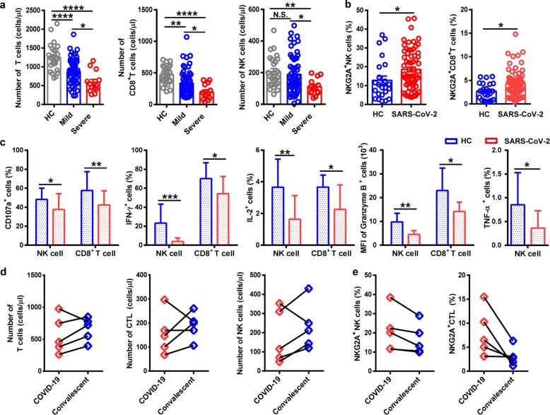
“These results suggest that the functional exhaustion of cytotoxic lymphocytes is associated with the acute phase of SARS-CoV-2 infection [33].”
BackgroundResultInfluentialA dynamic COVID-19 immune signature includes associations with poor prognosis
Laing, A. G., Lorenc, A., del Molino del Barrio, I., Das, A., Fish, M., Monin, L., Muñoz-Ruiz, M., McKenzie, D. R., Hayday, T. S., Francos-Quijorna, I., et al. (2020) · Nature Medicine · doi:10.1038/s41591-020-1038-6 · Cited by 946
COVID-19: immunopathogenesis and Immunotherapeutics
Li, Y., Liu, S., Liu, J., Zhang, Z., Wan, X., Huang, B., Chen, Y., & Zhang, Y. (2020) · Signal Transduction and Targeted Therapy · doi:10.1038/s41392-020-00243-2 · Cited by 962
“Moreover, killer cell lectin-like receptor subfamily C member 1 receptor expression on cytotoxic lymphocytes, including NK and CD8 T cells, is elevated.(34,35) Thus, elevated exhaustion levels and reduced functional diversity of T cells may predict severe progression in patients with COVID-19.”
BackgroundSelf-citationCodo, A. C., Davanzo, G. G., Monteiro, L. D. B., de Souza, G. F., Muraro, S. P., Virgilio-da-Silva, J. V., Prodonoff, J. S., Carregari, V. C., de Biagi Junior, C. A. O., Crunfli, F., et al. (2020) · Cell Metabolism · doi:10.1016/j.cmet.2020.07.007 · Cited by 733
“The cytokine storm is thought to be associated with lymphopenia in COVID-19 patients (Zheng et al., 2020).”
Biomarkers associated with COVID-19 disease progression
Ponti, G., Maccaferri, M., Ruini, C., Tomasi, A., & Özben, T. (2020) · Critical Reviews in Clinical Laboratory Sciences · doi:10.1080/10408363.2020.1770685 · Cited by 920
Proal, A. D., & VanElzakker, M. B. (2021) · Frontiers in Microbiology · doi:10.3389/fmicb.2021.698169 · Cited by 921
De Biasi, S., Meschiari, M., Gibellini, L., Bellinazzi, C., Borella, R., Fidanza, L., Gozzi, L., Iannone, A., Lo Tartaro, D., Mattioli, M., et al. (2020) · Nature Communications · doi:10.1038/s41467-020-17292-4 · Cited by 789
Lee, J. S., Park, S.-W., Jeong, H. W., Ahn, J. Y., Choi, S. J., Lee, H., Choi, B., Nam, S. K., Sa, M., Kwon, J., et al. (2020) · Science Immunology · doi:10.1126/sciimmunol.abd1554 · Cited by 817
COVID-19: Current understanding of its Pathophysiology, Clinical presentation and Treatment
Parasher, A. (2020) · Postgraduate Medical Journal · doi:10.1136/postgradmedj-2020-138577 · Cited by 810
COVID-19 and the human innate immune system
Schultze, J. L., & Aschenbrenner, A. C. (2021) · Cell · doi:10.1016/j.cell.2021.02.029 · Cited by 714
T cell responses in patients with COVID-19
Chen, Z., & John Wherry, E. (2020) · Nature Reviews Immunology · doi:10.1038/s41577-020-0402-6 · Cited by 797
Immune response, inflammation, and the clinical spectrum of COVID-19
García, L. F. (2020) · Frontiers in Immunology · doi:10.3389/fimmu.2020.01441 · Cited by 739
“(67) ↓ CD8, NK (NKG2CD107a IFNγgrzB) ↓↓ CD8, NK (NKG2CD107a IFNγgrzB)”
BackgroundInfluentialMulti-Omics Resolves a Sharp Disease-State Shift between Mild and Moderate COVID-19
Su, Y., Chen, D., Yuan, D., Lausted, C. G., Choi, J., Dai, C. L., Voillet, V., Duvvuri, V. R. S. K., Scherler, K., Troisch, P., et al. (2020) · Cell · doi:10.1016/j.cell.2020.10.037 · Cited by 605
“, 2020), lymphopenia (Cao, 2020), immune cell exhaustion (Zheng et al., 2020), and elevated levels of inflammatory cytokines (Del Valle et al.”
The signal pathways and treatment of cytokine storm in COVID-19
Yang, L., Xie, X., Tu, Z., Fu, J., Xu, D., & Zhou, Y. (2021) · Signal Transduction and Targeted Therapy · doi:10.1038/s41392-021-00679-0 · Cited by 618
“Other investigations showed that the PD-1- or NK group 2 member A-positive NK cells were also significantly increased in the peripheral blood of COVID-19 patients compared to healthy controls.(35,36) Several mechanisms may contribute to SARS-CoV-2-induced depletion and exhaustion of lymphocytes: (1) SARS-CoV-2 may dir…”
BackgroundImmunity, endothelial injury and complement-induced coagulopathy in COVID-19
Perico, L., Benigni, A., Casiraghi, F., Ng, L. F. P., Rénia, L., & Remuzzi, G. (2020) · Nature Reviews Nephrology · doi:10.1038/s41581-020-00357-4 · Cited by 616
Geers, D., Shamier, M. C., Bogers, S., den Hartog, G., Gommers, L., Nieuwkoop, N. N., Schmitz, K. S., Rijsbergen, L. C., van Osch, J. A. T., Dijkhuizen, E., et al. (2021) · Science Immunology · doi:10.1126/sciimmunol.abj1750 · Cited by 529
“In ex vivo studies, NK cells from COVID-19 patients displayed impaired cytotoxicity (64, 65), potentially due to cytokine dysregulation and high plasma levels of IL-6 and TNF-α (66).”
SARS-CoV-2 immunity: review and applications to phase 3 vaccine candidates
Poland, G. A., Ovsyannikova, I. G., & Kennedy, R. B. (2020) · The Lancet · doi:10.1016/s0140-6736(20)32137-1 · Cited by 572
Association of COVID-19 inflammation with activation of the C5a–C5aR1 axis
Carvelli, J., Demaria, O., Vély, F., Batista, L., Chouaki Benmansour, N., Fares, J., Carpentier, S., Thibult, M.-L., Morel, A., Remark, R., et al. (2020) · Nature · doi:10.1038/s41586-020-2600-6 · Cited by 526
Immunopathology of Hyperinflammation in COVID-19
Gustine, J. N., & Jones, D. (2021) · The American Journal of Pathology · doi:10.1016/j.ajpath.2020.08.009 · Cited by 516
“Serum cytokine levels are inversely correlated with CD4þ and CD8þ T-cell counts, and improved cytokine levels have been associated with the reversal of lymphopenia in the convalescent period of recovered patients.(4,8,12,61,63) In decedents who had lymphopenia, autopsy evaluations have revealed the extensive cell deat…”
InfluentialBajaj, V., Gadi, N., Spihlman, A. P., Wu, S. C., Choi, C. H., & Moulton, V. R. (2021) · Frontiers in Physiology · doi:10.3389/fphys.2020.571416 · Cited by 476
Zhao, Q., Meng, M., Kumar, R., Wu, Y., Huang, J., Deng, Y., Weng, Z., & Yang, L. (2020) · International Journal of Infectious Diseases · doi:10.1016/j.ijid.2020.04.086 · Cited by 519
Biochemical characterization of SARS-CoV-2 nucleocapsid protein
Zeng, W., Liu, G., Ma, H., Zhao, D., Yang, Y., Liu, M., Mohammed, A., Zhao, C., Yang, Y., Xie, J., et al. (2020) · Biochemical and Biophysical Research Communications · doi:10.1016/j.bbrc.2020.04.136 · Cited by 480
Self-citationDysregulation of type I interferon responses in COVID-19
Acharya, D., Liu, G., & Gack, M. U. (2020) · Nature Reviews Immunology · doi:10.1038/s41577-020-0346-x · Cited by 454
Pathophysiology of SARS-CoV-2: the Mount Sinai COVID-19 autopsy experience
Bryce, C., Grimes, Z., Pujadas, E., Ahuja, S., Beasley, M. B., Albrecht, R. A., Hernandez, T., Stock, A., Zhao, Z., AlRasheed, M. R., et al. (2021) · Modern Pathology · doi:10.1038/s41379-021-00793-y · Cited by 422
“impairment of NK-cell function, a feature of primary HLH, is described in association with COVID-19 disease [50].”
BackgroundChen, R., Sang, L., Jiang, M., Yang, Z., Jia, N., Fu, W., Xie, J., Guan, W., Liang, W., Ni, Z., et al. (2020) · Journal of Allergy and Clinical Immunology · doi:10.1016/j.jaci.2020.05.003 · Cited by 380
“onset to admission 13 (9-18) 13 (10-18)* 9 (6-17) 13 (9-16) 13 (9-16) 14 (7-18) 15 (8-21) 17 (11-24)* 9 (6-18) 9 (6-23) 23 (12-54)* 7 (3-9)”
Cytokine storm and COVID-19: a chronicle of pro-inflammatory cytokines
Fara, A. F., Mitrev, Z. K., Rosalia, R. A., & Assas, M. B. (2020) · Open Biology · doi:10.1098/rsob.200160 · Cited by 368
“Along with IL-6, IFN-γ has been a reliable indicator of COVID-19 patient deterioration and ICU admission [37,38,41].”
BackgroundBernardes, J. P., Mishra, N., Tran, F., Bahmer, T., Best, L., Blase, J. I., Bordoni, D., Franzenburg, J., Geisen, U. M., Josephs‐Spaulding, J., et al. (2020) · Immunity · doi:10.1016/j.immuni.2020.11.017 · Cited by 362
Detection of COVID-19 Infection from Routine Blood Exams with Machine Learning: A Feasibility Study
Brinati, D., Campagner, A., Ferrari, D., Locatelli, M., Banfi, G., & Cabitza, F. (2020) · Journal of Medical Systems · doi:10.1007/s10916-020-01597-4 · Cited by 365
“Also other studies confirm the relevance of these features and their association with the COVID-19 positivity [8, 30, 32, 44], compared to other kinds of pneumonia [43].”
ResultTwo X-linked agammaglobulinemia patients develop pneumonia as COVID-19 manifestation but recover
Soresina, A., Moratto, D., Chiarini, M., Paolillo, C., Baresi, G., Focà, E., Bezzi, M., Baronio, B., Giacomelli, M., & Badolato, R. (2020) · Pediatric Allergy and Immunology · doi:10.1111/pai.13263 · Cited by 327
COVID-19: towards understanding of pathogenesis
Cao, W., & Li, T. (2020) · Cell Research · doi:10.1038/s41422-020-0327-4 · Cited by 344
COVID-19 and the Immune System
Pačes, J., Strizova, Z. O., Smrž, D., & Černý, J. (2020) · Physiological Research · doi:10.33549/physiolres.934492 · Cited by 353
“In relation to CD8+ T cells, it has been revealed that these cells exhibit functional exhaustion patterns, such as the expression of NKG2A, PD-1, and TIM-3 (Zheng et al. 2020, Moon 2020).”
BackgroundResultInfluentialLiang, Y., Wang, M., Chien, C., Yarmishyn, A. A., Yang, Y.-P., Lai, W.-Y., Luo, Y., Lin, Y.-T., Chen, Y.-J., Chang, P., et al. (2020) · Frontiers in Immunology · doi:10.3389/fimmu.2020.01022 · Cited by 347
“As for adaptive immunity, it is known that the low levels of CD4+ and CD8+ T cells are related to the mortality of SARSCoV-2 patients (104, 105).”
BackgroundLymphopenia in COVID‐19: Therapeutic opportunities
Fathi, N., & Rezaei, N. (2020) · Cell Biology International · doi:10.1002/cbin.11403 · Cited by 324
“The study by M. Zheng et al. (2020) showed that the NK cells and CTLs were reduced significantly in patients with COVID‐19.”
BackgroundResultInfluentialAutoinflammatory and autoimmune conditions at the crossroad of COVID-19
Rodríguez, Y., Novelli, L., Rojas, M., Santis, M. D., Acosta‐Ampudia, Y., Monsalve, D. M., Ramírez-Santana, C., Costanzo, A., Ridgway, W. M., Ansari, A. A., et al. (2020) · Journal of Autoimmunity · doi:10.1016/j.jaut.2020.102506 · Cited by 319
“also been reported [71], as well as an increased expression of their inhibitory receptor NKG2A that likely contributes to their reduced cytolytic activity [72].”
Severe immunosuppression and not a cytokine storm characterizes COVID-19 infections
Remy, K. E., Mazer, M. B., Striker, D. A., Ellebedy, A. H., Walton, A. H., Unsinger, J., Blood, T. M., Mudd, P. A., Yi, D. J., Mannion, D. A., et al. (2020) · JCI Insight · doi:10.1172/jci.insight.140329 · Cited by 309
“6) n 19 17 8 8 10 10 10 10 4 IL-8 Mean (SEM) 118 (21) 327.”
COVID-19 and Solid Organ Transplantation: A Review Article
Azzi, Y., Bartash, R. M., Scalea, J. R., Loarte-Campos, P., & Akalin, E. (2020) · Transplantation · doi:10.1097/tp.0000000000003523 · Cited by 308
“Severe COVID19 patients have depleted peripheral NK cell counts and increased neutrophil-to-lymphocyte ratio compared with mild cases and healthy controls.(21,22) Cellular immunity to coronavirus could be more sustained, and T helper cell responses are critical in the generation of neutralizing antibodies.”
BackgroundEarly predictors of clinical outcomes of COVID-19 outbreak in Milan, Italy
Ciceri, F., Castagna, A., Rovere, P. Q., COBELLI, F. D., Ruggeri, A., Galli, L., Conte, C., LORENZO, R. D., Poli, A., Ambrosio, A. G., et al. (2020) · Clinical Immunology · doi:10.1016/j.clim.2020.108509 · Cited by 294
Meftahi, G. H., Jangravi, Z., Sahraei, H., & Bahari, Z. (2020) · Inflammation Research · doi:10.1007/s00011-020-01372-8 · Cited by 301
“Furthermore, exhaustion markers, such as NKG2A receptors on NK cells and CD8+ T cells, are up-regulated in patients with COVID-19 [28, 31].”
BackgroundFrederiksen, L. S. F., Zhang, Y., Foged, C., & Thakur, A. (2020) · Frontiers in Immunology · doi:10.3389/fimmu.2020.01817 · Cited by 295
“In another study, the function of NK and CD8 T cells was exhausted with the increased expression of natural killer group 2 member A (NKG2A) in COVID19 patients (36).”
BackgroundCOVID-19 and Cancer: a Comprehensive Review
Gosain, R., Abdou, Y. G., Singh, A., Rana, N. K., Puzanov, I. J., & Ernstoff, M. S. (2020) · Current Oncology Reports · doi:10.1007/s11912-020-00934-7 · Cited by 295
Current status of potential therapeutic candidates for the COVID-19 crisis
Zhang, J., Xie, B., & Hashimoto, K. (2020) · Brain Behavior and Immunity · doi:10.1016/j.bbi.2020.04.046 · Cited by 287
“…2020), immune injury (Chen et al., 2020b; Guan et al., 2020; Qin et al., 2020; Wang et al., 2020b; Xu et al., 2020; Yao et al., 2020; Liu et al., 2020b; Zheng et al., 2020), coagulation impairment (Han et al., 2020b; Tang et al., 2020a), and even death (Huang et al., 2020; Wang et al., 2020a) (Fig.”
Shi, L., Huang, H., Lu, X.-C., Yan, X., Jiang, X., Xu, R., Wang, S., Zhang, C., Yuan, X., Xu, Z., et al. (2021) · Signal Transduction and Targeted Therapy · doi:10.1038/s41392-021-00488-5 · Cited by 282
Body Localization of ACE-2: On the Trail of the Keyhole of SARS-CoV-2
Salamanna, F., Maglio, M., Landini, M. P., & Fini, M. (2020) · Frontiers in Medicine · doi:10.3389/fmed.2020.594495 · Cited by 298
“Nevertheless, lymphocytes have an exhaustion phenotype with programmed cell death protein-1 (PD1), T cell immunoglobulin domain and mucin domain-3 (TIM3), and killer cell lectin-like receptor subfamily C member 1 (NKG2A) upregulation (337).”
BackgroundTwo distinct immunopathological profiles in autopsy lungs of COVID-19
Nienhold, R., Ciani, Y., Koelzer, V. H., Tzankov, A., Haslbauer, J. D., Menter, T., Schwab, N., Henkel, M., Frank, A., Zsikla, V., et al. (2020) · Nature Communications · doi:10.1038/s41467-020-18854-2 · Cited by 246
Unique immunological profile in patients with COVID-19
Varchetta, S., Mele, D., Oliviero, B., Mantovani, S., Ludovisi, S., Cerino, A., Bruno, R., Castelli, A., Mosconi, M., Vecchia, M., et al. (2020) · Cellular and Molecular Immunology · doi:10.1038/s41423-020-00557-9 · Cited by 282
Arastehfar, A. A., Carvalho, A., Nguyen, M. H., Hedayati, M. T., Netea, M. G., Perlin, D. S., & Hoenigl, M. (2020) · Journal of Fungi · doi:10.3390/jof6040211 · Cited by 276
“Similarly, an increased peripheral neutrophil-to-lymphocyte ratio was also observed in severe cases of COVID-19, and was likely associated with unfavorable prognosis [35].”
BackgroundRobust T cell immunity in convalescent individuals with asymptomatic or mild COVID-19Preprint
Sekine, T., Perez‐Potti, A., Rivera‐Ballesteros, O., Strålin, K., Gorin, J., Olsson, A., Llewellyn‐Lacey, S., Kamal, H., Bogdanović, G., Muschiol, S., et al. (2020) · bioRxiv (Cold Spring Harbor Laboratory) · doi:10.1101/2020.06.29.174888 · Cited by 272
Untimely TGFβ responses in COVID-19 limit antiviral functions of NK cells
Witkowski, M., Tizian, C., Ferreira‐Gomes, M., Niemeyer, D., Jones, T. C., Heinrich, F., Frischbutter, S., Angermair, S., Hohnstein, T., Mattiola, I., et al. (2021) · Nature · doi:10.1038/s41586-021-04142-6 · Cited by 254
Sánchez‐Cerrillo, I., Landete, P., Orzaiz, B. A., Sánchez‐Alonso, S., Sánchez-Azofra, A., Marcos‐Jiménez, A., Ávalos, E., Alcaraz‐Serna, A., Santos, I. D. L., Mateu‐Albero, T., et al. (2020) · Journal of Clinical Investigation · doi:10.1172/jci140335 · Cited by 250
Guo, Q., Zhao, Y., Li, J., Liu, J., Yang, X., Guo, X., Kuang, M., Xia, H., Zhang, Z., Cao, L., et al. (2020) · Cell Host & Microbe · doi:10.1016/j.chom.2020.12.016 · Cited by 253
“In addition, T cell exhaustion or dysfunction has also been observed (Diao et al., 2020; Zheng et al., 2020a; Zheng et al., 2020b).”
Chemokines and chemokine receptors during COVID-19 infection
Khalil, B. A., Elemam, N. M., & Maghazachi, A. A. (2021) · Computational and Structural Biotechnology Journal · doi:10.1016/j.csbj.2021.01.034 · Cited by 248
“Additionally, circulating CD8 T and NK cells displayed abnormal function and exhaustion, where a negative correlation between perforin content and serum cytokine levels was detected [5,7].”
Cunha, L. L., Perazzio, S. F., Azzi, J., Cravedi, P., & Riella, L. V. (2020) · Frontiers in Immunology · doi:10.3389/fimmu.2020.01748 · Cited by 245
Belsky, J. A., Tullius, B. P., Lamb, M. G., Sayegh, R., Stanek, J. R., & Auletta, J. J. (2021) · Journal of Infection · doi:10.1016/j.jinf.2021.01.022 · Cited by 238
Deciphering the COVID‐19 cytokine storm: Systematic review and meta‐analysis
Mulchandani, R., Lyngdoh, T., & Kakkar, A. K. (2020) · European Journal of Clinical Investigation · doi:10.1111/eci.13429 · Cited by 251
Unraveling the Mystery Surrounding Post-Acute Sequelae of COVID-19
Ramakrishnan, R. K., Kashour, T., Hamid, Q. A., Halwani, R., & Tleyjeh, I. M. (2021) · Frontiers in Immunology · doi:10.3389/fimmu.2021.686029 · Cited by 246
“Notably, a marked decrease in the absolute number and functional exhaustion of anti-viral cytotoxic lymphocytes, including cytotoxic T lymphocytes (CTLs) and natural killer (NK) cells, were reported in patients with SARS-CoV-2 infection, particularly in those with severe disease (60, 61).”
BackgroundImpact of PD-1 Blockade on Severity of COVID-19 in Patients with Lung Cancers
Luo, J., Rizvi, H. A., Egger, J. V., Preeshagul, I. R., Wolchok, J. D., & Hellmann, M. D. (2020) · Cancer Discovery · doi:10.1158/2159-8290.cd-20-0596 · Cited by 236
Baricitinib: A Review of Pharmacology, Safety, and Emerging Clinical Experience in COVID‐19
Jorgensen, S. C. J., Tse, C. L. Y., Burry, L. D., & Dresser, L. (2020) · Pharmacotherapy The Journal of Human Pharmacology and Drug Therapy · doi:10.1002/phar.2438 · Cited by 233
“Our understanding of the pathogenesis of and immune response to COVID-19 is rapidly evolving and, like sepsis, relative immunodeficiency also appears to be at play.(9,44,46,58) At this time, we do not have a reliable way to gauge whether the overruling response is pro or antiinflammatory and this complicates deploymen…”
BackgroundRha, M., Jeong, H. W., Ko, J. H., Choi, S. J., Seo, I., Lee, J. S., Sa, M., Kim, A. R., Joo, E., Ahn, J. Y., et al. (2020) · Immunity · doi:10.1016/j.immuni.2020.12.002 · Cited by 227
“Although many studies have suggested exhausted phenotypes of CD8 T cells on the basis of increased PD-1 expression in patients with COVID-19 (De Biasi et al., 2020; Song et al., 2020; Zheng et al., 2020a; Zheng et al., 2020b), whether CD8 T cells truly become exhausted remains controversial.”
The Innate Immune System: Fighting on the Front Lines or Fanning the Flames of COVID-19?
McKechnie, J. L., & Blish, C. A. (2020) · Cell Host & Microbe · doi:10.1016/j.chom.2020.05.009 · Cited by 218
Intranasal vaccines for SARS-CoV-2: From challenges to potential in COVID-19 management
Chavda, V. P., Vora, L. K., Pandya, A., & Patravale, V. B. (2021) · Drug Discovery Today · doi:10.1016/j.drudis.2021.07.021 · Cited by 216
The Immune Response and Immunopathology of COVID-19
Mortaz, E., Tabarsi, P., Varahram, M., Folkerts, G., & Adcock, I. M. (2020) · Frontiers in Immunology · doi:10.3389/fimmu.2020.02037 · Cited by 210
T‐cell responses and therapies against SARS‐CoV‐2 infection
Toor, S. M., Saleh, R., Nair, V. S., Taha, R. Z., & Elkord, E. (2020) · Immunology · doi:10.1111/imm.13262 · Cited by 208
COVID-19 is a systemic vascular hemopathy: insight for mechanistic and clinical aspects
Smadja, D. M., Mentzer, S. J., Fontenay, M., Laffan, M., Ackermann, M., Helms, J., Jonigk, D. D., Chocron, R., Pier, G. B., Gendron, N., et al. (2021) · Angiogenesis · doi:10.1007/s10456-021-09805-6 · Cited by 204
Carsetti, R., Zaffina, S., Mortari, E. P., Terreri, S., Corrente, F., Capponi, C., Palomba, P., Mirabella, M., Cascioli, S., Palange, P., et al. (2020) · Frontiers in Immunology · doi:10.3389/fimmu.2020.610300 · Cited by 201
“Surviving T cells are functionally exhausted, and reduced T-cell count predicts an unfavorable clinical course (12, 13).”
BackgroundActivation or exhaustion of CD8+ T cells in patients with COVID-19
Rha, M., & Shin, E. (2021) · Cellular and Molecular Immunology · doi:10.1038/s41423-021-00750-4 · Cited by 201
“The functions of CD8+ T cells in patients with COVID-19 Several studies have reported that CD8+ T cells from patients with COVID-19 exhibit a decreased cytokine-producing capacity upon stimulation with PMA/ionomycin [23, 27].”
BackgroundWong, L.-Y. R., & Perlman, S. M. (2021) · Nature reviews. Immunology · doi:10.1038/s41577-021-00656-2 · Cited by 202
Viral persistence, reactivation, and mechanisms of long COVID
Chen, B. K., Jülg, B. D., Mohandas, S., Bradfute, S. B., & RECOVER Mechanistic Pathways Task Force (2023) · eLife · doi:10.7554/elife.86015 · Cited by 211
Could there be a link between oral hygiene and the severity of SARS-CoV-2 infections?
Sampson, V., Kamona, N., & Sampson, A. (2020) · BDJ · doi:10.1038/s41415-020-1747-8 · Cited by 205
The Impact of Pre-existing Comorbidities and Therapeutic Interventions on COVID-19
Callender, L. A., Curran, M., Bates, S. M., Mairesse, M., Weigandt, J., & Betts, C. J. (2020) · Frontiers in Immunology · doi:10.3389/fimmu.2020.01991 · Cited by 195
“For example, elevated programmed cell death protein-1 (PD-1), cytotoxic T-lymphocyte-associated protein-4 (CTLA-4) and T cell Ig and ITM domain (TIGIT) on CD8 T cells and increased NKG2A on NK cells (34, 35).”
BackgroundGadi, N., Wu, S., Spihlman, A. P., & Moulton, V. R. (2020) · Frontiers in Immunology · doi:10.3389/fimmu.2020.02147 · Cited by 188
“Their exhaustion is correlated with disease progression (27), as they are primary secretors of IFN-γ, TNF-α, colony stimulating factors (CSF), and many other cytokines (15, 27).”
BackgroundT Cells: Warriors of SARS-CoV-2 Infection
de Candia, P., Prattichizzo, F., Garavelli, S., & Matarese, G. (2020) · Trends in Immunology · doi:10.1016/j.it.2020.11.002 · Cited by 189
“Accordingly, protein expression of three exhaustion markers, that is, the CD28 family member programmed cell death marker 1 (PD-1), the receptor mucin domaincontaining protein-3 (TIM-3), and the ITIM-bearing receptor NKG2A [68–70] were increased in both peripheral CD4 and CD8 T cells from severe COVID-19 patients, a f…”
Innate immunological pathways in COVID-19 pathogenesis
Paludan, S. R., & Mogensen, T. H. (2022) · Science Immunology · doi:10.1126/sciimmunol.abm5505 · Cited by 188
“However, at the same time they also display markers of functional exhaustion and down-regulation of genes associated with cytoxicity (39, 41, 42), thus demonstrating a central similarity to HLH (43).”
ResultSelenium and selenoproteins in viral infection with potential relevance to COVID-19
Zhang, J., Saad, R., Taylor, E. W., & Rayman, M. P. (2020) · Redox Biology · doi:10.1016/j.redox.2020.101715 · Cited by 191
““Functional exhaustion” of natural killer (NK) Cells and cytotoxic T-cells has also been identified as a factor in severe COVID-19 [65].”
I mmunosenescence and Inflammaging: Risk Factors of Severe COVID-19 in Older People
Pietrobon, A. J., Teixeira, F. M. E., & Sato, M. N. (2020) · Frontiers in Immunology · doi:10.3389/fimmu.2020.579220 · Cited by 187
“reported impaired NK function in severe COVID-19 patients, expressing higher levels of the NKG2A receptor, a cellular exhaustion marker, indicating impaired antiviral immunity (150).”
BackgroundHeming, M., Li, X., Räuber, S. J., Mausberg, A. K., Börsch, A., Hartlehnert, M., Singhal, A., Lu, I., Fleischer, M., Szepanowski, F., et al. (2020) · Immunity · doi:10.1016/j.immuni.2020.12.011 · Cited by 181
Islam, H., Chamberlain, T. C., Mui, A., & Little, J. P. (2021) · Frontiers in Immunology · doi:10.3389/fimmu.2021.677008 · Cited by 191
“IL-10mediated hyperactivation of CD8+ T cells despite an overall reduced cell count may also explain why some studies report functional exhaustion of T cells in severe COVID-19 cases (25) and significant inverse associations between serum IL-10 levels and T cell count (26).”
BackgroundHoagland, D. A., Møller, R., Uhl, S. A., Oishi, K., Frere, J. J., Golynker, I., Horiuchi, S., Panis, M., Blanco-Melo, D., Sachs, D. M., et al. (2021) · Immunity · doi:10.1016/j.immuni.2021.01.017 · Cited by 177
“This juxtaposition of low IFN-I and high chemokines may be responsible for enhanced infiltration of neutrophils and monocytes to the respiratory tract, resulting in COVID-19 (Coperchini et al., 2020; Zheng et al., 2020a).”
Natural Killer Cell Dysfunction and Its Role in COVID-19
van Eeden, C., Khan, L., Osman, M. S., & Tervaert, J. W. C. (2020) · International Journal of Molecular Sciences · doi:10.3390/ijms21176351 · Cited by 179
“SARS-CoV-2 [34,35,84] infection in most cases is characterised by mild symptoms of fever, tiredness and dry cough.”
BackgroundInfluentialWang, X., Xu, W., Hu, G., Xia, S., Sun, Z., Liu, Z., Xie, Y., Zhang, R., Jiang, S., & Lu, L. (2020) · Cellular and Molecular Immunology · doi:10.1038/s41423-020-0498-4 · Cited by 175
“4, 5 Lymphocytopenia was also found in the Middle East respiratory syndrome (MERS) cases.”
BackgroundRetractedRethinking the role of hydroxychloroquine in the treatment of COVID‐19
Meyerowitz, E. A., Vannier, A., Friesen, M. G. N., Schoenfeld, S. R., Gelfand, J. A., Callahan, M. V., Kim, A. Y., Reeves, P. M., & Poznansky, M. C. (2020) · The FASEB Journal · doi:10.1096/fj.202000919 · Cited by 174
Potential benefits and risks of omega-3 fatty acids supplementation to patients with COVID-19
Rogero, M. M., Leão, M. D. C., Santana, T. M., Pimentel, M. V. D. M. B., Carlini, G. C. G., da Silveira, T. F. F., Gonçalves, R. C., & de Castro, I. A. (2020) · Free Radical Biology and Medicine · doi:10.1016/j.freeradbiomed.2020.07.005 · Cited by 181
“[28] observed lower levels of Cytotoxic CD8 lymphocytes in critically ill patients compared to mild ones.”
Is pregnancy a risk factor of COVID-19?
Phoswa, W. N., & Khaliq, O. P. (2020) · European Journal of Obstetrics & Gynecology and Reproductive Biology · doi:10.1016/j.ejogrb.2020.06.058 · Cited by 173
“Several studies have associated increased NKG2A expression with normal pregnancy development [24,29,33].”
InfluentialSokołowska, M., Łukasik, Z. M., Agache, I. O., Akdiş, C. A., Akdiş, D., Akdiş, M., Barcik, W., Brough, H. A., Eiwegger, T., Eljaszewicz, A., et al. (2020) · Allergy · doi:10.1111/all.14462 · Cited by 170
Wu, H., Zhu, H., Yuan, C., Yao, C., Luo, W. P., Shen, X., Wang, J., Shao, J., & Xiang, Y. (2020) · JAMA Network Open · doi:10.1001/jamanetworkopen.2020.10895 · Cited by 168
“048), C4 complement (median [IQR], 18 [13-23] mg/dL vs 22 [16-30] mg/dL; P = .”
BackgroundMicrostructure, pathophysiology, and potential therapeutics of COVID‐19: A comprehensive review
Singh, S. P., Pritam, M., Pandey, B., & Yadav, T. P. (2021) · Journal of Medical Virology · doi:10.1002/jmv.26254 · Cited by 166
Gyöngyösi, M., Alcaide, P., Asselbergs, F. W., Brundel, B. J. J. M., Camici, G. G., Martins, P. A. D. C., Ferdinandy, P., Fontana, M., Girão, H., Gnecchi, M., et al. (2022) · Cardiovascular Research · doi:10.1093/cvr/cvac115 · Cited by 175
Peripheral CD4+ T cell subsets and antibody response in COVID-19 convalescent individuals
Gong, F., Dai, Y., Zheng, T., Cheng, L., Zhao, D., Wang, H., Liu, M., Pei, H., Jin, T., Yu, D., et al. (2020) · Journal of Clinical Investigation · doi:10.1172/jci141054 · Cited by 160
“However, CD8+ T cells often exhibit exhausted phenotypes in this disease (7, 8), together with markedly reduced cell counts in some severe patients.”
BackgroundWang, X., Xu, W., Hu, G., Xia, S., Sun, Z., Liu, Z., Xie, Y., Zhang, R., Jiang, S., & Lu, L. (2020) · Cellular and Molecular Immunology · doi:10.1038/s41423-020-0424-9 · Cited by 162
“Some studies reported that lymphocytopenia might be related to mortality, especially in patients with low levels of CD3, CD4, and CD8 T lymphocytes.(4,5) Lymphocytopenia was also found in the Middle East respiratory syndrome (MERS) cases.”
BackgroundRetracted
Metadata
- Authors
- Zheng, M., Gao, Y., Wang, G., Song, G., Liu, S., Sun, D., Xu, Y., & Tian, Z.
- Affiliations
- First Affiliated Hospital of Anhui Medical University (CN)
- Anhui Medical University (CN)
- University of Science and Technology of China (CN)
- Corresponding author
- Zheng, Meijuan
- Xu, Yuanhong
- Tian, Zhigang
- Venue
- Cellular & Molecular Immunology
- Published
- 2020-03-19
- Volume
- 17
- Issue
- 5
- Pages
- 533-535
- ISSN
- 1672-7681 (ISSN-L)
- 2042-0226
- PMC
- PMC7091858
- OpenAlex
- W3011508332
- Cited by
- 1,768
- References
- 11
- Citations by year
- 2026: 26 · 2025: 63 · 2024: 119 · 2023: 186 · 2022: 329 · 2021: 514 · 2020: 531
- Type
- journal article
- Language
- en
- Open access
- hybrid
- doi.org
- Locations
- Cellular and Molecular Immunology (publishedVersion, cc-by)
- PubMed Central (publishedVersion, cc-by)
- PubMed (publishedVersion)
- Funders
- National Natural Science Foundation of China (81788101, 81771685)
- Chinese Academy of Sciences (XDB29030000, 81788101)
- Anhui Medical University (YJGG202002)
- Topics
- Health Sciences › Medicine › Infectious Diseases › SARS-CoV-2 and COVID-19 Research
- Health Sciences › Medicine › Infectious Diseases › COVID-19 Clinical Research Studies
- Life Sciences › Immunology and Microbiology › Immunology › T-cell and B-cell Immunology
- Keywords
- COVID-19 pandemic, SARS-CoV-2, T cell exhaustion, T cell dysfunction
- MeSH terms
- Betacoronavirus, COVID-19, SARS-CoV-2, Antiviral Agents, Hepatitis C, Humans, Killer Cells, Natural, Pneumonia, Viral, Coronavirus Infections, Pandemics
About these checks
Registry, retraction, Cites and statistics results are computed, not judged, with one exception: a printed reference that no index lists is read once by a model before it counts as a citation error. Claims are extracted by a model and kept only when their quote appears verbatim in the text; each cited source is then checked, with a model’s help, for whether it says what the claim relies on. Those verdicts can be wrong — read the claim’s quote and the cited source before relying on one.
Statistics and arithmetic are recomputed, not judged.
Confidence bands are shown only for fields calibrated on our annotated test set. None is calibrated yet, so no band is shown.
What counts as a claim ›How we measure confidence ›How we check this ›
Are you an author, or do you see a problem with this page? Request a review
