

The DOI matches its Crossref record; 62 of the 62 references it cites were found in a registry or an index; 4 of the 27 sources its claims cite were checked for support.
DRP1-mediated mitochondrial shape controls calcium homeostasis and muscle mass
Open accessAbstract
Mitochondrial quality control is essential in highly structured cells such as neurons and muscles. In skeletal muscle the mitochondrial fission proteins are reduced in different physiopathological conditions including ageing sarcopenia, cancer cachexia and chemotherapy-induced muscle wasting. However, whether mitochondrial fission is essential for muscle homeostasis is still unclear. Here we show that muscle-specific loss of the pro-fission dynamin related protein (DRP) 1 induces muscle wasting and weakness. Constitutive Drp1 ablation in muscles reduces growth and causes animal death while inducible deletion results in atrophy and degeneration. Drp1 deficient mitochondria are morphologically bigger and functionally abnormal. The dysfunctional mitochondria signals to the nucleus to induce the ubiquitin-proteasome system and an Unfolded Protein Response while the change of mitochondrial volume results in an increase of mitochondrial Ca 2+ uptake and myofiber death. Our findings reveal that morphology of mitochondrial network is critical for several biological processes that control nuclear programs and Ca 2+ handling.
Overview
AI snapshot
Written from the paper’s full text.
TL;DR
Muscle-specific loss of DRP1 in mice causes a lethal mitochondrial myopathy with atrophy, weakness, and altered calcium homeostasis. DRP1-deficient mitochondria are larger and dysfunctional, triggering UPR/UPS activation, blocked autophagy/mitophagy, and increased MCU-mediated Ca²⁺ uptake that contributes to myofiber death.
Key findings
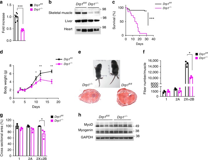
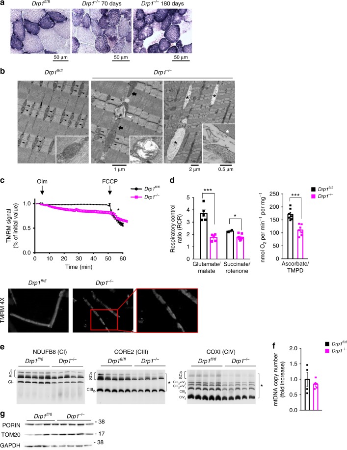
- Constitutive muscle-specific Drp1 deletion caused postnatal lethality within 30 days (WT n = 43; KO n = 14), reduced body growth, fewer glycolytic 2B/2X fibers, smaller cross-sectional area, and a 30% reduction in protein synthesis.
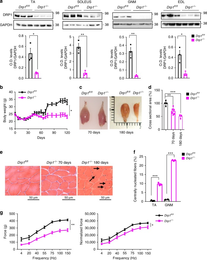
- Inducible adult Drp1 deletion reduced fiber size by 25% at 70 days and 50% at 180 days, decreased absolute and specific force, and produced center-nucleated fibers in 10% of tibialis anterior and 25% of gastrocnemius myofibers at 180 days.
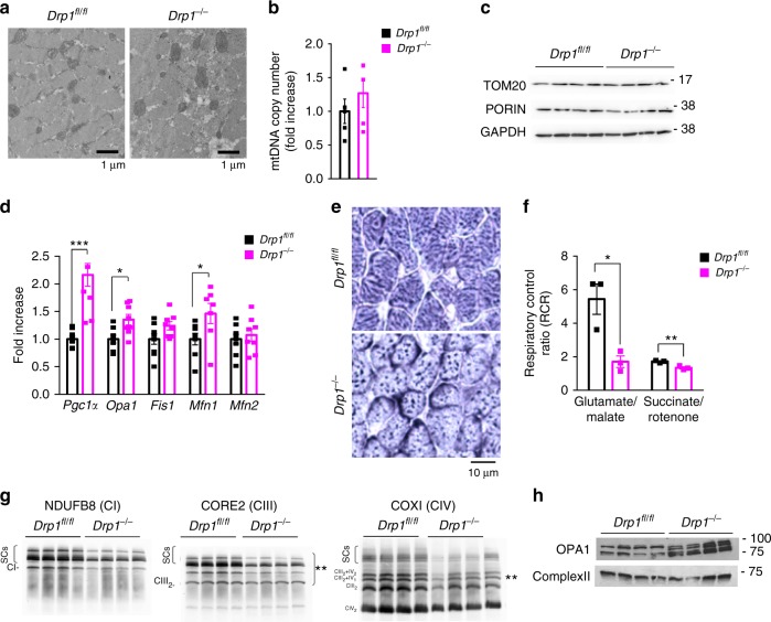
- Drp1-deficient mitochondria were larger and fewer with unchanged mtDNA content and mitochondrial mass, but showed reduced respiration (complex-I-driven respiration most affected), decreased respiratory chain supercomplexes, increased OPA1 cleavage, and in acute deletion about 10% damaged mitochondria versus 2% in WT.
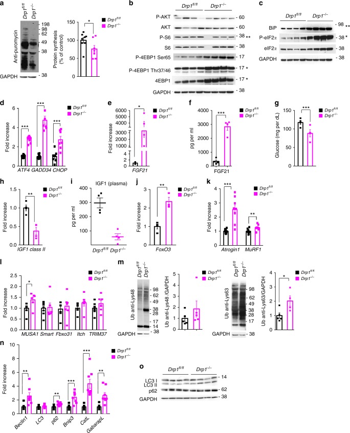
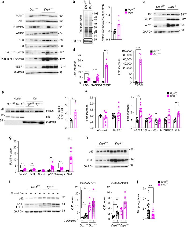
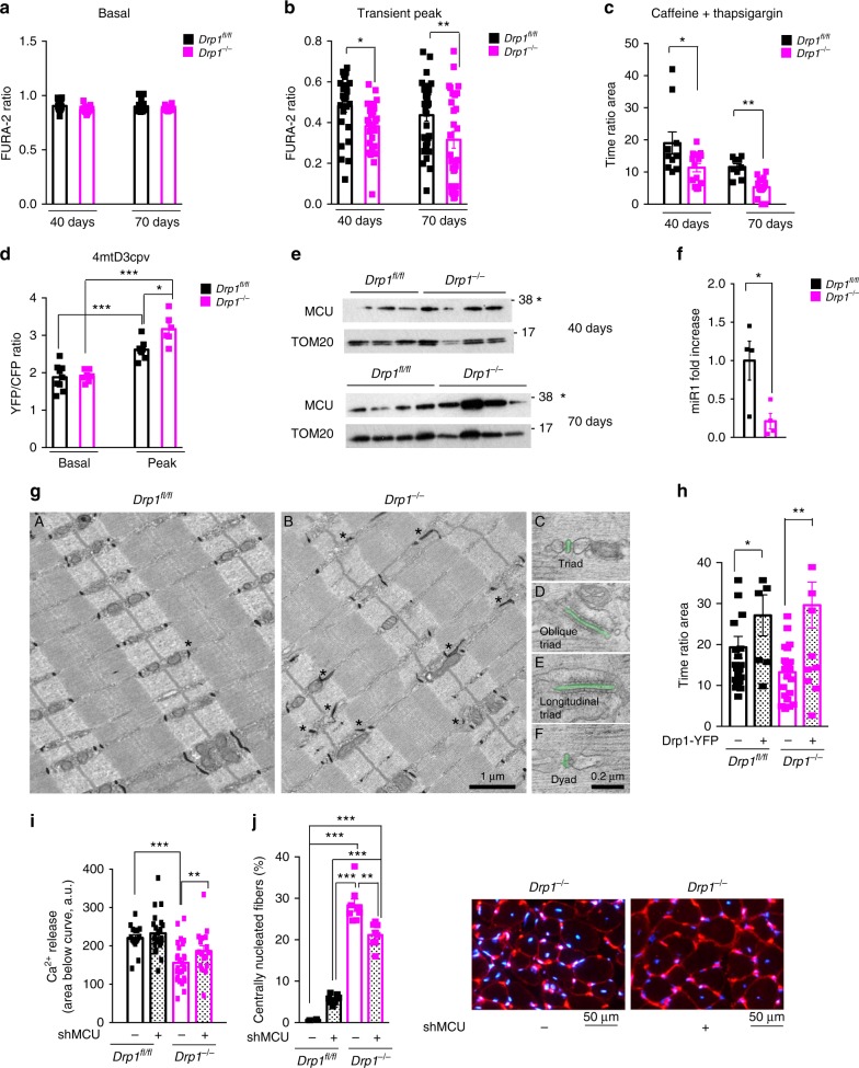
- DRP1 loss activated UPR (Bip/Grp78, p-eIF2α, ATF4, FGF21; FGF21 blood increased 20-fold in acute KO), induced FoxO3/UPS, blocked autophagy/mitophagy, and increased mitochondrial Ca²⁺ uptake via MCU upregulation and miR1 downregulation; MCU knockdown normalized cytosolic Ca²⁺ and reduced center-nucleated fibers.
Methods
- Two muscle-specific Drp1 knockout models: constitutive Drp1fl/fl × MLC1f-Cre mice analyzed at postnatal day 12, and tamoxifen-inducible Drp1fl/fl × HSA-Cre-ER mice treated at 5 months of age; Cre-negative littermates served as controls.
- Readouts included electron microscopy/stereology, histology and SDH staining, RT-qPCR, immunoblotting, mitochondrial respiration and Blue Native PAGE, SUnSET protein synthesis, TMRM membrane potential, Fura-2 and cameleon Ca²⁺ probes, in vivo/ex vivo force and fatigue measurements, mito-Keima mitophagy assay, MCU knockdown, and plasma FGF21/IGF1 measurements.
- Data are means ± SEM; statistical analysis used two-tailed unpaired Student’s t test or two-way ANOVA with Bonferroni’s multiple comparison test, with significance at p < 0.05.
Limitations
- Stated: MCU knockdown did not completely block degeneration/regeneration because transfection efficiency was low, so MCU was reduced only in some myofibers.
- Evident: all findings are from mouse models; no human patient validation is reported.
- Evident: some analyses used very small n (e.g., WT n = 2 for succinate/rotenone respiration in Fig. 5d), and the causal role of Ca²⁺ dysregulation is supported by rescue/knockdown experiments but not fully isolated from other DRP1-dependent defects.
Model-written summary. Check it against the paper before relying on it.
Integrity signals
Registry
Cites
Statistics
Figures
Verification
Claims the paper makes
36 Own result · 22 Own interpretation · 13 Not checked yet · 8 Own method · 5 No basis given · 1 Partly supported · accuracy not yet measured
The cited sources support only part of this claim — read each source’s verdict below.
In the paper
“In skeletal muscle the mitochondrial fission proteins are reduced in different physiopathological conditions including ageing sarcopenia, cancer cachexia and chemotherapy-induced muscle wasting.”
- Also stated in: § Introduction · plain
“In cancer cachexia and in chemotherapy-induced cachexia, DRP1, OPA1, and Mitofusin2 are dramatically suppressed16–18.”
§ DRP1-mediated mitochondrial shape controls calcium homeostasis and muscle mass > Abstract
What the sources say
- Partly supportedcites [13] Tezze, C. et al. Age-associated loss of OPA1 in muscle impacts muscle mass, metabolic homeostasis, systemic inflammation, and epithelial senescence. Cell Metab. 25, 1374–1389 e1376 (2017). sourceExperimental — accuracy not yet measured
The Results section, “Age-Related Muscle Loss in Humans and Mice Is Associated with Decreased OPA1 Expression” (page unavailable), reports reduced DRP1 transcripts and protein in muscle biopsies from older sedentary sarcopenic subjects. The passages from our copy do not establish reduced fission proteins in cancer cachexia or chemotherapy-induced muscle wasting. (LLM, conf=0.88)
- Partly supportedcites [14] Zhou, J. et al. Changes in macroautophagy, chaperone-mediated autophagy, and mitochondrial metabolism in murine skeletal and cardiac muscle during aging. Aging (Albany NY) 9, 583–599 (2017). sourceExperimental — accuracy not yet measured
The Results section ('Decreased markers for mitochondrial quality control in muscle but not heart in aged mice'; page unavailable) reports reduced Drp1, a mitochondrial fission protein, in skeletal muscle from aged mice. The paper does not report reduced fission proteins in cancer cachexia or chemotherapy-induced muscle wasting, and its finding in aged mice does not specifically establish sarcopenia. (LLM, conf=0.88)
- Partly supportedcites [16] Barreto, R. et al. Cancer and chemotherapy contribute to muscle loss by activating common signaling pathways. Front Physiol. 7, 472 (2016). sourceExperimental — accuracy not yet measured
The Discussion reports decreased expression of mitochondrial fusion and fission markers in skeletal muscle from C26 tumor-bearing mice and mice treated with chemotherapy. It does not report that fission proteins are reduced in ageing sarcopenia, so the claim's third condition is unsupported by this paper. (LLM, conf=0.88)
- Not checked yetcites [17] van der Ende, M. et al. Mitochondrial dynamics in cancer-induced cachexia. Biochimica et biophysica acta. Rev. cancer 1870, 137–150 (2018).
- Partly supportedcites [18] Brown, J. L. et al. Mitochondrial degeneration precedes the development of muscle atrophy in progression of cancer cachexia in tumour-bearing mice. J. Cachex-. Sarcopenia Muscle 8, 926–938 (2017). sourceExperimental — accuracy not yet measured
The Results section (‘Lewis lung carcinoma impairs mitochondrial quality control’; page not provided) reports that Drp1 was about 45% lower in cachectic mice, supporting a reduction in one fission protein in cancer cachexia. But the other measured fission protein, Fis1, was about 80% higher. The paper does not report fission-protein reductions in ageing sarcopenia or chemotherapy-induced muscle wasting. (LLM, conf=0.90)
plainstates an association- Asserted: passes — The authors put it forward as true.
- Checkable: passes — It can be checked against evidence.
- Load-bearing: unclear — It is unclear whether the paper’s conclusions depend on it.
- Based: passes — The paper gives a reason why it holds.
- Also stated in: § Introduction · plain
A result the paper reports from its own data — it cites no other source for it, so there is none to check. The quote shows it is in the paper.
In the paper
“FGF21 blood levels were also increased of 20-fold in DRP1 knockout mice when compared with controls”
§ DRP1-mediated mitochondrial shape controls calcium homeostasis and muscle mass > Results > Acute deletion of Drp1 induces ER stress and blocks autophagy
plainstates an association- Asserted: passes — The authors put it forward as true.
- Checkable: passes — It can be checked against evidence.
- Load-bearing: unclear — It is unclear whether the paper’s conclusions depend on it.
- Based: passes — The paper gives a reason why it holds.
A result the paper reports from its own data — it cites no other source for it, so there is none to check. The quote shows it is in the paper.
In the paper
“inducible deletion results in atrophy and degeneration”
§ DRP1-mediated mitochondrial shape controls calcium homeostasis and muscle mass > Abstract
plainstates a cause- Asserted: passes — The authors put it forward as true.
- Checkable: passes — It can be checked against evidence.
- Load-bearing: passes — The paper’s conclusions depend on it.
- Based: passes — The paper gives a reason why it holds.
A result the paper reports from its own data — it cites no other source for it, so there is none to check. The quote shows it is in the paper.
In the paper
“while atrogin1 and MuRF1 did not change”
§ DRP1-mediated mitochondrial shape controls calcium homeostasis and muscle mass > Results > Acute deletion of Drp1 induces ER stress and blocks autophagy
plain- Asserted: passes — The authors put it forward as true.
- Checkable: passes — It can be checked against evidence.
- Load-bearing: unclear — It is unclear whether the paper’s conclusions depend on it.
- Based: passes — The paper gives a reason why it holds.
A result the paper reports from its own data — it cites no other source for it, so there is none to check. The quote shows it is in the paper.
In the paper
“we found an upregulation of most of these genes”
§ DRP1-mediated mitochondrial shape controls calcium homeostasis and muscle mass > Results > Acute deletion of Drp1 induces ER stress and blocks autophagy
plainstates a cause- Asserted: passes — The authors put it forward as true.
- Checkable: passes — It can be checked against evidence.
- Load-bearing: unclear — It is unclear whether the paper’s conclusions depend on it.
- Based: passes — The paper gives a reason why it holds.
A result the paper reports from its own data — it cites no other source for it, so there is none to check. The quote shows it is in the paper.
In the paper
“protein synthesis was reduced by 30% in Drp1−/− mice”
§ DRP1-mediated mitochondrial shape controls calcium homeostasis and muscle mass > Results > Drp1 deletion reduces protein synthesis and induces atrophy
plainstates a cause- Asserted: passes — The authors put it forward as true.
- Checkable: passes — It can be checked against evidence.
- Load-bearing: passes — The paper’s conclusions depend on it.
- Based: passes — The paper gives a reason why it holds.
A result the paper reports from its own data — it cites no other source for it, so there is none to check. The quote shows it is in the paper.
In the paper
“Absence of DRP1 induces mitochondrial depolarization.”
§ DRP1-mediated mitochondrial shape controls calcium homeostasis and muscle mass > Results > Acute deletion of Drp1 causes mitochondrial dysfunction
plainstates a cause- Asserted: passes — The authors put it forward as true.
- Checkable: passes — It can be checked against evidence.
- Load-bearing: passes — The paper’s conclusions depend on it.
- Based: passes — The paper gives a reason why it holds.
A result the paper reports from its own data — it cites no other source for it, so there is none to check. The quote shows it is in the paper.
In the paper
“we initially measured cytosolic calcium of Drp1−/− and controls in basal condition and found no difference”
§ DRP1-mediated mitochondrial shape controls calcium homeostasis and muscle mass > Results > Inhibition of DRP1 alters normal calcium homeostasis
plain- Asserted: passes — The authors put it forward as true.
- Checkable: passes — It can be checked against evidence.
- Load-bearing: passes — The paper’s conclusions depend on it.
- Based: passes — The paper gives a reason why it holds.
A result the paper reports from its own data — it cites no other source for it, so there is none to check. The quote shows it is in the paper.
In the paper
“we showed an increase of MCU in mitochondria of DRP1-deficient mice”
§ DRP1-mediated mitochondrial shape controls calcium homeostasis and muscle mass > Results > Inhibition of DRP1 alters normal calcium homeostasis
boostedstates an association- Asserted: passes — The authors put it forward as true.
- Checkable: passes — It can be checked against evidence.
- Load-bearing: passes — The paper’s conclusions depend on it.
- Based: passes — The paper gives a reason why it holds.
A result the paper reports from its own data — it cites no other source for it, so there is none to check. The quote shows it is in the paper.
In the paper
“we found no increase of p62 and LC3II upon colchicine treatment in DRP1-deficient mice”
§ DRP1-mediated mitochondrial shape controls calcium homeostasis and muscle mass > Results > Acute deletion of Drp1 induces ER stress and blocks autophagy
plain- Asserted: passes — The authors put it forward as true.
- Checkable: passes — It can be checked against evidence.
- Load-bearing: passes — The paper’s conclusions depend on it.
- Based: passes — The paper gives a reason why it holds.
- It may state two separate claims as one. 36
- Probably not a substantial claim. 14
A result the paper reports from its own data — it cites no other source for it, so there is none to check. The quote shows it is in the paper.
In the paper
“we found an activation of the UPR pathway. Bip/Grp78 and p-eIF2α (Fig. 6c; Supplementary Fig. 7a) and the transcripts of ATF4 and downstream genes, including FGF21, were induced in DRP1-null muscles”
§ DRP1-mediated mitochondrial shape controls calcium homeostasis and muscle mass > Results > Acute deletion of Drp1 induces ER stress and blocks autophagy
plainstates a cause- Asserted: passes — The authors put it forward as true.
- Checkable: passes — It can be checked against evidence.
- Load-bearing: passes — The paper’s conclusions depend on it.
- Based: passes — The paper gives a reason why it holds.
It may state two separate claims as one.
A result the paper reports from its own data — it cites no other source for it, so there is none to check. The quote shows it is in the paper.
In the paper
“we did not detect an increase of lipidated LC3II, but instead we found an accumulation of LC3I”
§ DRP1-mediated mitochondrial shape controls calcium homeostasis and muscle mass > Results > Acute deletion of Drp1 induces ER stress and blocks autophagy
plain- Asserted: passes — The authors put it forward as true.
- Checkable: passes — It can be checked against evidence.
- Load-bearing: passes — The paper’s conclusions depend on it.
- Based: passes — The paper gives a reason why it holds.
It may state two separate claims as one.
A result the paper reports from its own data — it cites no other source for it, so there is none to check. The quote shows it is in the paper.
In the paper
“Ablation of DRP1 in adulthood modifies mitochondrial shape and reduces RCS assembly, Complex-I-, Complex-II-, and complex-IV-dependent respiration.”
§ DRP1-mediated mitochondrial shape controls calcium homeostasis and muscle mass > Results > Acute deletion of Drp1 causes mitochondrial dysfunction
plainstates a causen = 5- Asserted: passes — The authors put it forward as true.
- Checkable: passes — It can be checked against evidence.
- Load-bearing: passes — The paper’s conclusions depend on it.
- Based: passes — The paper gives a reason why it holds.
It may state two separate claims as one.
A result the paper reports from its own data — it cites no other source for it, so there is none to check. The quote shows it is in the paper.
In the paper
“Here we show that muscle-specific loss of the pro-fission dynamin related protein (DRP) 1 induces muscle wasting and weakness.”
§ DRP1-mediated mitochondrial shape controls calcium homeostasis and muscle mass > Abstract
plainstates a cause- Asserted: passes — The authors put it forward as true.
- Checkable: passes — It can be checked against evidence.
- Load-bearing: passes — The paper’s conclusions depend on it.
- Based: passes — The paper gives a reason why it holds.
It may state two separate claims as one.
A result the paper reports from its own data — it cites no other source for it, so there is none to check. The quote shows it is in the paper.
In the paper
“Drp1 deficient mitochondria are morphologically bigger and functionally abnormal.”
§ DRP1-mediated mitochondrial shape controls calcium homeostasis and muscle mass > Abstract
plainstates a cause- Asserted: passes — The authors put it forward as true.
- Checkable: passes — It can be checked against evidence.
- Load-bearing: passes — The paper’s conclusions depend on it.
- Based: passes — The paper gives a reason why it holds.
It may state two separate claims as one.
A result the paper reports from its own data — it cites no other source for it, so there is none to check. The quote shows it is in the paper.
In the paper
“SDH staining indicating the presence of bigger mitochondria in KO muscles compared with control.”
- Also stated in: § Acute deletion of Drp1 causes mitochondrial dysfunction · plain
“Ablation of DRP1 in adulthood modifies mitochondrial shape and reduces RCS assembly, Complex-I-, Complex-II-, and complex-IV-dependent respiration.”
§ DRP1-mediated mitochondrial shape controls calcium homeostasis and muscle mass > Results > Acute deletion of Drp1 causes mitochondrial dysfunction
plainstates a cause- Asserted: passes — The authors put it forward as true.
- Checkable: passes — It can be checked against evidence.
- Load-bearing: passes — The paper’s conclusions depend on it.
- Based: passes — The paper gives a reason why it holds.
It may state two separate claims as one.
- Also stated in: § Acute deletion of Drp1 causes mitochondrial dysfunction · plain
A result the paper reports from its own data — it cites no other source for it, so there is none to check. The quote shows it is in the paper.
In the paper
“Mitochondrial matrix-free calcium concentration is not affected at rest but is higher in Drp1−/− compared with control during a train of electrical stimulation at 60 Hz, 2 s duration”
- Also stated in: § Inhibition of DRP1 alters normal calcium homeostasis · plain
“the peak of Ca2+ concentration reached during high-frequency electrical stimulation was higher in DRP1-deficient myofibers”
§ DRP1-mediated mitochondrial shape controls calcium homeostasis and muscle mass > Results > Inhibition of DRP1 alters normal calcium homeostasis
plainstates an association- Asserted: passes — The authors put it forward as true.
- Checkable: passes — It can be checked against evidence.
- Load-bearing: passes — The paper’s conclusions depend on it.
- Based: passes — The paper gives a reason why it holds.
It may state two separate claims as one.
- Also stated in: § Inhibition of DRP1 alters normal calcium homeostasis · plain
A result the paper reports from its own data — it cites no other source for it, so there is none to check. The quote shows it is in the paper.
In the paper
“Constitutive Drp1 ablation in muscles reduces growth and causes animal death”
§ DRP1-mediated mitochondrial shape controls calcium homeostasis and muscle mass > Abstract
plainstates a cause- Asserted: passes — The authors put it forward as true.
- Checkable: passes — It can be checked against evidence.
- Load-bearing: passes — The paper’s conclusions depend on it.
- Based: passes — The paper gives a reason why it holds.
It may state two separate claims as one.
A result the paper reports from its own data — it cites no other source for it, so there is none to check. The quote shows it is in the paper.
In the paper
“the Akt-mTOR axis appeared inconsistent with the atrophic phenotype being 4EBP1 robustly hyper-phosphorylated in two mTOR-dependent sites and S6 unchanged”
§ DRP1-mediated mitochondrial shape controls calcium homeostasis and muscle mass > Results > Acute deletion of Drp1 induces ER stress and blocks autophagy
hedgedstates a cause- Asserted: passes — The authors put it forward as true.
- Checkable: passes — It can be checked against evidence.
- Load-bearing: passes — The paper’s conclusions depend on it.
- Based: passes — The paper gives a reason why it holds.
It may state two separate claims as one.
A result the paper reports from its own data — it cites no other source for it, so there is none to check. The quote shows it is in the paper.
In the paper
“These changes occurred early after 40 days from tamoxifen treatment when animals have not yet lost muscle mass and progressively increased at 70 days.”
§ DRP1-mediated mitochondrial shape controls calcium homeostasis and muscle mass > Results > Inhibition of DRP1 alters normal calcium homeostasis
plainstates an association- Asserted: passes — The authors put it forward as true.
- Checkable: passes — It can be checked against evidence.
- Load-bearing: passes — The paper’s conclusions depend on it.
- Based: passes — The paper gives a reason why it holds.
It may state two separate claims as one.
A result the paper reports from its own data — it cites no other source for it, so there is none to check. The quote shows it is in the paper.
In the paper
“the inflammatory response is not present and oxidative stress is not increased in DRP1 knockout mice (Supplementary Figs. 9f, 6e).”
§ DRP1-mediated mitochondrial shape controls calcium homeostasis and muscle mass > Discussion
plain- Asserted: passes — The authors put it forward as true.
- Checkable: passes — It can be checked against evidence.
- Load-bearing: passes — The paper’s conclusions depend on it.
- Based: passes — The paper gives a reason why it holds.
It may state two separate claims as one.
A result the paper reports from its own data — it cites no other source for it, so there is none to check. The quote shows it is in the paper.
In the paper
“DRP1 overexpression reverted or induced UPR in DRP1 knockout and control mice, respectively”
§ DRP1-mediated mitochondrial shape controls calcium homeostasis and muscle mass > Results > DRP1 rescue or MCU inhibition normalizes Ca2+ homeostasis
plainstates a cause- Asserted: passes — The authors put it forward as true.
- Checkable: passes — It can be checked against evidence.
- Load-bearing: passes — The paper’s conclusions depend on it.
- Based: passes — The paper gives a reason why it holds.
It may state two separate claims as one.
The paper states this without a source or data behind it here, so there is nothing to check it against. The quote shows it is in the paper.
In the paper
“Under these assumptions, the resting cytosolic calcium concentration will be 71 nM at R = 0.87 and 87 nM at R = 0.92.”
§ DRP1-mediated mitochondrial shape controls calcium homeostasis and muscle mass > Methods > Free calcium concentration in the cytosol
hedgedstates an association- Asserted: passes — The authors put it forward as true.
- Checkable: passes — It can be checked against evidence.
- Load-bearing: fails — The paper’s conclusions may not depend on it.
- Based: passes — The paper gives a reason why it holds.
Probably not a substantial claim. The paper’s conclusions may not depend on it. It may state two separate claims as one.
- The authors state this as an assumption: The calibration equation and constants (KD = 145 nM, Rmin, Rmax, β) used to convert Fura-2 fluorescence ratios to free cytosolic calcium concentration are valid.
A result the paper reports from its own data — it cites no other source for it, so there is none to check. The quote shows it is in the paper.
In the paper
“we performed EM analyses and found some modification in CRUs compared with WT fibers”
- Also stated in: § Inhibition of DRP1 alters normal calcium homeostasis · boosted
“we showed an increased frequency of oblique/longitudinal triads (inserts D and E) and a decrease in the number/area of CRUs compared with WT fibers”
§ DRP1-mediated mitochondrial shape controls calcium homeostasis and muscle mass > Results > Inhibition of DRP1 alters normal calcium homeostasis
boostedstates a cause- Asserted: passes — The authors put it forward as true.
- Checkable: passes — It can be checked against evidence.
- Load-bearing: passes — The paper’s conclusions depend on it.
- Based: passes — The paper gives a reason why it holds.
It may state two separate claims as one.
- Also stated in: § Inhibition of DRP1 alters normal calcium homeostasis · boosted
A result the paper reports from its own data — it cites no other source for it, so there is none to check. The quote shows it is in the paper.
In the paper
“By reducing MCU expression for two months, we reduced the amount of center-nucleated fibers (Fig. 7j) and suppressed UPR (Supplementary Fig. 9e).”
§ DRP1-mediated mitochondrial shape controls calcium homeostasis and muscle mass > Results > DRP1 rescue or MCU inhibition normalizes Ca2+ homeostasis
plainstates a cause- Asserted: passes — The authors put it forward as true.
- Checkable: passes — It can be checked against evidence.
- Load-bearing: passes — The paper’s conclusions depend on it.
- Based: passes — The paper gives a reason why it holds.
It may state two separate claims as one.
In the paper
“Another peculiarity of the loss-of-function approach is the decrease of the ER-mitochondria tethering protein mitofusin2 and the down- and upregulation of the ER chaperones calnexin and Bip/Grp78, respectively11.”
§ DRP1-mediated mitochondrial shape controls calcium homeostasis and muscle mass > Discussion
What the sources say
- Not checked yetcites [11] Touvier, T. et al. Muscle-specific Drp1 overexpression impairs skeletal muscle growth via translational attenuation. Cell Death Dis. 6, e1663 (2015). source
plainstates a cause- Asserted: passes — The authors put it forward as true.
- Checkable: passes — It can be checked against evidence.
- Load-bearing: passes — The paper’s conclusions depend on it.
- Based: passes — The paper gives a reason why it holds.
It may state two separate claims as one.
A result the paper reports from its own data — it cites no other source for it, so there is none to check. The quote shows it is in the paper.
In the paper
“we found a downregulation in their expression”
§ DRP1-mediated mitochondrial shape controls calcium homeostasis and muscle mass > Results > Inhibition of DRP1 alters normal calcium homeostasis
plainstates a cause- Asserted: passes — The authors put it forward as true.
- Checkable: passes — It can be checked against evidence.
- Load-bearing: passes — The paper’s conclusions depend on it.
- Based: passes — The paper gives a reason why it holds.
It may state two separate claims as one.
A conclusion the paper draws from its own results — it cites no other source for it, so there is none to check. The quote shows it is in the paper.
In the paper
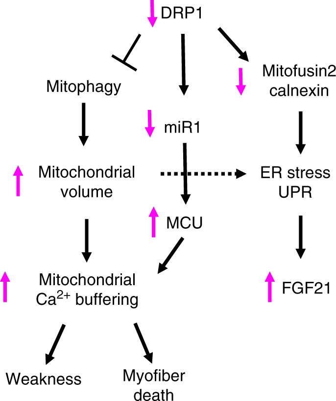
“the change of mitochondrial volume results in an increase of mitochondrial Ca2+ uptake and myofiber death”
§ DRP1-mediated mitochondrial shape controls calcium homeostasis and muscle mass > Abstract
plainstates a cause- Asserted: passes — The authors put it forward as true.
- Checkable: passes — It can be checked against evidence.
- Load-bearing: passes — The paper’s conclusions depend on it.
- Based: passes — The paper gives a reason why it holds.
It may state two separate claims as one.
A conclusion the paper draws from its own results — it cites no other source for it, so there is none to check. The quote shows it is in the paper.
In the paper
“Thus, acute inhibition of DRP1 causes muscle loss and important weakness, but ameliorates fatigue.”
§ DRP1-mediated mitochondrial shape controls calcium homeostasis and muscle mass > Results > Acute deletion of Drp1 causes muscle wasting and weakness
plainstates a cause- Asserted: passes — The authors put it forward as true.
- Checkable: passes — It can be checked against evidence.
- Load-bearing: passes — The paper’s conclusions depend on it.
- Based: passes — The paper gives a reason why it holds.
It may state two separate claims as one.
In the paper
“loss of DRP1 in adult muscle is sufficient to induce a catabolic condition that involves activation of the UPS system and inhibition of autophagy and mitophagy pathways34.”
§ DRP1-mediated mitochondrial shape controls calcium homeostasis and muscle mass > Results > Acute deletion of Drp1 induces ER stress and blocks autophagy
What the sources say
- Not checked yetcites [34] Masiero, E. et al. Autophagy is required to maintain muscle mass. Cell Metab. 10, 507–515 (2009). source
plainstates a cause- Asserted: passes — The authors put it forward as true.
- Checkable: passes — It can be checked against evidence.
- Load-bearing: passes — The paper’s conclusions depend on it.
- Based: passes — The paper gives a reason why it holds.
It may state two separate claims as one.
A conclusion the paper draws from its own results — it cites no other source for it, so there is none to check. The quote shows it is in the paper.
In the paper
“Therefore, DRP1 ablation alters mitochondrial morphology and function.”
§ DRP1-mediated mitochondrial shape controls calcium homeostasis and muscle mass > Results > Drp1 deletion impairs mitochondrial function
plainstates a cause- Asserted: passes — The authors put it forward as true.
- Checkable: passes — It can be checked against evidence.
- Load-bearing: passes — The paper’s conclusions depend on it.
- Based: passes — The paper gives a reason why it holds.
It may state two separate claims as one.
A conclusion the paper draws from its own results — it cites no other source for it, so there is none to check. The quote shows it is in the paper.
In the paper
“supporting the concept that UPR is strongly activated in Drp1−/− muscles and can account for the translation impairment”
§ DRP1-mediated mitochondrial shape controls calcium homeostasis and muscle mass > Results > Drp1 deletion reduces protein synthesis and induces atrophy
boostedstates a cause- Asserted: passes — The authors put it forward as true.
- Checkable: passes — It can be checked against evidence.
- Load-bearing: passes — The paper’s conclusions depend on it.
- Based: passes — The paper gives a reason why it holds.
It may state two separate claims as one.
In the paper
“We monitored autophagic flux in basal condition using colchicine62.”
§ DRP1-mediated mitochondrial shape controls calcium homeostasis and muscle mass > Methods > Autophagic flux quantification
What the sources say
- Not checked yetcites [62] Ju, J. S., Varadhachary, A. S., Miller, S. E. & Weihl, C. C. Quantitation of “autophagic flux” in mature skeletal muscle. Autophagy 6, 929–935 (2010). source
plain- Asserted: passes — The authors put it forward as true.
- Checkable: passes — It can be checked against evidence.
- Load-bearing: fails — The paper’s conclusions may not depend on it.
- Based: passes — The paper gives a reason why it holds.
Probably not a substantial claim. The paper’s conclusions may not depend on it.
None of the sources cited for this claim have been checked yet.
In the paper
“it has been shown that inhibition of PTP opening is beneficial in dystrophic muscles45–47.”
§ DRP1-mediated mitochondrial shape controls calcium homeostasis and muscle mass > Discussion
What the sources say
- Not checked yetcites [45] Millay, D. P. et al. Genetic and pharmacologic inhibition of mitochondrial-dependent necrosis attenuates muscular dystrophy. Nat. Med. 14, 442–447 (2008). source
- Not checked yetcites [46] Irwin, W. A. et al. Mitochondrial dysfunction and apoptosis in myopathic mice with collagen VI deficiency. Nat. Genet. 35, 367–371 (2003). source
- Not checked yetcites [47] Angelin, A. et al. Mitochondrial dysfunction in the pathogenesis of Ullrich congenital muscular dystrophy and prospective therapy with cyclosporins. Proc. Natl Acad. Sci. USA 104, 991–996 (2007). source
boostedstates a cause- Asserted: passes — The authors put it forward as true.
- Checkable: passes — It can be checked against evidence.
- Load-bearing: fails — The paper’s conclusions may not depend on it.
- Based: passes — The paper gives a reason why it holds.
Probably not a substantial claim. The paper’s conclusions may not depend on it.
A conclusion the paper draws from its own results — it cites no other source for it, so there is none to check. The quote shows it is in the paper.
In the paper
“In conclusion, even though both fusion and fission machinery inhibitions induce mitochondrial dysfunction, revealed by decreased respiration, supercomplex formation, and mitochondrial membrane potential, the signaling pathways and the biological effects downstream mitochondria are peculiar for fusion or fission deficiency.”
§ DRP1-mediated mitochondrial shape controls calcium homeostasis and muscle mass > Discussion
plainstates a cause- Asserted: passes — The authors put it forward as true.
- Checkable: passes — It can be checked against evidence.
- Load-bearing: passes — The paper’s conclusions depend on it.
- Based: passes — The paper gives a reason why it holds.
It may state two separate claims as one.
A conclusion the paper draws from its own results — it cites no other source for it, so there is none to check. The quote shows it is in the paper.
In the paper
“The finding that two different knockouts mice, muscle-specific and inducible muscle-specific, result in striking phenotypes (Supplementary Table 3) underlines the critical role of DRP1 in mitochondria function and muscle homeostasis.”
§ DRP1-mediated mitochondrial shape controls calcium homeostasis and muscle mass > Discussion
boostedstates a causen = 2- Asserted: passes — The authors put it forward as true.
- Checkable: passes — It can be checked against evidence.
- Load-bearing: passes — The paper’s conclusions depend on it.
- Based: passes — The paper gives a reason why it holds.
It may state two separate claims as one.
What the paper did — it cites no source for this, so there is none to check. The quote shows it is in the paper.
In the paper
“Force was normalized to the muscle mass as an estimate of specific force.”
§ DRP1-mediated mitochondrial shape controls calcium homeostasis and muscle mass > Methods > Contractile force measurements
plain- Asserted: passes — The authors put it forward as true.
- Checkable: passes — It can be checked against evidence.
- Load-bearing: fails — The paper’s conclusions may not depend on it.
- Based: passes — The paper gives a reason why it holds.
Probably not a substantial claim. The paper’s conclusions may not depend on it.
In the paper
“ER stress and UPR activation are well-known triggers of FGF21 production in different organs, such as the liver, pancreas, or adipose tissue39.”
§ DRP1-mediated mitochondrial shape controls calcium homeostasis and muscle mass > Discussion
What the sources say
- Not checked yetcites [39] Gimeno, R. E. & Moller, D. E. FGF21-based pharmacotherapy-potential utility for metabolic disorders. Trends Endocrinol. Metab. 25, 303–311 (2014). source
boostedstates a cause- Asserted: passes — The authors put it forward as true.
- Checkable: passes — It can be checked against evidence.
- Load-bearing: fails — The paper’s conclusions may not depend on it.
- Based: passes — The paper gives a reason why it holds.
Probably not a substantial claim. The paper’s conclusions may not depend on it.
A conclusion the paper draws from its own results — it cites no other source for it, so there is none to check. The quote shows it is in the paper.
In the paper
“Therefore, the shape of the mitochondria is critical for mitochondrial function, but triggers different signaling pathways and biological processes that are peculiar of fusion or fission impairment.”
§ DRP1-mediated mitochondrial shape controls calcium homeostasis and muscle mass > Discussion
plainstates a cause- Asserted: passes — The authors put it forward as true.
- Checkable: passes — It can be checked against evidence.
- Load-bearing: passes — The paper’s conclusions depend on it.
- Based: passes — The paper gives a reason why it holds.
It may state two separate claims as one.
None of the sources cited for this claim have been checked yet.
In the paper
“Thus, the impairment of the SR–mitochondria tethering and ER chaperon proteins would explain the abnormalities of CRUs and the presence of ER stress36,37.”
§ DRP1-mediated mitochondrial shape controls calcium homeostasis and muscle mass > Results > Inhibition of DRP1 alters normal calcium homeostasis
What the sources say
- Not checked yetcites [36] Gutierrez, T. & Simmen, T. Endoplasmic reticulum chaperones tweak the mitochondrial calcium rheostat to control metabolism and cell death. Cell Calcium 70, 64–75 (2018). source
- Not checked yetcites [37] Debattisti, V., Pendin, D., Ziviani, E., Daga, A. & Scorrano, L. Reduction of endoplasmic reticulum stress attenuates the defects caused by Drosophila mitofusin depletion. J. Cell Biol. 204, 303–312 (2014). source
hedgedstates a possible cause- Asserted: passes — The authors put it forward as true.
- Checkable: passes — It can be checked against evidence.
- Load-bearing: passes — The paper’s conclusions depend on it.
- Based: passes — The paper gives a reason why it holds.
It may state two separate claims as one.
What the paper did — it cites no source for this, so there is none to check. The quote shows it is in the paper.
In the paper
“The mtDNA copy number was calculated using 18S amplification as a reference for nuclear DNA content.”
§ DRP1-mediated mitochondrial shape controls calcium homeostasis and muscle mass > Methods > Measurement of mitochondrial DNA copy number
plain- Asserted: passes — The authors put it forward as true.
- Checkable: passes — It can be checked against evidence.
- Load-bearing: fails — The paper’s conclusions may not depend on it.
- Based: passes — The paper gives a reason why it holds.
Probably not a substantial claim. The paper’s conclusions may not depend on it.
What the paper did — it cites no source for this, so there is none to check. The quote shows it is in the paper.
In the paper
“We did not completely block the degeneration/regeneration process because the efficiency of transfection was low, and consequently, MCU was reduced only in some myofibers.”
§ DRP1-mediated mitochondrial shape controls calcium homeostasis and muscle mass > Discussion
plainstates a cause- Asserted: passes — The authors put it forward as true.
- Checkable: passes — It can be checked against evidence.
- Load-bearing: unclear — It is unclear whether the paper’s conclusions depend on it.
- Based: passes — The paper gives a reason why it holds.
It may state two separate claims as one.
A conclusion the paper draws from its own results — it cites no other source for it, so there is none to check. The quote shows it is in the paper.
In the paper
“Like other systems, such as autophagy, both chronic inhibition or acute hyperactivation of mitochondrial fission result in detrimental effects on muscle mass and function, but with different phenotypes and mechanisms.”
§ DRP1-mediated mitochondrial shape controls calcium homeostasis and muscle mass > Discussion
plainstates a cause- Asserted: passes — The authors put it forward as true.
- Checkable: passes — It can be checked against evidence.
- Load-bearing: passes — The paper’s conclusions depend on it.
- Based: passes — The paper gives a reason why it holds.
It may state two separate claims as one.
A conclusion the paper draws from its own results — it cites no other source for it, so there is none to check. The quote shows it is in the paper.
In the paper
“Therefore, DRP1 inhibition causes increased MCU expression via miR1 downregulation and enhanced Ca2+ uptake of the mitochondria.”
§ DRP1-mediated mitochondrial shape controls calcium homeostasis and muscle mass > Results > Inhibition of DRP1 alters normal calcium homeostasis
plainstates a cause- Asserted: passes — The authors put it forward as true.
- Checkable: passes — It can be checked against evidence.
- Load-bearing: passes — The paper’s conclusions depend on it.
- Based: passes — The paper gives a reason why it holds.
It may state two separate claims as one.
A conclusion the paper draws from its own results — it cites no other source for it, so there is none to check. The quote shows it is in the paper.
In the paper
“However, while the initial outcome of genetic DRP1 ablation is identical to its overexpression, i.e., muscle atrophy via FoxO, our loss-of-function study also reveals the appearance of center-nucleated fibers, a sign of myopathy, and the involvement of different signaling pathways, including MCU upregulation, ER-stress, UPR activation, and great FGF21 induction (Fig. 8).”
§ DRP1-mediated mitochondrial shape controls calcium homeostasis and muscle mass > Discussion
plainstates a cause- Asserted: passes — The authors put it forward as true.
- Checkable: passes — It can be checked against evidence.
- Load-bearing: passes — The paper’s conclusions depend on it.
- Based: passes — The paper gives a reason why it holds.
It may state two separate claims as one.
What the paper did — it cites no source for this, so there is none to check. The quote shows it is in the paper.
In the paper
“No evidence of necrosis or inflammation were observed after the transfection procedure.”
§ DRP1-mediated mitochondrial shape controls calcium homeostasis and muscle mass > Methods > In vivo FDB electroporation
plain- Asserted: passes — The authors put it forward as true.
- Checkable: passes — It can be checked against evidence.
- Load-bearing: fails — The paper’s conclusions may not depend on it.
- Based: passes — The paper gives a reason why it holds.
Probably not a substantial claim. The paper’s conclusions may not depend on it. It may state two separate claims as one.
A conclusion the paper draws from its own results — it cites no other source for it, so there is none to check. The quote shows it is in the paper.
In the paper
“Altogether, these findings confirm that inhibition of the fission machinery alters the SR–mitochondria tethering, decreases CRU units, increases mitochondrial Ca2+ uptake that, altogether, reduces Ca2+ availability for contraction, and simultaneously induces myofiber death, due to mitochondrial Ca2+ overload.”
§ DRP1-mediated mitochondrial shape controls calcium homeostasis and muscle mass > Results > DRP1 rescue or MCU inhibition normalizes Ca2+ homeostasis
boostedstates a cause- Asserted: passes — The authors put it forward as true.
- Checkable: passes — It can be checked against evidence.
- Load-bearing: passes — The paper’s conclusions depend on it.
- Based: passes — The paper gives a reason why it holds.
It may state two separate claims as one.
A conclusion the paper draws from its own results — it cites no other source for it, so there is none to check. The quote shows it is in the paper.
In the paper
“The increased expression of MCU in Drp1−/− fibers could induce mitochondrial Ca2+ overload, and possibly explains the increased presence of the swollen and damaged mitochondria (Fig. 5 b; Supplementary Table 1).”
§ DRP1-mediated mitochondrial shape controls calcium homeostasis and muscle mass > Discussion
hedgedstates a possible cause- Asserted: passes — The authors put it forward as true.
- Checkable: passes — It can be checked against evidence.
- Load-bearing: passes — The paper’s conclusions depend on it.
- Based: passes — The paper gives a reason why it holds.
It may state two separate claims as one.
The paper states this without a source or data behind it here, so there is nothing to check it against. The quote shows it is in the paper.
In the paper
“ATP-synthase can reversely transport protons across the inner mitochondrial membrane, so maintaining the potential also in dysfunctional mitochondria.”
§ DRP1-mediated mitochondrial shape controls calcium homeostasis and muscle mass > Methods > Mitochondrial membrane potential determination
plainstates a cause- Asserted: passes — The authors put it forward as true.
- Checkable: passes — It can be checked against evidence.
- Load-bearing: passes — The paper’s conclusions depend on it.
- Based: fails — The paper may give no reason why it holds.
Probably not a substantial claim. The paper may give no reason why it holds. It may state two separate claims as one.
None of the sources cited for this claim have been checked yet.
In the paper
“Free Ca2+ concentrations in the mitochondrial matrix and SR lumen were determined using cameleon Ca2+ probes, 4mtD3cpv54 for mitochondria and D1-ER 55 for sarcoplasmic reticulum, kindly donated by Dr. R.Y. Tsien (University of California, San Diego).”
§ DRP1-mediated mitochondrial shape controls calcium homeostasis and muscle mass > Methods > Detection of free Ca2+ in the SR and mitochondrial matrix
What the sources say
- Not checked yetcites [54] Giacomello, M. et al. Ca2+ hot spots on the mitochondrial surface are generated by Ca2+ mobilization from stores, but not by activation of store-operated Ca2+ channels. Mol. Cell 38, 280–290 (2010). source
- Not checked yetcites [55] Palmer, A. E., Jin, C., Reed, J. C. & Tsien, R. Y. Bcl-2-mediated alterations in endoplasmic reticulum Ca2+ analyzed with an improved genetically encoded fluorescent sensor. Proc. Natl Acad. Sci. USA 101, 17404–17409 (2004). source
plain- Asserted: passes — The authors put it forward as true.
- Checkable: passes — It can be checked against evidence.
- Load-bearing: passes — The paper’s conclusions depend on it.
- Based: passes — The paper gives a reason why it holds.
Probably not a substantial claim.
In the paper
“it is well known that Ca2+ overload triggers permeability transition pore (PTP) opening and mitochondrial swelling44.”
§ DRP1-mediated mitochondrial shape controls calcium homeostasis and muscle mass > Discussion
What the sources say
- Not checked yetcites [44] Giorgio, V., Guo, L., Bassot, C., Petronilli, V. & Bernardi, P. Calcium and regulation of the mitochondrial permeability transition. Cell Calcium 70, 56–63 (2017). source
boostedstates a cause- Asserted: passes — The authors put it forward as true.
- Checkable: passes — It can be checked against evidence.
- Load-bearing: passes — The paper’s conclusions depend on it.
- Based: passes — The paper gives a reason why it holds.
It may state two separate claims as one.
A conclusion the paper draws from its own results — it cites no other source for it, so there is none to check. The quote shows it is in the paper.
In the paper
“The apparent paradox could be explained by the lower force developed by DRP1-deficient muscles compared with control muscles. The reduction of force due to less contractile proteins and of cytosolic Ca2+ level leads to a decreased ATP consumption during contraction by myosin and SERCA ATPase pump, delaying ATP drop and consequently, improving fatigue.”
§ DRP1-mediated mitochondrial shape controls calcium homeostasis and muscle mass > Discussion
hedgedstates a cause- Asserted: passes — The authors put it forward as true.
- Checkable: passes — It can be checked against evidence.
- Load-bearing: passes — The paper’s conclusions depend on it.
- Based: passes — The paper gives a reason why it holds.
It may state two separate claims as one.
None of the sources cited for this claim have been checked yet.
In the paper
“Mitochondrial volume (Supplementary Table 1, column A and Supplementary Fig. 2a) was determined using the well-established stereology point-counting technique49,50 in micrographs taken from transversal sections at ×7.100 magnifications.”
§ DRP1-mediated mitochondrial shape controls calcium homeostasis and muscle mass > Methods > Quantitative analyses of the mitochondria
What the sources say
- Not checked yetcites [49] Loud, A. V., Barany, W. C. & Pack, B. A. Quantitative evaluation of cytoplasmic structures in electron micrographs. Lab. Invest. 14, 996–1008 (1965).
- Not checked yetcites [50] Mobley, B. A. & Eisenberg, B. R. Sizes of components in frog skeletal muscle measured by methods of stereology. J. Gen. Physiol. 66, 31–45 (1975). source
boosted- Asserted: passes — The authors put it forward as true.
- Checkable: passes — It can be checked against evidence.
- Load-bearing: passes — The paper’s conclusions depend on it.
- Based: passes — The paper gives a reason why it holds.
Probably not a substantial claim.
What the paper did — it cites no source for this, so there is none to check. The quote shows it is in the paper.
In the paper
“Only mitochondria which were entirely visualized in the micrograph were measured.”
§ DRP1-mediated mitochondrial shape controls calcium homeostasis and muscle mass > Methods > Quantitative analyses of the mitochondria
plain- Asserted: passes — The authors put it forward as true.
- Checkable: passes — It can be checked against evidence.
- Load-bearing: fails — The paper’s conclusions may not depend on it.
- Based: passes — The paper gives a reason why it holds.
Probably not a substantial claim. The paper’s conclusions may not depend on it.
None of the sources cited for this claim have been checked yet.
In the paper
“In vivo protein synthesis was measured by using the SUnSET technique25,60.”
§ DRP1-mediated mitochondrial shape controls calcium homeostasis and muscle mass > Methods > In vivo protein synthesis measurements
What the sources say
- Not checked yetcites [25] Schmidt, E. K., Clavarino, G., Ceppi, M. & Pierre, P. SUnSET, a nonradioactive method to monitor protein synthesis. Nat. Methods 6, 275–277 (2009). source
- Not checked yetcites [60] Goodman, C. A. et al. Novel insights into the regulation of skeletal muscle protein synthesis as revealed by a new nonradioactive in vivo technique. FASEB J. 25, 1028–1039 (2011). source
plain- Asserted: passes — The authors put it forward as true.
- Checkable: passes — It can be checked against evidence.
- Load-bearing: fails — The paper’s conclusions may not depend on it.
- Based: passes — The paper gives a reason why it holds.
Probably not a substantial claim. The paper’s conclusions may not depend on it.
What the paper did — it cites no source for this, so there is none to check. The quote shows it is in the paper.
In the paper
“The results were normalized to citrate synthase enzymatic activity.”
§ DRP1-mediated mitochondrial shape controls calcium homeostasis and muscle mass > Methods > Mitochondrial assays
plain- Asserted: passes — The authors put it forward as true.
- Checkable: passes — It can be checked against evidence.
- Load-bearing: fails — The paper’s conclusions may not depend on it.
- Based: passes — The paper gives a reason why it holds.
Probably not a substantial claim. The paper’s conclusions may not depend on it.
What the paper did — it cites no source for this, so there is none to check. The quote shows it is in the paper.
In the paper
“Micrographs of non-overlapping regions were randomly collected from transversal and longitudinal sections of internal fiber areas of EDL muscles.”
§ DRP1-mediated mitochondrial shape controls calcium homeostasis and muscle mass > Methods > Quantitative analyses of the mitochondria
plain- Asserted: passes — The authors put it forward as true.
- Checkable: passes — It can be checked against evidence.
- Load-bearing: passes — The paper’s conclusions depend on it.
- Based: passes — The paper gives a reason why it holds.
Probably not a substantial claim.
The paper relies on this without support.
Mitochondrial quality control is essential in highly structured cells such as neurons and muscles.
“Mitochondrial quality control is essential in highly structured cells such as neurons and muscles.”
The paper relies on this without support.
Insulin signaling through mTOR plays a critical role in controlling protein synthesis.
“Insulin signaling, through mTOR, plays a critical role in controlling protein synthesis.”
The paper relies on this without support.
FGF21 causes GH resistance.
“Because FGF21 causes GH resistance”
The authors state this as an assumption.
The calibration equation and constants (KD = 145 nM, Rmin, Rmax, β) used to convert Fura-2 fluorescence ratios to free cytosolic calcium concentration are valid.
“Under these assumptions, the resting cytosolic calcium concentration will be 71 nM at R = 0.87 and 87 nM at R = 0.92.”
Claims other papers make about this one
Each is checked against this paper’s own text: does it say what the citing paper says it does?
The cited source doesn’t support the claim as written.
In the citing paper
“Conversely, DRP1 ablation alters calcium homeostasis and mitochondria–ER tethering resulting in myofiber degeneration but with normal animal survival78.”
From Mechanisms of muscle atrophy and hypertrophy: implications in health and disease (2021) · cites this paper as [78]
Checked by a reasoning model — may be wrong.
Experimental — accuracy not yet measuredThe source backs part of the claim, not all of it — or backs it for a different population, condition or comparison.
In the citing paper
“Interestingly, the stress-response myokine FGF21 is dramatically increased in muscle and serum of all these three models, even if with a different degree.”
From The connection between the dynamic remodeling of the mitochondrial network and the regulation of muscle mass (2021) · cites this paper as [196]
Experimental — accuracy not yet measuredThe cited source says what the claim attributes to it.
In the citing paper
“In rodent genetic models, these findings indicate that mitophagy, mitochondrial fusion, and fission are essential for skeletal muscle health (Dulac et al., 2020; Favaro et al., 2019; Tezze et al., 2017).”
From Autophagy gene expression in skeletal muscle of older individuals is associated with physical performance, muscle volume and mitochondrial function in the study of muscle, mobility and aging ( SOMMA ) (2024) · cites this paper as Favaro et al., 2019
The cited source says what the claim attributes to it.
In the citing paper
“There is strong evidence in skeletal muscle that supports a causal link between the dysregulation of mitochondrial fission and alterations in muscle maintenance [152, 196, 197, 210, 211].”
From The connection between the dynamic remodeling of the mitochondrial network and the regulation of muscle mass (2021) · cites this paper as [196]
The cited source says what the claim attributes to it.
In the citing paper
“Accordingly, the failure in mitochondrial-shaping processes has detrimental consequences for the maintenance of muscle mass and function [4,5,6,7,8,13,29].”
From Inhibition of the Fission Machinery Mitigates OPA1 Impairment in Adult Skeletal Muscles (2019) · cites this paper as [8]
In the citing paper
“Consistently, knockout mice for the fusion protein OPA1 or the fission component DRP1 display muscle wasting77,78.”
From Mechanisms of muscle atrophy and hypertrophy: implications in health and disease (2021) · cites this paper as [78]
In the citing paper
“Impairment of fission leads to the disruption of selective mitochondrial autophagic degradation, followed by dysfunctional organelles accumulation [58, 196–200].”
From The connection between the dynamic remodeling of the mitochondrial network and the regulation of muscle mass (2021) · cites this paper as [196]
In the citing paper
“Consistent with this crucial role, conditional ablation of DRP1 in the heart, brain, and skeletal muscle causes lethality [196, 198–200, 208, 209].”
From The connection between the dynamic remodeling of the mitochondrial network and the regulation of muscle mass (2021) · cites this paper as [196]
In the citing paper
“Early deletion of DRP1 in skeletal muscle during embryogenesis resulted in reduced postnatal growth and premature lethality, while its acute ablation in adulthood causes muscle loss and degeneration [196].”
From The connection between the dynamic remodeling of the mitochondrial network and the regulation of muscle mass (2021) · cites this paper as [196]
In the citing paper
“FGF21 serum levels are lower in DRP1 knockout mice than OPA1-null mice.”
From The connection between the dynamic remodeling of the mitochondrial network and the regulation of muscle mass (2021) · cites this paper as [196]
In the citing paper
“OPA1 inhibition triggers IL6 and IL1 upregulation via ROS [187], while the ablation of DRP1 does not alter the expression of inflammatory cytokines [196].”
From The connection between the dynamic remodeling of the mitochondrial network and the regulation of muscle mass (2021) · cites this paper as [196]
In the citing paper
“In DRP1-null muscles, the sarcoplasmic reticulum's calcium stores are unchanged, while abnormal elongated mitochondria display increased MCU-dependent mitochondrial Ca2 + uptake capacity, leading to myofiber death and muscle regeneration [196].”
From The connection between the dynamic remodeling of the mitochondrial network and the regulation of muscle mass (2021) · cites this paper as [196]
In the citing paper
“Early deletion of DRP1 in skeletal muscle during embryogenesis resulted in reduced postnatal growth and premature lethality, while its acute ablation in adulthood causes muscle loss and degeneration [196].”
From The connection between the dynamic remodeling of the mitochondrial network and the regulation of muscle mass (2021) · cites this paper as [196]
In the citing paper
“Mechanistically, DRP1 inhibition induced autophagy and mitophagy impairment, MCU upregulation and mitochondrial calcium overload, ER stress, UPR activation, and FGF21 induction.”
From The connection between the dynamic remodeling of the mitochondrial network and the regulation of muscle mass (2021) · cites this paper as [196]
In the citing paper
“In muscles, the Drp1 inhibition causes muscle weakness and wasting that is consequent to an abnormal calcium homeostasis [8].”
From Inhibition of the Fission Machinery Mitigates OPA1 Impairment in Adult Skeletal Muscles (2019) · cites this paper as [8]
In the citing paper
“Emerging evidence suggested that apart from energy production, mitochondria perform other critical functions to maintain homeostasis and function in the skeletal myocytes, including regulation of intracellular Ca2+ homeostasis, modulation of cell proliferation, and integration of apoptotic signaling (17–19).”
From From mitochondria to sarcopenia: role of 17β-estradiol and testosterone (2023) · cites this paper as [18]
In the citing paper
“Additionally, muscle-specific ablation of DRP1 induces severe muscle wasting and weakness, as well as abnormal morphology, function, and Ca2+ homeostasis in mitochondria (18).”
From From mitochondria to sarcopenia: role of 17β-estradiol and testosterone (2023) · cites this paper as [18]
In the citing paper
“indicating that Drp1 is a key player in regulating skeletal muscle development and maintenance.”
From Mitochondrial Dynamics and Mitophagy in Skeletal Muscle Health and Aging (2021) · cites this paper as [42]
In the citing paper
“Muscle-specific Drp1 knockout in mice leads to a severe myopathic phenotype including muscle wasting, weakness, and signs of muscle degeneration and regeneration [42].”
From Mitochondrial Dynamics and Mitophagy in Skeletal Muscle Health and Aging (2021) · cites this paper as [42]
In the citing paper
“Taken altogether, the findings discussed above clearly indicate that mitochondrial fission is essential for skeletal muscle health.”
From Mitochondrial Dynamics and Mitophagy in Skeletal Muscle Health and Aging (2021) · cites this paper as [42]
In the citing paper
“deletion or insufficient expression of Drp1 in skeletal muscle led to 40–50% atrophy [42,43]”
From Mitochondrial Dynamics and Mitophagy in Skeletal Muscle Health and Aging (2021) · cites this paper as [42]
Self-check
Percentages, sums and reported differences re-computed from the paper’s own numbers.
Each contradiction between two passages listed here was confirmed by an independent check.
Rated major: it would touch a result — a value a result rests on, reported and recomputed p-values on opposite sides of the significance level, two passages that conflict with no sign which is right, or a cited work retracted before this paper was published.
In the paper
§ DRP1-mediated mitochondrial shape controls calcium homeostasis and muscle mass > Results > Muscle-specific Drp1 deletion results in postnatal lethality
“Muscles displayed a reduction in the total number of fibers (Supplementary Fig. 1e) because glycolytic type 2B/2X fibers are less than controls (Fig. 1f)”
§ DRP1-mediated mitochondrial shape controls calcium homeostasis and muscle mass > Results > Muscle-specific Drp1 deletion results in postnatal lethality
“The cross-sectional area analysis of fibers (g) and the total number of different fiber types in hindlimb muscles (f) indicate a reduction of DRP1 KO fiber size without hypoplasia.”
Rated minor: it would not touch a result — a value no result rests on, a p-value that stays on the same side of the significance level, or a conflict that is clerical, resolved elsewhere in the paper, or outside its own findings.
In the paper
§ DRP1-mediated mitochondrial shape controls calcium homeostasis and muscle mass > Methods > Free calcium concentration in the cytosol
“Ca2+ measurements were expressed as fluorescence ratio of the emission at 480 nm with reference to the excitation wavelengths of 360 and 380 nm, respectively.”
§ DRP1-mediated mitochondrial shape controls calcium homeostasis and muscle mass > Methods > Free calcium concentration in the cytosol
“where R is the ratio of fluorescence excited at 340 nm to that excited at 380 nm”
Citations
Cites
Fiji: an open-source platform for biological-image analysis
Schindelin, J., Arganda-Carreras, I., Frise, E., Kaynig, V., Longair, M., Pietzsch, T., Preibisch, S., Rueden, C., Saalfeld, S., Schmid, B., et al. (2012) · Nature Methods · doi:10.1038/nmeth.2019 · Cited by 66,890
Sandri, M., Sandri, C., Gilbert, A., Skurk, C., Calabria, E., Picard, A., Walsh, K., Schiaffino, S., Lecker, S. H., & Goldberg, A. L. (2004) · Cell · doi:10.1016/s0092-8674(04)00400-3 · Cited by 3,012
Self-citationA forty-kilodalton protein of the inner membrane is the mitochondrial calcium uniporter
Stefani, D. D., Raffaello, A., Teardo, E., Szabó, I., & Rizzuto, R. (2011) · Nature · doi:10.1038/nature10230 · Cited by 1,957
FoxO3 Controls Autophagy in Skeletal Muscle In Vivo
Mammucari, C., Milan, G., Romanello, V., Masiero, E., Rudolf, R., Del Piccolo, P., Burden, S. J., Di Lisi, R., Sandri, C., Zhao, J., et al. (2007) · Cell Metabolism · doi:10.1016/j.cmet.2007.11.001 · Cited by 1,953
Self-citationDynamin-related Protein Drp1 Is Required for Mitochondrial Division in Mammalian Cells
Smirnova, E., Griparić, L., Shurland, D., & van der Bliek, A. M. (2001) · Molecular Biology of the Cell · doi:10.1091/mbc.12.8.2245 · Cited by 1,938
SUnSET, a nonradioactive method to monitor protein synthesis
Schmidt, E. K., Clavarino, G., Ceppi, M., & Pierre, P. (2009) · Nature Methods · doi:10.1038/nmeth.1314 · Cited by 1,706
Mechanisms regulating skeletal muscle growth and atrophy
Schiaffino, S., Dyar, K. A., Ciciliot, S., Blaauw, B., & Sandri, M. (2013) · The FEBS Journal · doi:10.1111/febs.12253 · Cited by 1,492
Self-citationZhao, J., Brault, J. J., Schild, A., Cao, P., Sandri, M., Schiaffino, S., Lecker, S. H., & Goldberg, A. L. (2007) · Cell Metabolism · doi:10.1016/j.cmet.2007.11.004 · Cited by 1,489
Self-citationAutophagy is required to maintain muscle mass.
Masiero, E., Agatea, L., Mammucari, C., Blaauw, B., Loro, E., Komatsu, M., Metzger, D., Reggiani, C., Schiaffino, S., & Sandri, M. (2009) · Cell Metabolism · doi:10.1016/j.cmet.2009.10.008 · Cited by 1,282
Self-citationCogliati, S., Frezza, C., Soriano, M. E., Varanita, T., Quintana-Cabrera, R., Corrado, M., Cipolat, S., Costa, V., Casarin, A., Gomes, L. C., et al. (2013) · Cell · doi:10.1016/j.cell.2013.08.032 · Cited by 1,265
Self-citation
Cited by
Sorted by citations
The cell biology of mitochondrial membrane dynamics
Giacomello, M., Pyakurel, A., Glytsou, C., & Scorrano, L. (2020) · Nature Reviews Molecular Cell Biology · doi:10.1038/s41580-020-0210-7 · Cited by 1,550
Self-citationMechanisms of muscle atrophy and hypertrophy: implications in health and disease
Sartori, R., Romanello, V., & Sandri, M. (2021) · Nature Communications · doi:10.1038/s41467-020-20123-1 · Cited by 940
Self-citationQuality control of the mitochondrion
Ng, M. Y. W., Wai, T., & Simonsen, A. (2021) · Developmental Cell · doi:10.1016/j.devcel.2021.02.009 · Cited by 468
Function and regulation of the divisome for mitochondrial fission
Kraus, F., Roy, K., Pucadyil, T. J., & Ryan, M. T. (2021) · Nature · doi:10.1038/s41586-021-03214-x · Cited by 445
Mitochondrial dysfunction: roles in skeletal muscle atrophy
Chen, X., Ji, Y., Liu, R., Zhu, X., Wang, K., Yang, X., Liu, B., Gao, Z., Huang, Y. H., Shen, Y., et al. (2023) · Journal of Translational Medicine · doi:10.1186/s12967-023-04369-z · Cited by 362
The role of mitochondrial fission in cardiovascular health and disease
Quiles, J. M., & Gustafsson, Å. B. (2022) · Nature Reviews Cardiology · doi:10.1038/s41569-022-00703-y · Cited by 315
Mitochondrial Dynamics and Mitophagy in Skeletal Muscle Health and Aging
Leduc-Gaudet, J.-P., Hussain, S. N. A., Barreiro, E., & Gouspillou, G. (2021) · International Journal of Molecular Sciences · doi:10.3390/ijms22158179 · Cited by 268
“As mentioned above, overexpression of Drp1 impaired skeletal muscle growth [39], whereas deletion or insufficient expression of Drp1 in skeletal muscle led to 40–50% atrophy [42,43], indicating that Drp1 is a key player in regulating skeletal muscle development and maintenance.”
BackgroundInfluentialThe Functional Impact of Mitochondrial Structure Across Subcellular Scales
Glancy, B. C., Kim, Y., Katti, P., & Willingham, T. B. (2020) · Frontiers in Physiology · doi:10.3389/fphys.2020.541040 · Cited by 270
Hematopoietic Stem Cell Metabolism during Development and Aging
Nakamura‐Ishizu, A., Ito, K., & Suda, T. (2020) · Developmental Cell · doi:10.1016/j.devcel.2020.06.029 · Cited by 245
Mitochondrial Structure and Function in Human Heart Failure
Hinton, A. O., Jr., Claypool, S. M., Neikirk, K., Senoo, N., Wanjalla, C. N., Kirabo, A., & Williams, C. R. (2024) · Circulation Research · doi:10.1161/circresaha.124.323800 · Cited by 239
Mitochondrial ROS-Modulated mtDNA: A Potential Target for Cardiac Aging
Quan, Y., Xin, Y., Tian, G., Zhou, J., & Liu, X. (2020) · Oxidative Medicine and Cellular Longevity · doi:10.1155/2020/9423593 · Cited by 226
Mitochondria, energy, and metabolism in neuronal health and disease
Trigo, D., Avelar, C., Fernandes, M., Sá, J., & Silva, O. A. B. D. C. E. (2022) · FEBS Letters · doi:10.1002/1873-3468.14298 · Cited by 225
Signaling Pathways That Control Muscle Mass
Vainshtein, A., & Sandri, M. (2020) · International Journal of Molecular Sciences · doi:10.3390/ijms21134759 · Cited by 222
Self-citationRomanello, V., & Sandri, M. (2021) · Cellular and Molecular Life Sciences · doi:10.1007/s00018-020-03662-0 · Cited by 197
Self-citationDysregulated Mitochondrial Dynamics and Metabolism in Obesity, Diabetes, and Cancer
Dai, W., & Jiang, L. (2019) · Frontiers in Endocrinology · doi:10.3389/fendo.2019.00570 · Cited by 186
Zeng, Z., Liang, J., Wu, L., Zhang, H., Lv, J., & Chen, N. (2020) · Frontiers in Physiology · doi:10.3389/fphys.2020.583478 · Cited by 200
Mitochondrial quality control in human health and disease
Liu, B., Xu, C.-Z., Liu, Y., Lu, Z., Fu, T.-L., Li, G., Deng, Y., Luo, G., Ding, S. Z., Li, N., et al. (2024) · Military Medical Research · doi:10.1186/s40779-024-00536-5 · Cited by 211
Chronic Stress Weakens Connectivity in the Prefrontal Cortex: Architectural and Molecular Changes.
Woo, E., Sansing, L. H., Arnsten, A. F. T., & Datta, D. (2021) · Chronic Stress · doi:10.1177/24705470211029254 · Cited by 159
Experimental Models of Sarcopenia: Bridging Molecular Mechanism and Therapeutic Strategy
Mankhong, S., Kim, S., Moon, S., Kwak, H., Park, D., & Kang, J. (2020) · Cells · doi:10.3390/cells9061385 · Cited by 150
Mechanisms and roles of mitochondrial localisation and dynamics in neuronal function
Seager, R., Lee, L. J., Henley, J. M., & Wilkinson, K. A. (2020) · Neuronal Signaling · doi:10.1042/ns20200008 · Cited by 152
Mitochondrial morphology regulates organellar Ca 2+ uptake and changes cellular Ca 2+ homeostasis
Kowaltowski, A. J., Menezes‐Filho, S. L., Assali, E. A., Gonçalves, I. G., Cabral‐Costa, J. V., Abreu, P., Miller, N., Nolasco, P., Laurindo, F. R. M., Bruni‐Cardoso, A., et al. (2019) · The FASEB Journal · doi:10.1096/fj.201901136r · Cited by 137
Mitochondrial dysfunction and skeletal muscle atrophy: Causes, mechanisms, and treatment strategies
Kubat, G. B., Bouhamida, E., Ülger, Ö., Türkel, İ., Pedriali, G., Ramaccini, D., Ekinci, Ö., Özerkliğ, B., Atalay, Ö., Patergnani, S., et al. (2023) · Mitochondrion · doi:10.1016/j.mito.2023.07.003 · Cited by 134
The Role of Autophagy in Skeletal Muscle Diseases
Xia, Q., Huang, X., Huang, J., Zheng, Y., March, M., Li, J., & Wei, Y. (2021) · Frontiers in Physiology · doi:10.3389/fphys.2021.638983 · Cited by 137
da Silva Rosa, S. C., Martens, M. D., Field, J. T., Nguyen, L., Kereliuk, S. M., Hai, Y., Chapman, D., Diehl-Jones, W., Aliani, M., West, A. R., et al. (2021) · Autophagy · doi:10.1080/15548627.2020.1821548 · Cited by 117
Neuropathic Pain: the Dysfunction of Drp1, Mitochondria, and ROS Homeostasis
Dai, C., Guo, Y., & Chu, X.-Y. (2020) · Neurotoxicity Research · doi:10.1007/s12640-020-00257-2 · Cited by 111
Ježek, P., Jabůrek, M., Holendová, B., Engstová, H., & Dlasková, A. (2023) · Antioxidants and Redox Signaling · doi:10.1089/ars.2022.0173 · Cited by 117
Choi, E.-H., Kim, M.-H., & Park, S.-J. (2024) · International Journal of Molecular Sciences · doi:10.3390/ijms25147952 · Cited by 116
Liu, H., Ho, P. W., Leung, C.-T., Pang, S. Y.-Y., Chang, E. E. S., Choi, Z. Y.-K., Kung, M. H.-W., Ramsden, D. B., & Ho, S. (2020) · Autophagy · doi:10.1080/15548627.2020.1850008 · Cited by 113
Neurological disorders and mitochondria
Cabral‐Costa, J. V., & Kowaltowski, A. J. (2019) · Molecular Aspects of Medicine · doi:10.1016/j.mam.2019.10.003 · Cited by 104
Irazoki, A., Gordaliza‐Alaguero, I., Frank, E., Giakoumakis, N.-N., Seco, J., Palacı́n, M., Gumà, A., Sylow, L., Sebastián, D., & Antonio Zorzano, A. (2023) · Nature Communications · doi:10.1038/s41467-022-35732-1 · Cited by 105
Impact of Melatonin on Skeletal Muscle and Exercise
Stacchiotti, A., Favero, G., & Rodella, L. F. (2020) · Cells · doi:10.3390/cells9020288 · Cited by 116
Xiao, Y., Li, Z., Bianco, A., & Ma, B. (2022) · Advanced Functional Materials · doi:10.1002/adfm.202209291 · Cited by 104
The Interplay between Mitochondrial Morphology and Myomitokines in Aging Sarcopenia
Romanello, V. (2020) · International Journal of Molecular Sciences · doi:10.3390/ijms22010091 · Cited by 101
Self-citationLei, Y., Gan, M., Qiu, Y., Chen, Q., Wang, X., Liao, T., Zhao, M., Chen, L., Zhang, S., Zhao, Y., et al. (2024) · Cellular & Molecular Biology Letters · doi:10.1186/s11658-024-00572-y · Cited by 111
Mitochondrial dynamics during spermatogenesis
Varuzhanyan, G., & Chan, D. C. (2020) · Journal of Cell Science · doi:10.1242/jcs.235937 · Cited by 106
Mitochondrial Impairment in Sarcopenia
Bellanti, F., Buglio, A. L., & Vendemiale, G. (2021) · Biology · doi:10.3390/biology10010031 · Cited by 104
Mitochondrial Properties in Skeletal Muscle Fiber
Dong, H., & Tsai, S. (2023) · Cells · doi:10.3390/cells12172183 · Cited by 102
Avellaneda, J., Rodier, C., Daian, F., Brouilly, N., Rival, T., Luis, N. M., & Schnorrer, F. (2021) · Nature Communications · doi:10.1038/s41467-021-22058-7 · Cited by 92
Inhibition of the Fission Machinery Mitigates OPA1 Impairment in Adult Skeletal Muscles
Romanello, V., Scalabrin, M., Albiero, M., Blaauw, B., Scorrano, L., & Sandri, M. (2019) · Cells · doi:10.3390/cells8060597 · Cited by 91
Self-citationMitochondrial Dysfunction as an Underlying Cause of Skeletal Muscle Disorders
Chen, T., Koh, K.-Y., Lin, K. M.-C., & Chou, C. (2022) · International Journal of Molecular Sciences · doi:10.3390/ijms232112926 · Cited by 95
Dulac, M., Leduc‐Gaudet, J., Reynaud, O., Ayoub, M., Guérin, A., Finkelchtein, M., Hussain, S. N., & Gouspillou, G. (2020) · The Journal of Physiology · doi:10.1113/jp279802 · Cited by 90
Mitochondria transplant therapy improves regeneration and restoration of injured skeletal muscle
Alway, S. E., Paez, H. G., Pitzer, C. R., Ferrandi, P. J., Khan, M. M., Mohamed, J. S., Carson, J. A., & Deschenes, M. R. (2023) · Journal of Cachexia Sarcopenia and Muscle · doi:10.1002/jcsm.13153 · Cited by 87
Lima, A., Lubatti, G., Burgstaller, J. P., Hu, D., Green, A. P., Gregorio, A. D., Zawadzki, T., Pernaute, B., Mahammadov, E., Pérez-Montero, S., et al. (2021) · Nature Metabolism · doi:10.1038/s42255-021-00422-7 · Cited by 80
Energy metabolism design of the striated muscle cell
Glancy, B. C., & Balaban, R. S. (2021) · Physiological Reviews · doi:10.1152/physrev.00040.2020 · Cited by 86
Gómez-Valadés, A. G., Pozo, M., Varela, L., Boudjadja, M. B., Ramírez, S., Chivite, Í., Eyre, E., Haddad-Tóvolli, R., Obri, A., Milà‐Guasch, M., et al. (2021) · Cell Metabolism · doi:10.1016/j.cmet.2021.07.008 · Cited by 83
Mitochondrial heterogeneity in diseases
Chen, L., Zhou, M., Li, H., Liu, D., Liao, P., Yao, Z., Zhang, C., Zou, W., & Gao, J. (2023) · Signal Transduction and Targeted Therapy · doi:10.1038/s41392-023-01546-w · Cited by 88
Goljanek‐Whysall, K., Soriano‐Arroquia, A., McCormick, R., Chinda, C., & McDonagh, B. (2020) · Aging Cell · doi:10.1111/acel.13140 · Cited by 81
“Similarly, inducible muscle-specific deletion of the mitochondrial fission protein, DRP1, resulted in the formation of functionally abnormally large mitochondria, activation of the ER stress response, an increase in Ca2+ uptake and myofibre death (Favaro et al., 2019).”
BackgroundMitochondrial Mechanisms of Neuromuscular Junction Degeneration with Aging
Anagnostou, M., & Hepple, R. T. (2020) · Cells · doi:10.3390/cells9010197 · Cited by 84
MYTHO is a novel regulator of skeletal muscle autophagy and integrity
Leduc‐Gaudet, J., Franco‐Romero, A., Cefis, M., Moamer, A., Broering, F. E., Milan, G., Sartori, R., Chaffer, T. J., Dulac, M., Marcangeli, V., et al. (2023) · Nature Communications · doi:10.1038/s41467-023-36817-1 · Cited by 75
Self-citationRahman, F. A., & Quadrilatero, J. (2021) · Cellular and Molecular Life Sciences · doi:10.1007/s00018-021-03807-9 · Cited by 77
Mitochondrial calcium at the synapse
Datta, S., & Jaiswal, M. (2021) · Mitochondrion · doi:10.1016/j.mito.2021.04.006 · Cited by 73
Mitochondrial fragmentation enables localized signaling required for cell repair
Horn, A. J., Raavicharla, S., Shah, S., Cox, D., & Jaiswal, J. K. (2020) · The Journal of Cell Biology · doi:10.1083/jcb.201909154 · Cited by 73
Lv, J., Li, Y., Shi, S., Xu, X., Wu, H., Zhang, B., & Song, Q. (2022) · Biomedicine & Pharmacotherapy · doi:10.1016/j.biopha.2022.113833 · Cited by 74
Michelucci, A., Chen, L., Protasi, F., & Dirksen, R. T. (2021) · Metabolites · doi:10.3390/metabo11070424 · Cited by 75
Self-citationChen, S., Liu, S., Wang, J., Wu, Q., Wang, A., Guan, H., Zhang, Q. Y., Zhang, D., Wang, X., Song, H., et al. (2020) · Molecular Cell · doi:10.1016/j.molcel.2020.10.018 · Cited by 74
Vue, Z., Lopez, E. G., Neikirk, K., Katti, P., Vang, L., Beasley, H. K., Shao, J., Marshall, A. G., Crabtree, A., Murphy, A. C., et al. (2023) · Aging Cell · doi:10.1111/acel.14009 · Cited by 69
Mechanobiology of muscle and myofibril morphogenesis
Luis, N. M., & Schnorrer, F. (2021) · Cells and Development · doi:10.1016/j.cdev.2021.203760 · Cited by 69
Neikirk, K., Vue, Z., Katti, P., Rodriguez, B. I., Omer, S., Shao, J., Christensen, T. A., Lopez, E. G., Marshall, A. G., Palavicino‐Maggio, C. B., et al. (2023) · Advanced Biology · doi:10.1002/adbi.202200221 · Cited by 66
Castillo, Í. M. P., Argilés, J. M. A., Rueda, R., Ramírez, M., & Pedrosa, J. M. L. (2025) · Reviews in Endocrine and Metabolic Disorders · doi:10.1007/s11154-025-09954-9 · Cited by 75
Zhen, Y., Yuan, Z., Zhang, J., Chen, Y., Fu, Y., Liu, Y., Fu, L., Zhang, L., & Zhou, X. (2022) · Cell Death and Disease · doi:10.1038/s41419-022-04823-8 · Cited by 61
Lacombe, A., & Scorrano, L. (2024) · Seminars in Cell and Developmental Biology · doi:10.1016/j.semcdb.2024.02.001 · Cited by 65
Self-citationMitochondrial dynamics regulators: implications for therapeutic intervention in cancer
Kumar, S., Ashraf, R., & Aparna, C. K. (2021) · Cell Biology and Toxicology · doi:10.1007/s10565-021-09662-5 · Cited by 61
Yan, X., Shen, Z.-L., Yu, D., Zhao, C., Zou, H., Ma, B., Dong, W.-X., Chen, W., Huang, D., & Yu, Z. (2021) · Free Radical Biology and Medicine · doi:10.1016/j.freeradbiomed.2021.11.030 · Cited by 60
Chen, Y., Hamidu, S., Yang, X., Yan, Y., Wang, Q., Li, L., Oduro, P. K., & Li, Y. (2022) · Frontiers in Genetics · doi:10.3389/fgene.2022.880421 · Cited by 62
Confounding Roles of ER Stress and the Unfolded Protein Response in Skeletal Muscle Atrophy
Gallot, Y. S., & Bohnert, K. R. (2021) · International Journal of Molecular Sciences · doi:10.3390/ijms22052567 · Cited by 61
Role of autophagy in muscle disease
Franco‐Romero, A., & Sandri, M. (2021) · Molecular Aspects of Medicine · doi:10.1016/j.mam.2021.101041 · Cited by 65
Self-citationMitochondrial Quality Control in Sarcopenia: Updated Overview of Mechanisms and Interventions
Liu, D., Fan, Y., Tao, X., Pan, W., Wu, Y., Wang, X., He, Y., Xiao, W., & Li, Y. (2021) · Aging and Disease · doi:10.14336/ad.2021.0427 · Cited by 62
Mitochondrial dynamics define muscle fiber type by modulating cellular metabolic pathways
Yasuda, T., Ishihara, T., Ichimura, A., & Ishihara, N. (2023) · Cell Reports · doi:10.1016/j.celrep.2023.112434 · Cited by 64
Skeletal muscle mitochondrial network dynamics in metabolic disorders and aging
Fealy, C. E., Grevendonk, L., Hoeks, J., & Hesselink, M. K. C. (2021) · Trends in Molecular Medicine · doi:10.1016/j.molmed.2021.07.013 · Cited by 58
The influence of aerobic exercise on mitochondrial quality control in skeletal muscle
Philp, A. M., Saner, N. J., Lazarou, M., Ganley, I. G., & Philp, A. (2020) · The Journal of Physiology · doi:10.1113/jp279411 · Cited by 62
Valera‐Alberni, M., Joffraud, M., Miró, J., Capellades, J., Junza, A., Dayon, L., Galindo, A. N., Sanchez-Garcia, J. L., Valsesia, A., Cercillieux, A., et al. (2021) · Cell Reports · doi:10.1016/j.celrep.2021.109565 · Cited by 61
The impact of bed rest on human skeletal muscle metabolism
Eggelbusch, M., Charlton, B. T., Bosutti, A., Ganse, B., Giakoumaki, I., Grootemaat, A. E., Hendrickse, P. W., Jaspers, Y. R. J., Kemp, S., Kerkhoff, T. J., et al. (2024) · Cell Reports Medicine · doi:10.1016/j.xcrm.2023.101372 · Cited by 61
Liao, J., He, W., Li, L., Wang, J., Gong, L., Zhang, Q., & Lin, Z. (2025) · Cell Biomaterials · doi:10.1016/j.celbio.2025.100016 · Cited by 63
Cisplatin resistance can be curtailed by blunting Bnip3-mediated mitochondrial autophagy
Vianello, C., Cocetta, V., Catanzaro, D., Dorn, G. W., Milito, A. D., Rizzolio, F., Canzonieri, V., Cecchin, E., Roncato, R., Toffoli, G., et al. (2022) · Cell Death and Disease · doi:10.1038/s41419-022-04741-9 · Cited by 58
Self-citationManifestations of Age on Autophagy, Mitophagy and Lysosomes in Skeletal Muscle
Triolo, M., & Hood, D. A. (2021) · Cells · doi:10.3390/cells10051054 · Cited by 60
Manipulating calcium homeostasis with nanoplatforms for enhanced cancer therapy
Feng, Y., Wang, J., Cao, J.-M., Cao, F., & Chen, X. S. (2023) · Exploration · doi:10.1002/exp.20230019 · Cited by 55
From mitochondria to sarcopenia: role of 17β-estradiol and testosterone
Tian, X., Lou, S., & Shi, R. (2023) · Frontiers in Endocrinology · doi:10.3389/fendo.2023.1156583 · Cited by 54
Parkin Overexpression Attenuates Sepsis-Induced Muscle Wasting
Leduc-Gaudet, J.-P., Mayaki, D., Reynaud, O., Broering, F. E., Chaffer, T. J., Hussain, S. N. A., & Gouspillou, G. (2020) · Cells · doi:10.3390/cells9061454 · Cited by 54
Anzell, A. R., Fogo, G. M., Gurm, Z., Raghunayakula, S., Wider, J. M., Maheras, K. J., Emaus, K. J., Bryson, T. D., Wang, M., Neumar, R. W., et al. (2021) · Cell Death and Disease · doi:10.1038/s41419-021-03752-2 · Cited by 55
Hinton, A. O., Jr., Katti, P., Christensen, T. A., Mungai, M., Shao, J., Zhang, L., Trushin, S., Alghanem, A. F., Jaspersen, A. L., Geroux, R. E., et al. (2023) · Advanced Biology · doi:10.1002/adbi.202200202 · Cited by 62
Chen, W., Shen, Z.-L., Dong, W.-X., Huang, G., Yu, D., Chen, W., Xialin, Y., & Yu, Z. (2024) · Phytomedicine · doi:10.1016/j.phymed.2024.155567 · Cited by 54
Li, J., Wang, Z., Li, C., Song, Y., Wang, Y., Hai, B., & Zhang, Y. (2022) · Cells · doi:10.3390/cells11132086 · Cited by 56
Sarcomere maturation: function acquisition, molecular mechanism, and interplay with other organelles
Ahmed, R. E., Tokuyama, T., Anzai, T., Chanthra, N., & Uosaki, H. (2022) · Philosophical Transactions of the Royal Society B Biological Sciences · doi:10.1098/rstb.2021.0325 · Cited by 49
The Role of Mitophagy in Skeletal Muscle Damage and Regeneration
Chatzinikita, E., Maridaki, M. D., Palikaras, K., Koutsilieris, M., & Philippou, Α. (2023) · Cells · doi:10.3390/cells12050716 · Cited by 63
The emerging role of skeletal muscle mitochondrial dynamics in exercise and ageing
Casuso, R. A., & Huertas, J. R. (2020) · Ageing Research Reviews · doi:10.1016/j.arr.2020.101025 · Cited by 56
Regulation of muscle and mitochondrial health by the mitochondrial fission protein Drp1 in aged mice
Dulac, M., Leduc‐Gaudet, J., Cefis, M., Ayoub, M., Reynaud, O., Shams, A., Moamer, A., Nery Ferreira, M. F., Hussain, S. N., & Gouspillou, G. (2021) · The Journal of Physiology · doi:10.1113/jp281752 · Cited by 51
Signatures of muscle disuse in spaceflight and bed rest revealed by single muscle fiber proteomics
Murgia, M., Ciciliot, S., Nagaraj, N., Reggiani, C., Schiaffino, S., Franchi, M. V., Pišot, R., Šimunič, B., Toniolo, L., Blaauw, B., et al. (2022) · PNAS Nexus · doi:10.1093/pnasnexus/pgac086 · Cited by 53
Self-citationLiu, J., Wang, X., Xue, F., Zheng, M., & Luan, Q. (2021) · Journal of Periodontal Research · doi:10.1111/jre.12941 · Cited by 50
Bile Acids Induce Alterations in Mitochondrial Function in Skeletal Muscle Fibers
Ábrigo, J., Olguín, H. C., Gutiérrez, D., Tacchi, F., Arrese, M. A., Cabrera, D., Valero‐Breton, M., Elorza, Á. A., Simón, F., & Cabello‐Verrugio, C. (2022) · Antioxidants · doi:10.3390/antiox11091706 · Cited by 50
C5a-C5aR1 axis controls mitochondrial fission to promote podocyte injury in lupus nephritis
Ye, B., Chen, B., Guo, C., Xiong, N., Huang, Y., Li, M., Lai, Y., Li, J., Zhou, M., Wang, S., et al. (2024) · Molecular Therapy · doi:10.1016/j.ymthe.2024.03.003 · Cited by 48
Arora, S., Singh, P., Tabassum, G., Dohare, R., & Syed, M. A. (2022) · Free Radical Biology and Medicine · doi:10.1016/j.freeradbiomed.2022.07.001 · Cited by 47
Intracellular to Interorgan Mitochondrial Communication in Striated Muscle in Health and Disease
Boardman, N. T., Trani, G., Scalabrin, M., Romanello, V., & Wüst, R. C. I. (2023) · Endocrine Reviews · doi:10.1210/endrev/bnad004 · Cited by 51
Self-citationCalcium-mediated regulation of mitophagy: implications in neurodegenerative diseases
Borbolis, F., Ploumi, C., & Palikaras, K. (2025) · npj Metabolic Health and Disease · doi:10.1038/s44324-025-00049-2 · Cited by 50
Bjorkman, S. H., & Pereira, R. O. (2021) · Antioxidants and Redox Signaling · doi:10.1089/ars.2020.8220 · Cited by 46
Song, H., Tian, X., Liu, D., Liu, M., Liu, Y., Liu, J., Mei, Z., Yan, C., & Han, Y. (2021) · Autophagy · doi:10.1080/15548627.2021.1904488 · Cited by 46
Molecular and biochemical regulation of skeletal muscle metabolism
Zumbaugh, M. D., Johnson, S., Shi, T. H., & Gerrard, D. E. (2022) · Journal of Animal Science · doi:10.1093/jas/skac035 · Cited by 56
Muscle aging and sarcopenia: The pathology, etiology, and most promising therapeutic targets
Grima-Terrén, M., Campanario, S., Ramírez‐Pardo, I., Cisneros, A., Hong, X., Perdiguero, E., Serrano, A. L., Isern, J., & Muñoz‐Cánoves, P. (2024) · Molecular Aspects of Medicine · doi:10.1016/j.mam.2024.101319 · Cited by 52
Tumor Cell–Derived Angiopoietin-2 Promotes Metastasis in Melanoma
Pari, A. A. A., Singhal, M., Hübers, C., Mogler, C., Schieb, B., Gampp, A., Gengenbacher, N., Reynolds, L. E., Terhardt, D., Géraud, C., et al. (2020) · Cancer Research · doi:10.1158/0008-5472.can-19-2660 · Cited by 46
Qin, Y. H., Li, A., Liu, B., Jiang, W., Gao, M., Tian, X., & Gong, G. (2019) · The FASEB Journal · doi:10.1096/fj.201901671r · Cited by 46
A Selective Look at Autophagy in Pancreatic β-Cells
Pearson, G. L., Gingerich, M. A., Walker, E. M., Biden, T. J., & Soleimanpour, S. A. (2021) · Diabetes · doi:10.2337/dbi20-0014 · Cited by 41
Metadata
- Authors
- Favaro, G., Romanello, V., Varanita, T., Andrea Desbats, M., Morbidoni, V., Tezze, C., Albiero, M., Canato, M., Gherardi, G., De Stefani, D., Mammucari, C., Blaauw, B., Boncompagni, S., Protasi, F., Reggiani, C., Scorrano, L., Salviati, L., & Sandri, M.
- Affiliations
- University of Padua (IT)
- University of Chieti-Pescara (IT)
- Corresponding author
- Salviati, Leonardo
- Sandri, Marco
- ORCID
- Venue
- Nature Communications
- Published
- 2019-06-12
- Volume
- 10
- Issue
- 1
- Pages
- 2576-2576
- ISSN
- 2041-1723 (ISSN-L)
- PMC
- PMC6561930
- OpenAlex
- W2950169360
- Semantic Scholar
- f4a2921dcb6e0705b3925fc5a9a47319adb62545
- Cited by
- 398
- References
- 62
- Citations by year
- 2026: 30 · 2025: 44 · 2024: 53 · 2023: 62 · 2022: 53 · 2021: 78 · 2020: 67 · 2019: 10
- FWCI
- 17.20
- Type
- journal article
- Language
- en
- Open access
- gold
- doi.org
- Locations
- Nature Communications (publishedVersion, cc-by)
- Europe PMC (PubMed Central) (publishedVersion, cc-by)
- PubMed Central (publishedVersion, cc-by)
- PubMed (publishedVersion)
- RePEc: Research Papers in Economics (submittedVersion)
- DOAJ (DOAJ: Directory of Open Access Journals) (submittedVersion, cc-by-sa)
- DOAJ (DOAJ: Directory of Open Access Journals) (submittedVersion)
- Padua Research Archive (University of Padova) (submittedVersion, cc-by)
- Funders
- Fondazione Cassa di Risparmio di Padova e Rovigo
- European Commission (MSCA-RISE-2014, H2020, 645648, H2020‐MSCA‐RISE‐2014, H2020-MSCA, H2020 MSCA Rise)
- Schweizerischer Nationalfonds zur Förderung der Wissenschaftlichen Forschung (133130)
- Ministero della Salute (GR-2011-02352681)
- Associazione Italiana per la Ricerca sul Cancro (PRIN 2015, 17388, PRIN 2010)
- Horizon 2020 Framework Programme (H2020-MSCA-RISE-2014, H2020-MSCA, H2020-MSCA-RISE, 645648)
- FP7 Ideas: European Research Council (282310)
- Topics
- Life Sciences › Biochemistry, Genetics and Molecular Biology › Molecular Biology › Mitochondrial Function and Pathology
- Life Sciences › Biochemistry, Genetics and Molecular Biology › Molecular Biology › Muscle Physiology and Disorders
- Life Sciences › Biochemistry, Genetics and Molecular Biology › Cell Biology › Autophagy in Disease and Therapy
- Keywords
- Drp1, mitochondrial fission, mitochondrial morphology, calcium homeostasis, muscle atrophy, skeletal muscle, mitochondrial quality control, ubiquitin-proteasome system, unfolded protein response, myofiber degeneration, mitochondrial calcium uptake, sarcopenia, cancer cachexia
- MeSH terms
- Animals, Calcium, Cell Nucleus, Disease Models, Animal, Homeostasis, Humans, Mitochondria, Muscle, Ubiquitins, Mitochondrial Myopathies, Mice, Knockout, Muscle, Skeletal, Dynamins, Proteasome Endopeptidase Complex, Mice, Unfolded Protein Response, Proteolysis, Mitochondrial Dynamics
About these checks
Registry, retraction, Cites and statistics results are computed, not judged, with one exception: a printed reference that no index lists is read once by a model before it counts as a citation error. Claims are extracted by a model and kept only when their quote appears verbatim in the text; each cited source is then checked, with a model’s help, for whether it says what the claim relies on. Those verdicts can be wrong — read the claim’s quote and the cited source before relying on one.
A language model drew the claims from the paper on 2026-10-06, under version 3 of the claim definition (model deepseek-v4.1-flash, prompt extract-v3).
A second model labelled each claim on 2026-10-06 (model jev-1.13-20260917, definition revision gold-guideline-v0.2).
Cited sources are checked with a language model’s help (gpt-6-sol).
Statistics and arithmetic are recomputed, not judged.
Confidence bands are shown only for fields calibrated on our annotated test set. None is calibrated yet, so no band is shown.
What counts as a claim ›How we measure confidence ›How we check this ›
Are you an author, or do you see a problem with this page? Request a review
