

The DOI matches its Crossref record; 40 of the 40 references it cites were found in a registry or an index.
Gut microbiota confers host resistance to obesity by metabolizing dietary polyunsaturated fatty acids
Open accessAbstract
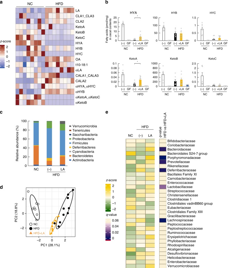
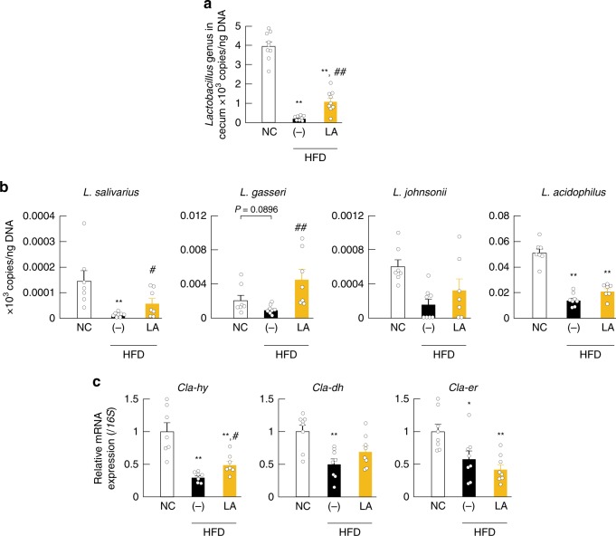
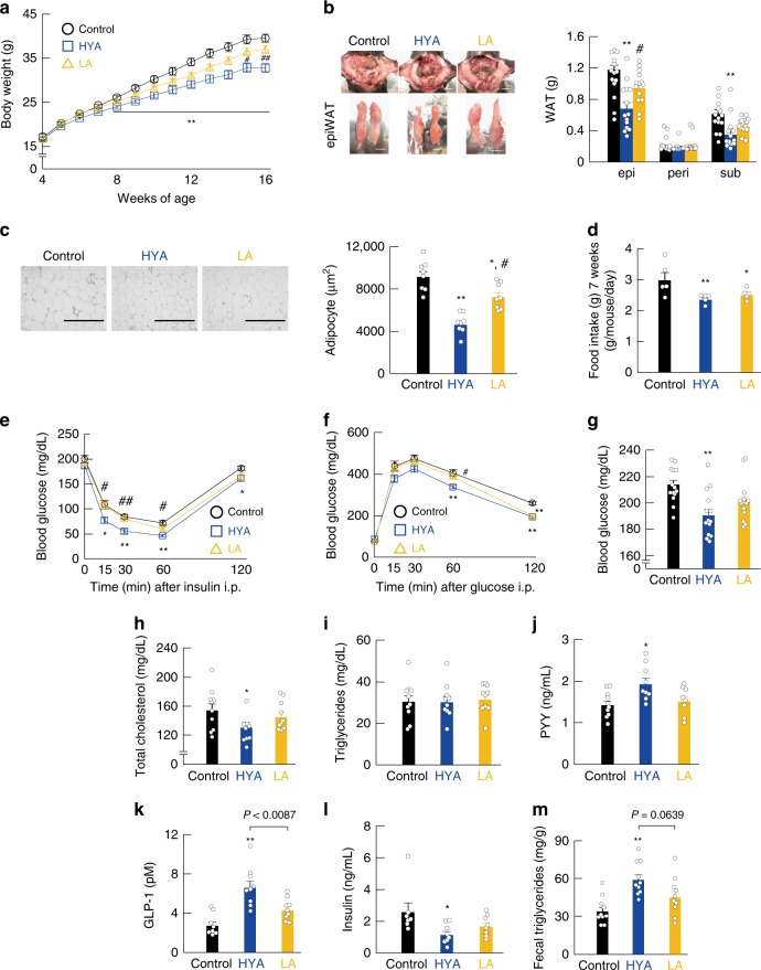
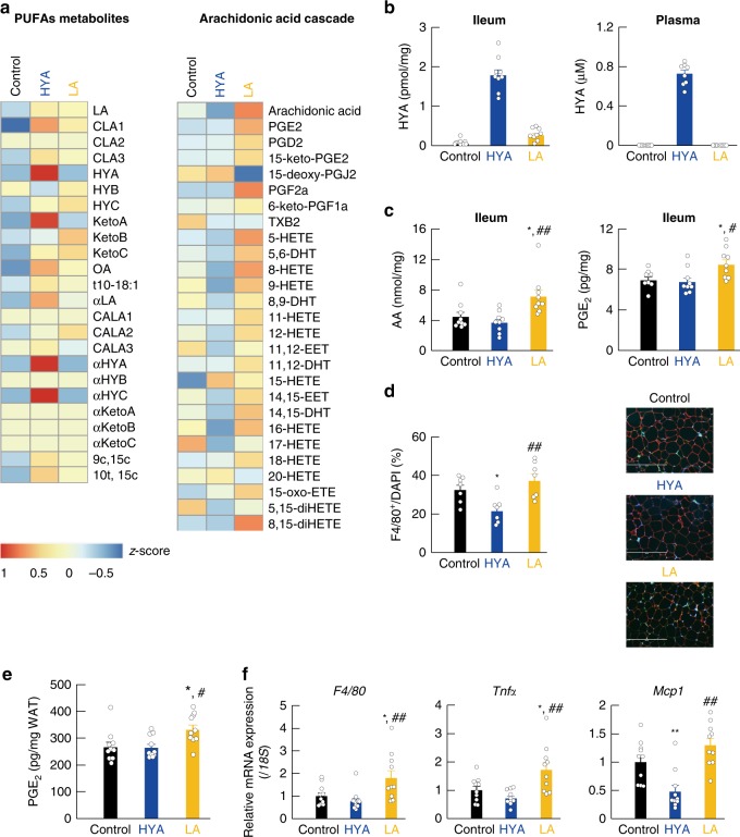
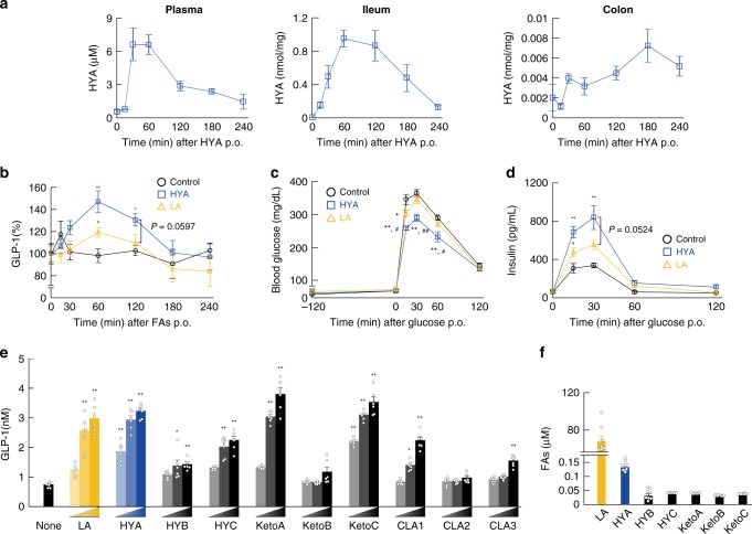
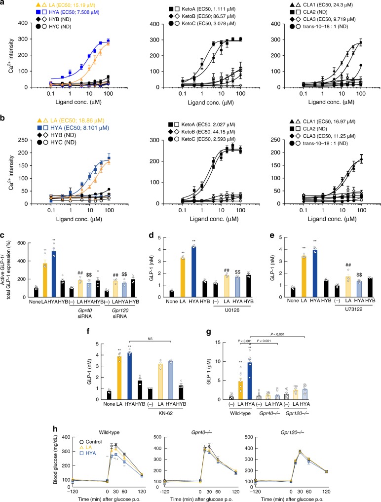
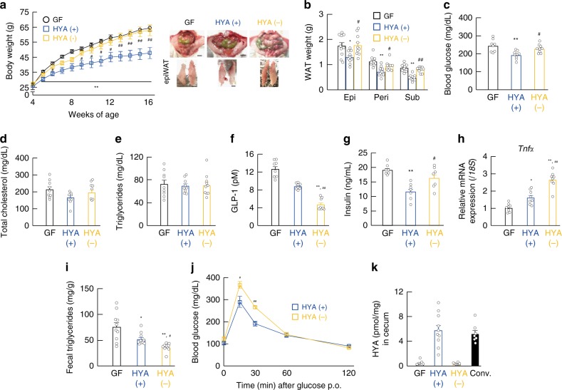
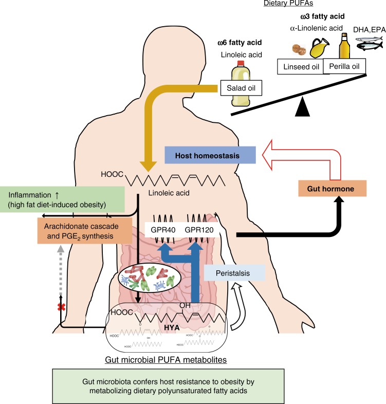
Gut microbiota mediates the effects of diet, thereby modifying host metabolism and the incidence of metabolic disorders. Increased consumption of omega-6 polyunsaturated fatty acid (PUFA) that is abundant in Western diet contributes to obesity and related diseases. Although gut-microbiota-related metabolic pathways of dietary PUFAs were recently elucidated, the effects on host physiological function remain unclear. Here, we demonstrate that gut microbiota confers host resistance to high-fat diet (HFD)-induced obesity by modulating dietary PUFAs metabolism. Supplementation of 10-hydroxy- cis -12-octadecenoic acid (HYA), an initial linoleic acid-related gut-microbial metabolite, attenuates HFD-induced obesity in mice without eliciting arachidonic acid-mediated adipose inflammation and by improving metabolic condition via free fatty acid receptors. Moreover, Lactobacillus -colonized mice show similar effects with elevated HYA levels. Our findings illustrate the interplay between gut microbiota and host energy metabolism via the metabolites of dietary omega-6-FAs thereby shedding light on the prevention and treatment of metabolic disorders by targeting gut microbial metabolites.
Overview
The claims and statistics of this paper haven’t been checked yet.
Run the check — free, no account needed. It reads the open-access text and checks the sources it cites.
Integrity signals
Registry
Cites
Statistics
Figures
Verification
Claims the paper makes
The claims and statistics of this paper haven’t been checked yet.
Run the check — free, no account needed. It reads the open-access text and checks the sources it cites.
Claims other papers make about this one
Each is checked against this paper’s own text: does it say what the citing paper says it does?
In the citing paper
“The secretion of GLP-1 is modulated by the gut microbiota”
From Microbiota–gut–brain axis and its therapeutic applications in neurodegenerative diseases (2024) · cites this paper as [437]
Self-check
Percentages, sums and reported differences re-computed from the paper’s own numbers.
The self-check needs the paper’s full text.
Citations
Cites
An obesity-associated gut microbiome with increased capacity for energy harvest
Turnbaugh, P. J., Ley, R. E., Mahowald, M. A., Magrini, V., Mardis, E. R., & Gordon, J. I. (2006) · Nature · doi:10.1038/nature05414 · Cited by 12,444
Human gut microbes associated with obesity
Ley, R. E., Turnbaugh, P. J., Klein, S., & Gordon, J. I. (2006) · Nature · doi:10.1038/4441022a · Cited by 8,785
A core gut microbiome in obese and lean twins
Turnbaugh, P. J., Hamady, M., Yatsunenko, T., Cantarel, B. L., Duncan, A., Ley, R. E., Sogin, M. L., Jones, W. J., Roe, B. A., Affourtit, J. P., et al. (2009) · Nature · doi:10.1038/nature07540 · Cited by 7,823
Mechanisms linking obesity to insulin resistance and type 2 diabetes
Kahn, S. E., Hull, R. L., & Utzschneider, K. M. (2006) · Nature · doi:10.1038/nature05482 · Cited by 5,448
Turnbaugh, P. J., Bäckhed, F., Fulton, L., & Gordon, J. I. (2008) · Cell Host & Microbe · doi:10.1016/j.chom.2008.02.015 · Cited by 2,915
Oh, D. Y., Talukdar, S., Bae, E. J., Imamura, T., Morinaga, H., Fan, W., Li, P., Lu, W. J., Watkins, S. M., & Olefsky, J. M. (2010) · Cell · doi:10.1016/j.cell.2010.07.041 · Cited by 2,428
An Increase in the Omega-6/Omega-3 Fatty Acid Ratio Increases the Risk for Obesity
Simopoulos, A. P. (2016) · Nutrients · doi:10.3390/nu8030128 · Cited by 1,673
Free fatty acids regulate gut incretin glucagon-like peptide-1 secretion through GPR120
Hirasawa, A., Tsumaya, K., Awaji, T., Katsuma, S., Adachi, T., Yamada, M., Sugimoto, Y., Miyazaki, S., & Tsujimoto, G. (2004) · Nature Medicine · doi:10.1038/nm1168 · Cited by 1,580
Self-citationFree fatty acids regulate insulin secretion from pancreatic β cells through GPR40
Itoh, Y., Kawamata, Y., Harada, M., Kobayashi, M., Fujii, R., Fukusumi, S., Ogi, K., Hosoya, M., Tanaka, Y., Uejima, H., et al. (2003) · Nature · doi:10.1038/nature01478 · Cited by 1,538
Kimura, I., Ozawa, K., Inoue, D., Imamura, T., Kimura, K., Maeda, T., Terasawa, K., Kashihara, D., Hirano, K., Tani, T., et al. (2013) · Nature Communications · doi:10.1038/ncomms2852 · Cited by 1,512
Self-citation
Cited by
Sorted by citations
Items 101–200 of 392
N-3 Polyunsaturated Fatty Acids and Gut Microbiota
Lim, S., & Jayapala, H. P. S. (2022) · Combinatorial Chemistry & High Throughput Screening · doi:10.2174/1386207325666220701121025 · Cited by 36
Olajide, T. M., & Cao, W. (2022) · Trends in Food Science & Technology · doi:10.1016/j.tifs.2022.11.005 · Cited by 33
Targeting gut-liver axis by dietary lignans ameliorate obesity: evidences and mechanisms
Chu, Z., Hu, Z., Luo, Y., Zhou, Y., Yang, F., & Luo, F. (2023) · Critical Reviews in Food Science and Nutrition · doi:10.1080/10408398.2023.2272269 · Cited by 35
“However, the gut microbiota also secrete potentially hazardous substances such as neurotoxins, carcinogens, and immu-notoxins (Miyamoto et al. 2019; Canfora et al. 2019).”
Obesity and the gut microbiota: implications of neuroendocrine and immune signaling
Romaní‐Pérez, M., Liébana-García, R., Flor‐Duro, A., Bonillo‐Jiménez, D., Bullich‐Vilarrubias, C., Olivares, M., & Sanz, Y. (2024) · FEBS Journal · doi:10.1111/febs.17249 · Cited by 36
“…acid, which is a LCFAs-C18 derived from linoleic acid produced by Lactobacillus salivarius strain JCM1044, JCM1045, and JCM1231, increases the plasma levels of both GLP-1 and PYY in mice and was shown to protect against DIO via GLP-1 secretion through a GPR40/120-dependent mechanism [62] (Fig.”
Sharma, V. K., Prateeksha, Gupta, S. C., Singh, B. N., Rao, C. V., & Barik, S. K. (2022) · Materials Today Bio · doi:10.1016/j.mtbio.2022.100204 · Cited by 33
Chen, Q., Yin, Q., Xie, Q., Liu, S., Guo, Z., & Li, B. (2023) · Food Chemistry · doi:10.1016/j.foodchem.2023.135413 · Cited by 32
Gut microbiota in alcohol-related liver disease: pathophysiology and gut-brain cross talk
Zhu, L., Wang, Y., Pan, C. Q., & Xing, H. (2023) · Frontiers in Pharmacology · doi:10.3389/fphar.2023.1258062 · Cited by 32
“Some gut microbe-derived metabolites involving SCFAs, LCFAs, mucus, indole derivatives, and vitamin B had protective effects on the gut and liver (Miyamoto et al., 2019; Zheng et al., 2020).”
BackgroundPolyunsaturated Fatty Acids as Prebiotics: Innovation or Confirmation?
Rinninella, E., & Costantini, L. (2022) · Foods · doi:10.3390/foods11020146 · Cited by 31
“Lipids are absorbed in the small intestine, and amazingly Lactobacillus and the higher concentration of the metabolite HYA are found in the small intestine, although most of the microbiota normally resides in the colon [5].”
BackgroundResultInfluentialRodrigues, M., Silva, C. B., Joaquim, A. G., Machado, C. D., Ramalho, L. N. Z., Carlos, D., Fassini, P. G., & Suen, V. M. M. (2022) · Food & Function · doi:10.1039/d1fo03619j · Cited by 29
New Advances in Improving Bone Health Based on Specific Gut Microbiota
Yan, Q., Cai, L., & Guo, W. (2022) · Frontiers in Cellular and Infection Microbiology · doi:10.3389/fcimb.2022.821429 · Cited by 33
Kim, S., Lee, S., Kim, T.-Y., Lee, S.-H., Seo, S., & Kweon, M. (2023) · Microbiome · doi:10.1186/s40168-023-01687-8 · Cited by 32
“Interestingly, metabolites associated with linoleic acid, produced by Lactobacillus species, have been shown to modulate host metabolic conditions and inflammation by binding to Gpr40 and Gpr120 [18].”
BackgroundGao, J., Mao, K., Wang, X., Mi, S., Fu, M., Li, X., Xiao, J., Simal‐Gándara, J., & Sang, Y. (2021) · Journal of Agricultural and Food Chemistry · doi:10.1021/acs.jafc.1c02430 · Cited by 31
Self-citationRong, B., Wu, Q., Reiter, R. J., & Sun, C. (2021) · Cellular and Molecular Gastroenterology and Hepatology · doi:10.1016/j.jcmgh.2021.06.024 · Cited by 30
“testines, numerous signaling pathways and microbial metabolites involved in their crosstalk also affect distal organs/ tissues such as adipose tissue.(8,9) Previous studies indicate the pattern of the microbial community in gut is changed in response to dietary intake, which influences host metabolism, including nutri…”
BackgroundImpact of gut microbiota on plasma oxylipins profile under healthy and obesogenic conditions
Ávila‐Román, J., Arreaza‐Gil, V., Cortés-Espinar, A. J., Soliz‐Rueda, J. R., Mulero, M., Muguerza, B., Arola‐Arnal, A., Arola, L., & Torres‐Fuentes, C. (2021) · Clinical Nutrition · doi:10.1016/j.clnu.2021.02.035 · Cited by 30
Polyunsaturated fatty acids-rich dietary lipid prevents high fat diet-induced obesity in mice
Haneishi, Y., Furuya, Y., Hasegawa, M., Takemae, H., Tanioka, Y., Mizutani, T., Rossi, M., & Miyamoto, J. (2023) · Scientific Reports · doi:10.1038/s41598-023-32851-7 · Cited by 28
Self-citationFuchs, D., Tang, X. Y., Johnsson, A., Dahlén, S., Hámberg, M., & Wheelock, C. E. (2020) · Biochimica et Biophysica Acta (BBA) - Molecular and Cell Biology of Lipids · doi:10.1016/j.bbalip.2020.158611 · Cited by 28
Microbiota-driven interleukin-17 production provides immune protection against invasive candidiasis
Li, M., Li, C., Wu, X., Chen, T., Ren, L., Xu, B., & Cao, J. (2020) · Critical Care · doi:10.1186/s13054-020-02977-5 · Cited by 29
Ortega‐Hernández, A., Martínez‐Martínez, E., Gómez-Gordo, R., López‐Andrés, N., Fernández‐Celis, A., Gutiérrez-Miranda, B., Nieto, M. L., Alarcón, T., Alba, C., Gómez‐Garré, D., et al. (2020) · Antioxidants · doi:10.3390/antiox9070640 · Cited by 28
“Different data have shown that GM can promote storage of calories as fat, thereby influencing the development and maintenance of obesity through different mechanisms, which includes the production of bacterial metabolites [11,16].”
BackgroundInvestigating the potential of fish oil as a nutraceutical in an animal model of early life stress
Egerton, S., Donoso, F., Fitzgerald, P., Gite, S., Fouhy, F., Whooley, J., Dinan, T. G., Cryan, J. F., Culloty, S. C., Ross, R. P., et al. (2020) · Nutritional Neuroscience · doi:10.1080/1028415x.2020.1753322 · Cited by 30
“FFAR4 or through PUFA-derived intermediate metabolites produced by certain gut microbiota [76].”
BackgroundLapidot, Y., Reshef, L., Goldsmith, R., Na’amnih, W., Kassem, E., Ornoy, A., Gophna, U., & Muhsen, K. (2021) · Nutrients · doi:10.3390/nu13082645 · Cited by 29
“The consumption of PUFAs has been widely investigated in relation to cardiovascular diseases, and excessive intake of dietary PUFAs, especially omega-6 fatty acids, such as linoleic acids, and imbalances of the omega-6/omega-3 ratio contribute to metabolic disease along with chronic inflammation [64].”
BackgroundGut microbiota-derived metabolites as key mucosal barrier modulators in obesity
Wei, Y., Zheng, K., & Wang, Y.-G. (2021) · World Journal of Gastroenterology · doi:10.3748/wjg.v27.i33.5555 · Cited by 29
Mundula, T., Russo, E., Curini, L., Giudici, F., Piccioni, A., Franceschi, F., & Amedei, A. (2022) · Current Medicinal Chemistry · doi:10.2174/0929867329666220430131018 · Cited by 29
Probiotic potential of Lactobacillus sakei L-7 in regulating gut microbiota and metabolism
Zou, X., Pan, L., Xu, M., Wang, X., Wang, Q., & Han, Y. (2023) · Microbiological Research · doi:10.1016/j.micres.2023.127438 · Cited by 28
Zhang, T., Liu, W., Lu, H., Cheng, T., Wang, L., Wang, G., Zhang, H., & Chen, W. (2023) · Critical Reviews in Food Science and Nutrition · doi:10.1080/10408398.2023.2278155 · Cited by 30
“Dietary linoleic acid is metabolized by Lactobacillus salivar-ius and Lactobacillus griffithi to form hydroxy fatty acids, which can enhance intestinal motility by binding to the prostaglandin 3 (EP3) receptor (Miyamoto et al. 2019).”
Liang, B., Shi, L., Du, D., Li, H., Ning, Y., Xi, Y., Cui, J., Li, P., Kang, H., Noda, M., et al. (2023) · Antioxidants · doi:10.3390/antiox12061245 · Cited by 27
Ren, Y., Shi, X., Mu, J., Liu, S., Qian, X., Pei, W., Ni, S., Zhang, Z., Li, L., & Zhang, Z. (2024) · Food & Function · doi:10.1039/d3fo04347a · Cited by 31
Xiao, Y., Yang, C., Xu, H., Wu, Q., Zhou, Y., Zhou, X., & Miao, J. (2020) · Journal of Functional Foods · doi:10.1016/j.jff.2020.104285 · Cited by 27
Role of Maternal Microbiota and Nutrition in Early-Life Neurodevelopmental Disorders
Daliry, A., & Pereira, E. N. G. D. S. (2021) · Nutrients · doi:10.3390/nu13103533 · Cited by 31
Oleate Hydratase (OhyA) Is a Virulence Determinant in Staphylococcus aureus
Radka, C. D., Batte, J. L., Frank, M. W., Rosch, J. W., & Rock, C. O. (2021) · Microbiology Spectrum · doi:10.1128/spectrum.01546-21 · Cited by 27
“are engaged by h18:1 (3, 25), but it is not obvious how these nutrient sensors (26)”
BackgroundNoguchi, M., Shimizu, M., Lü, P., Takahashi, Y., Yamauchi, Y., Sato, S., Kiyono, H., Kishino, S., Ogawa, J., Nagata, K., et al. (2022) · Journal of Biological Chemistry · doi:10.1016/j.jbc.2022.102534 · Cited by 26
“Although bacterial fatty acid metabolites exists lower levels than original compounds, these metabolites produced by lactic acid bacteria were detected in the intestine (13, 46), implying that substantial levels of these metabolites are present in host tissues.”
BackgroundSelf-citationLiu, G., Gibson, R. A., Callahan, D. L., Guo, X.-F., Li, D., & Sinclair, A. J. (2020) · Food & Function · doi:10.1039/c9fo02440a · Cited by 26
Fu, Q., Huang, H., Ding, A., Yu, Z., Huang, Y., Fu, G., Huang, Y., & Huang, X. B. (2022) · Frontiers in Nutrition · doi:10.3389/fnut.2022.965653 · Cited by 25
“The metabolites levels derived from LA, such as 10-hydroxy-cis-12-octadecenoic acid, were significantly reduced in the intestinal tract of rats fed a high-fat diet for 2 weeks, but were reversed by supplementation with LA (52).”
MethodThe Role of Diet and Gut Microbiota Interactions in Metabolic Homeostasis
Guan, L., & Liu, R. (2023) · Advanced Biology · doi:10.1002/adbi.202300100 · Cited by 27
Wang, B., He, Y., Tang, J., Ou, Q., & Lin, J. (2020) · International Journal of Rheumatic Diseases · doi:10.1111/1756-185x.13802 · Cited by 25
Gao, R., Qi, Z., Lin, J., Wang, G., Chen, G., Yuan, L., & Sun, Q. (2023) · Journal of Agricultural and Food Chemistry · doi:10.1021/acs.jafc.3c02642 · Cited by 24
The Relationship Between Dietary Patterns and the Epidemiology of Food Allergy
Leung, A. S. Y., Xing, Y., Fernández‐Rivas, M., & Wong, G. (2024) · Allergy · doi:10.1111/all.16455 · Cited by 25
“Although the mechanism by which diet impacts the gut microbiome is beyond the scope of this review, there is evidence that consuming an animal-based diet [119, 120] and ome- ga-6 polyunsaturated fatty acids [121] can negatively impact gut health.”
Hu, D., Yang, W., Mao, P., & Cheng, M. (2020) · Nutrition and Cancer · doi:10.1080/01635581.2020.1767166 · Cited by 25
Abnormal n-6 fatty acid metabolism in cystic fibrosis contributes to pulmonary symptoms
Wheelock, C. E., & Strandvik, B. (2020) · Prostaglandins Leukotrienes and Essential Fatty Acids · doi:10.1016/j.plefa.2020.102156 · Cited by 24
“transfer of bacterial metabolic products or immunological factors [87].”
BackgroundPal, A., Sun, S., Armstrong, M., Manke, J., Reisdorph, N., Adams, V. R., Kennedy, A. J., Zu, Y., Moustaïd‐Moussa, N., Carroll, I. M., et al. (2021) · Biochimica et Biophysica Acta (BBA) - Molecular and Cell Biology of Lipids · doi:10.1016/j.bbalip.2021.159059 · Cited by 25
Igarashi, M., Hayakawa, T., Tanabe, H., Watanabe, K., Nishida, A., & Kimura, I. (2022) · Molecular Metabolism · doi:10.1016/j.molmet.2022.101649 · Cited by 24
“OEA generated from OA by gut microbiota modulates meal behavior Our previous study showed that microorganisms could convert unabsorbed fatty acids reaching in the distal intestine to unique fatty acid metabolites, such as conjugated fatty acids and hydroxy fatty acids, which have physiological functions [3].”
BackgroundMethodInfluentialSelf-citationLi, K., Xiao, X., Li, Y., Lu, S., Zi, J., Sun, X., Xu, J., Liu, H., Li, X., Song, T., et al. (2024) · Journal of Animal Science and Biotechnology/Journal of animal science and biotechnology · doi:10.1186/s40104-024-01073-w · Cited by 30
“By producing metabolites such as HYA, the gut microbiota helps to regulate the host’s energy balance and insulin sensitivity, thereby enhancing resistance to metabolic disorders [148].”
BackgroundSelf-citationHou, T., Dai, H., Wang, Q., Hou, Y., Zhang, X., Lin, H., Wang, S., Li, M., Zhao, Z., Lu, J., et al. (2023) · Frontiers in Immunology · doi:10.3389/fimmu.2023.1148591 · Cited by 22
Wang, D., Deng, Y., Zhao, L., Wang, K., Wu, D., Hu, Z., & Liu, X. (2023) · Food & Function · doi:10.1039/d2fo04038g · Cited by 25
Dietary fat disrupts a commensal-host lipid network that promotes metabolic health
Klag, K. A., Ott, D., Tippetts, T. S., Nicholson, R. J., Tatum, S. M., Bauer, K. M., Victor, E. S., Weis, A. M., Bell, R. S., Weagley, J. S., et al. (2025) · Cell Metabolism · doi:10.1016/j.cmet.2025.10.007 · Cited by 22
Targeting the Gut Microbiota for Remediating Obesity and Related Metabolic Disorders
Wang, B., Wang, L., Wang, H., Dai, H., Lu, X., Lee, Y. K., Gu, Z., Zhao, J., Zhang, H., Chen, W., et al. (2021) · Journal of Nutrition · doi:10.1093/jn/nxab103 · Cited by 26
Kwek, E., Zhu, H., Ding, H., He, Z., Hao, W., Liu, J., Ma, K. Y., & Chen, Z. (2022) · European Journal of Nutrition · doi:10.1007/s00394-021-02785-9 · Cited by 23
Involvement of Gut Microbial Metabolites Derived from Diet on Host Energy Homeostasis
Nishida, A., Ando, Y., Kimura, I., & Miyamoto, J. (2022) · International Journal of Molecular Sciences · doi:10.3390/ijms23105562 · Cited by 23
“trimethylamine N-oxide (TMAO) increased cardiovascular disease [21,22] branched-chain amino acids (BCAAs) increased obesity, type II diabetes [19] imidazole propionate increased type II diabetes [23,24] tauro-β-muricholic acid increased obesity [25–28] short-chain fatty acids (SCFAs) decreased obesity [29–38] 10-hydro…”
BackgroundSelf-citationGut flora in multiple sclerosis: implications for pathogenesis and treatment
Zhang, W., Wang, Y., Zhu, M., Liu, K., & Zhang, H. (2023) · Neural Regeneration Research · doi:10.4103/1673-5374.387974 · Cited by 24
“Multiple Sclerosis and the Hygiene Hypothesis MS and hygiene have been linked to the increasing incidence of immune-associated disorders in modern living environments.”
Zhang, J., Zhang, R., Chen, Y., Guo, X., Ren, Y., Wang, M., Li, X., Huang, Z., Zhu, W., & Yu, K. (2024) · Journal of Agricultural and Food Chemistry · doi:10.1021/acs.jafc.4c02381 · Cited by 22
Self-citationTrimethylamine-N-oxide accelerates osteoporosis by PERK activation of ATF5 unfolding
Lin, Y.-H., Lian, W., Wu, R., Chen, Y., Wu, S.-L., Ko, J., Wang, S., Jahr, H., & Wang, F. (2024) · Cellular and Molecular Life Sciences · doi:10.1007/s00018-024-05501-y · Cited by 21
“2d), including Muriculum, Lactobacillus, Akkermansia, and Ruminococcaceae [28, 29].”
Microbiota in Health and Disease—Potential Clinical Applications
Laudes, M., Geisler, C., Rohmann, N., Bouwman, J., Pischon, T., & Schlicht, K. (2021) · Nutrients · doi:10.3390/nu13113866 · Cited by 22
“Of interest, gut microbes are not only able to produce small fatty acids, but also complex polyunsaturated once (PUFA) via the metabolite 10-hydroxy-cis-12-octadecenoic acid (HYA), which has been shown to induce host resistance to HFD-induced obesity [98].”
BackgroundDysbiosis: The first hit for digestive system cancer
Mei, S., Deng, Z., Chen, Y., Ning, D.-M., Guo, Y., Fan, X., Wang, R., Meng, Y., Zhou, Q., & Tian, X. (2022) · Frontiers in Physiology · doi:10.3389/fphys.2022.1040991 · Cited by 22
“, 2020; Poland and Flynn, 2021), energy consumption (Coelho et al., 2019; Miyamoto et al., 2019), and neurotransmitter biosynthesis (Yano et al.”
BackgroundSebag, S. C., Hao, M., Qian, Q., Upara, C., Ding, Q. J., Zhu, M., Banas, J. A., Cao, H., Hong, L., & Yang, L. (2024) · Acta Pharmaceutica Sinica B · doi:10.1016/j.apsb.2024.01.002 · Cited by 23
Pan, R., Guo, M., Chen, Y., Lin, G., Tian, P., Wang, L., Zhao, J., Chen, W., & Wang, G. (2024) · Nutrients · doi:10.3390/nu16040483 · Cited by 22
“On the other hand, the gut microbiota can metabolize polyunsaturated fatty acids to confer host resistance to obesity [52].”
BackgroundInteractions between lipid metabolism and the microbiome in aquatic organisms: A review
Yoon, D.-S., Kim, D., Kim, J., Sakakura, Y., Hagiwara, A., Park, H. G., Lee, M., & Lee, J. (2024) · Marine Pollution Bulletin · doi:10.1016/j.marpolbul.2024.116858 · Cited by 20
Zhang, Y., Wang, C., Chen, C., Yu, J., Yang, Y., & Wang, S. (2025) · Carbohydrate Polymers · doi:10.1016/j.carbpol.2025.123445 · Cited by 20
Recent insights into the role of microbiome in the pathogenesis of obesity
van der Vossen, E. W. J., de Goffau, M. C., Levin, E., & Nieuwdorp, M. (2022) · Therapeutic Advances in Gastroenterology · doi:10.1177/17562848221115320 · Cited by 20
Wang, J., Chen, J., Gao, M., Ouyang, Z., Li, Y., Liu, D., Zhu, M., & Sun, H. (2025) · Foods · doi:10.3390/foods14091583 · Cited by 21
“Among these, t10,c12-CLA is the second most abundant after c9,t11-CLA and plays a significant role in regulating lipid metabolism. t10,c12-CLA can promote the secretion of GLP-1 through G protein-coupled receptors 40/120 (GPR40/120) and enhance lipid breakdown [34].”
Arruda, H. S., Geraldi, M. V., Cedran, M. F., Bicas, J. L., Maróstica, M. R., & Pastore, G. M. (2021) · Elsevier eBooks · doi:10.1016/b978-0-12-823569-0.00006-0 · Cited by 20
Structures and functions of the gut microbial lipidome
Morozumi, S., Ueda, M., Okahashi, N., & Arita, M. (2022) · Biochimica et Biophysica Acta (BBA) - Molecular and Cell Biology of Lipids · doi:10.1016/j.bbalip.2021.159110 · Cited by 20
Self-citationYi, S., Zhang, C., Yin, P., Yu, L., Tian, F., Chen, W., & Zhai, Q. (2023) · Food & Function · doi:10.1039/d3fo02182c · Cited by 19
Lee, C.-T., & Tribble, G. D. (2023) · Frontiers in Oral Health · doi:10.3389/froh.2023.1217088 · Cited by 20
“These bacteria have been shown to produce enzymes that modify omega-6 linoleic acid to a conjugated linoleic acid (CLA) that has anti-inflammatory effects and blocks the production of ARA-derived lipids (67).”
BackgroundHost–diet–microbiota interplay in intestinal nutrition and health
Ignatiou, A., & Pitsouli, C. (2024) · FEBS Letters · doi:10.1002/1873-3468.14966 · Cited by 21
“Noteworthily, the ideal dietary ratio of omega-6 to omega-3 PUFAs is 1 – 4 : 1; nonetheless, the modern WD contains a ratio of ~ 20 : 1, which increases the risk for obesity, CVD, and cancer [155].”
Matcha alleviates obesity by modulating gut microbiota and its metabolites
Luo, Y., Wen, Y., Huang, J., Chen, B., Lv, S., Qiu, H., Li, S., Liu, S., Yang, Q., He, L., et al. (2024) · Current Research in Food Science · doi:10.1016/j.crfs.2024.100823 · Cited by 19
“…the gut microbiota has been shown to regulate metabolic pathways such as branched-chain amino acid metabolism and bile acid metabolism in the gut, thus playing a significant role in the onset and progression of obesity (Allegretti et al., 2020; Miyamoto et al., 2019; Zeng et al., 2020).”
Watanabe, A., Balas, L., Saigusa, D., Ogura, J., Durand, T., Mano, N., & Yamaguchi, H. (2022) · Food Chemistry Advances · doi:10.1016/j.focha.2022.100040 · Cited by 18
Attaye, I., Lassen, P. B., Adriouch, S., Steinbach, E., Patiño‐Navarrete, R., Davids, M., Alili, R., Jacques, F., Benzeguir, S., Belda, E., et al. (2023) · iScience · doi:10.1016/j.isci.2023.107471 · Cited by 21
Impact of the lipase inhibitor orlistat on the human gut microbiota
Uehira, Y., Ueno, H., Miyamoto, J., Kimura, I., Ishizawa, Y., Iijima, H., Muroga, S., Fujita, T., Sakai, S., Samukawa, Y., et al. (2023) · Obesity Research & Clinical Practice · doi:10.1016/j.orcp.2023.08.005 · Cited by 18
Self-citationQassab, M. A., Chaarani, N., Hamou, A., Harb, R. M., Jradi, A., Zeineddine, M., Ghadieh, H. E., Khattar, Z. A., Azar, S. T., Kanaan, A., et al. (2026) · FASEB BioAdvances · doi:10.1096/fba.2025-00218 · Cited by 28
“It also affects energy homeostasis systemically, impacting metabolic processes in the brain, liver, and adipose tissue [49, 54–55].”
Enterocyte superoxide dismutase 2 deletion drives obesity
García‐Irigoyen, O., Bovenga, F., Piglionica, M., Piccinin, E., Cariello, M., Arconzo, M., Peres, C., Corsetto, P. A., Rizzo, A. M., Ballanti, M., et al. (2021) · iScience · doi:10.1016/j.isci.2021.103707 · Cited by 17
Kang, M., Feng, K., Dai, W., Miao, J., Liu, G., Fang, H., & Cao, Y. (2023) · Food & Function · doi:10.1039/d2fo03755f · Cited by 17
Chen, X., Gu, J., & Huang, Y. (2023) · Lipids in Health and Disease · doi:10.1186/s12944-023-01982-1 · Cited by 17
“Miyamoto and coworkers [27] reported that PUFA affected energy regulation in obese mice and reduced their body weight, CHO, insulin resistance, and glucose tolerance to levels lower than that measured in obese mice eating a regular diet.”
BackgroundGut microbiome and metabolomics in systemic sclerosis: feature, link and mechanisms
Yao, Q., Tan, W., & Bai, F. (2024) · Frontiers in Immunology · doi:10.3389/fimmu.2024.1475528 · Cited by 17
Adipose tissue–gut microbiome crosstalk in inflammation and thermogenesis
Mauney, E. E., Wibowo, M. C., Tseng, Y., & Kostic, A. D. (2024) · Trends in Endocrinology and Metabolism · doi:10.1016/j.tem.2024.10.004 · Cited by 18
Zeng, Y., Wu, R., He, Y., Zhang, Q., Wang, Z., Qin, P., Yang, F., Han, Y., Hao, M., Zheng, Y., et al. (2025) · Cell Host & Microbe · doi:10.1016/j.chom.2025.04.011 · Cited by 19
Intestinal HIF-2α Regulates GLP-1 Secretion via Lipid Sensing in L-Cells
Mooli, R. G. R., Mukhi, D., Pasupulati, A. K., Evers, S. S., SIPULA, I. J., Jurczak, M. J., Seeley, R. J., Shah, Y. M., & Ramakrishnan, S. K. (2021) · Cellular and Molecular Gastroenterology and Hepatology · doi:10.1016/j.jcmgh.2021.12.004 · Cited by 16
Liu, N., Song, Z., Jin, W., Yang, Y., Sun, S., Zhang, Y., Zhang, S., Liu, S., Ren, F., & Wang, P. (2022) · Journal of Functional Foods · doi:10.1016/j.jff.2022.105234 · Cited by 18
Yang, H., Wu, C., Chen, L., Chang, X., Luo, G., Wu, K., & Tian, W. (2023) · Microbial Pathogenesis · doi:10.1016/j.micpath.2023.106084 · Cited by 18
Qin, Y., Fan, R., Liu, Y.-X., Qiu, S., & Wang, L. (2023) · Food & Function · doi:10.1039/d3fo02653a · Cited by 16
Ando, M., Nagata, K., Takeshita, R., Ito, N., Noguchi, S., Minamikawa, N., Kodama, N., Yamamoto, A., Yashiro, T., Hachisu, M., et al. (2024) · Frontiers in Immunology · doi:10.3389/fimmu.2024.1374425 · Cited by 16
Self-citationApproaching precision medicine by tailoring the microbiota
Ryu, G., Kim, H., & Koh, A. (2021) · Mammalian Genome · doi:10.1007/s00335-021-09859-3 · Cited by 17
Chemoproteomic Analysis of Microbiota Metabolite–Protein Targets and Mechanisms
Zhao, X., Yang, X., & Hang, H. C. (2022) · Biochemistry · doi:10.1021/acs.biochem.1c00758 · Cited by 16
Xu, J., Zhang, Q., Wang, Y., Cheng, Z., Zhu, H., Zhao, H., Yao, Y., Hua, L., Qiao, B., Zhao, L., et al. (2025) · Journal of Hazardous Materials · doi:10.1016/j.jhazmat.2025.138335 · Cited by 15
Fatty Acids as Prebiotics and Their Role in Antibiofilm Activity
Nazzaro, F., Coppola, F., Fratianni, F., & Coppola, R. (2026) · Antibiotics · doi:10.3390/antibiotics15010057 · Cited by 15
“Dietary fatty acids play significant roles in energy metabolism and influence the microbiota and intestinal physiology by modulating intestinal permeability and exerting anti-inflammatory activity, rather than through direct fermentation [27].”
Gut microbiome in liver pathophysiology and cholestatic liver disease
Yan, S., & Yin, X. (2021) · Liver Research · doi:10.1016/j.livres.2021.08.001 · Cited by 16
Xu, H., Jurado‐Fasoli, L., Ortiz-Alvarez, L., Osuna‐Prieto, F. J., Köhler, I., Di, X., Vilchez‐Vargas, R., Link, A., Plaza‐Díaz, J., Gil, Á., et al. (2022) · Nutrients · doi:10.3390/nu14234991 · Cited by 15
Zhou, Q., Qu, Z., Wang, N., Liu, H., Hongmei, Y., & Wang, H. (2022) · Food Science & Nutrition · doi:10.1002/fsn3.3136 · Cited by 14
Honda, T., Kabashima, K., & Kunisawa, J. (2023) · Immunological Reviews · doi:10.1111/imr.13193 · Cited by 15
Chen, Q., Cheng, W., Zhang, J., Chi, C., Lin, M., He, C., Liao, Z., & Gong, F. (2024) · Free Radical Biology and Medicine · doi:10.1016/j.freeradbiomed.2024.09.017 · Cited by 17
Cao, W., Fang, L., Li, R. W., Chin, Y., Wang, Y., Xue, C., & Tang, Q. (2021) · Food Science and Human Wellness · doi:10.1016/j.fshw.2021.07.018 · Cited by 16
Watanabe, Y., Fujisaka, S., Ikeda, K., Ishikawa, M., Yamada, T., Nawaz, A., Kado, T., Kuwano, T., Nishimura, A., Bilal, M., et al. (2021) · iScience · doi:10.1016/j.isci.2021.102445 · Cited by 15
Wang, M., Guo, W., Yang, Z., Chen, F., Lin, T., Li, W., Lv, X., Rao, P., Ai, L., & Ni, L. (2022) · Current Research in Food Science · doi:10.1016/j.crfs.2022.08.015 · Cited by 16
Nagahisa, T., Kosugi, S., & Yamaguchi, S. (2023) · Nutrients · doi:10.3390/nu15061494 · Cited by 14
Staphylococcus aureus oleate hydratase produces ligands that activate host PPARα
Radka, C. D., Frank, M. W., Simmons, T. S., Johnson, C. N., Rosch, J. W., & Rock, C. O. (2024) · Frontiers in Cellular and Infection Microbiology · doi:10.3389/fcimb.2024.1352810 · Cited by 14
Chewing the Fat with Microbes: Lipid Crosstalk in the Gut
Lemons, J. M. S., & Liu, L. (2022) · Nutrients · doi:10.3390/nu14030573 · Cited by 14
“However, some PUFAs are partially metabolized in the distal intestine by Bifidobacteria and Lactobacilli, which facilitates their ability to outcompete pathogenic Enterobacteria [112,113].”
BackgroundGu, S., Chen, C., Wang, J., Wang, Y., Zhao, L., Xiong, Z., Zhang, H., Deng, T., Pan, Q., Zheng, Y., et al. (2025) · Phytomedicine · doi:10.1016/j.phymed.2025.156806 · Cited by 13
Czauderna, A., Kulkarni, G. V., Bianchi, N., Cheng, L., Sim, M., Realini, N. R., Gach, J., Caillon, A., Kloehn, J., Lambeau, G. J., et al. (2025) · Cell Host & Microbe · doi:10.1016/j.chom.2025.08.011 · Cited by 18
Yu, H., Fang, C., Li, P., Wu, M., & Shen, S. (2021) · European Journal of Pharmacology · doi:10.1016/j.ejphar.2021.173885 · Cited by 13
Yong, G. J. M., Porsche, C. E., Sitarik, A. R., Fujimura, K. E., McCauley, K. E., Nguyen, D., Levin, A. M., Woodcroft, K. J., Ownby, D. R., Rundle, A., et al. (2023) · Gut Microbes · doi:10.1080/19490976.2023.2290661 · Cited by 14
“20–23 Mode of delivery and nutrition in infancy have been associated with alterations to the infant gut microbiome and with obesity in childhood, 24,25 though the mechanisms by which they contribute to disease remain unclear.”
BackgroundZhang, L., Liu, Z., Zhang, W., Wang, J., Kang, H., Jing, J., Lin, H., & Gao, A. (2024) · Gut Microbes · doi:10.1080/19490976.2024.2323227 · Cited by 14
“11,41 In addition, LCFAs are also essential energy providers, coming from FAO that produces more than twice as much ATP as glucose or amino acids.”
BackgroundLi, B., Jin, Y., Zhang, B., Lu, T., Li, J., Zhang, J., Zhou, Y., Wang, Y., Zhang, C., Zhao, Y., et al. (2024) · Clinical and Translational Medicine · doi:10.1002/ctm2.70029 · Cited by 14
“44,45 To further confirm that obesity contributes to the development of TMJ OA, we established a TMJ OA mouse model using UAC and an obesity model using HFD as previously described 26,27 (Figure 1D).”
Method
Metadata
- Authors
- Miyamoto, J., Igarashi, M., Watanabe, K., Karaki, S.-I., Mukouyama, H., Kishino, S., Li, X., Ichimura, A., Irie, J., Sugimoto, Y., Mizutani, T., Sugawara, T., Miki, T., Ogawa, J., Drucker, D. J., Arita, M., Itoh, H., & Kimura, I.
- Affiliations
- Japan Agency for Medical Research and Development (JP)
- Tokyo University of Agriculture and Technology (JP)
- Shizuoka University (JP)
- Kyoto University (JP)
- Keio University (JP)
- Chiba University (JP)
- Mount Sinai Hospital (CA)
- Corresponding author
- Kimura, Ikuo
- ORCID
- Venue
- Nature Communications
- Published
- 2019-09-05
- Volume
- 10
- Issue
- 1
- Pages
- 4007-4007
- ISSN
- 2041-1723 (ISSN-L)
- PMC
- PMC6728375
- OpenAlex
- W2971602650
- Cited by
- 392
- References
- 40
- Citations by year
- 2026: 46 · 2025: 60 · 2024: 65 · 2023: 62 · 2022: 64 · 2021: 51 · 2020: 40 · 2019: 1
- FWCI
- 12.91
- Type
- journal article
- Language
- en
- Open access
- gold
- doi.org
- Locations
- Nature Communications (publishedVersion, cc-by)
- Europe PMC (PubMed Central) (publishedVersion, cc-by)
- PubMed Central (publishedVersion, cc-by)
- PubMed (publishedVersion)
- RePEc: Research Papers in Economics (submittedVersion)
- DOAJ (DOAJ: Directory of Open Access Journals) (submittedVersion, cc-by-sa)
- DOAJ (DOAJ: Directory of Open Access Journals) (submittedVersion)
- Funders
- MEXT | Japan Society for the Promotion of Science (JP15H05344, JP16H01355)
- Japan Agency for Medical Research and Development (JP18gm1010007)
- Topics
- Life Sciences › Biochemistry, Genetics and Molecular Biology › Molecular Biology › Gut microbiota and health
- Health Sciences › Nursing › Nutrition and Dietetics › Fatty Acid Research and Health
- Health Sciences › Medicine › Endocrinology, Diabetes and Metabolism › Adipokines, Inflammation, and Metabolic Diseases
- Keywords
- gut microbiota, obesity, omega-6 fatty acids, high-fat diet, linoleic acid metabolites, Lactobacillus, free fatty acid receptors, adipose tissue inflammation, arachidonic acid, energy metabolism, metabolic disorders
- MeSH terms
- Gastrointestinal Microbiome, Adipose Tissue, Animals, Cell Line, Dietary Fats, Unsaturated, Energy Metabolism, Fatty Acids, Unsaturated, Humans, Inflammation, Lactobacillus, Metabolic Diseases, Mice, Inbred C57BL, Obesity, Oleic Acids, Dietary Supplements, Linoleic Acid, Models, Animal, Fatty Acids, Omega-6, Mice, Diet, High-Fat, Diet, Western
About these checks
Registry, retraction, Cites and statistics results are computed, not judged, with one exception: a printed reference that no index lists is read once by a model before it counts as a citation error. Claims are extracted by a model and kept only when their quote appears verbatim in the text; each cited source is then checked, with a model’s help, for whether it says what the claim relies on. Those verdicts can be wrong — read the claim’s quote and the cited source before relying on one.
Statistics and arithmetic are recomputed, not judged.
Confidence bands are shown only for fields calibrated on our annotated test set. None is calibrated yet, so no band is shown.
What counts as a claim ›How we measure confidence ›How we check this ›
Are you an author, or do you see a problem with this page? Request a review
