

The DOI matches its Crossref record; 76 of the 76 references it cites were found in a registry or an index.
Maturation of the human striatal dopamine system revealed by PET and quantitative MRI
Open accessAbstract
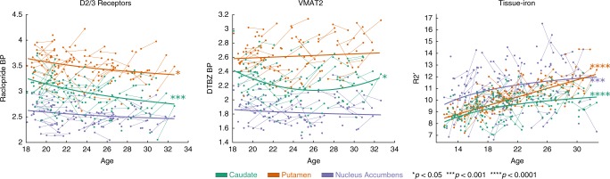
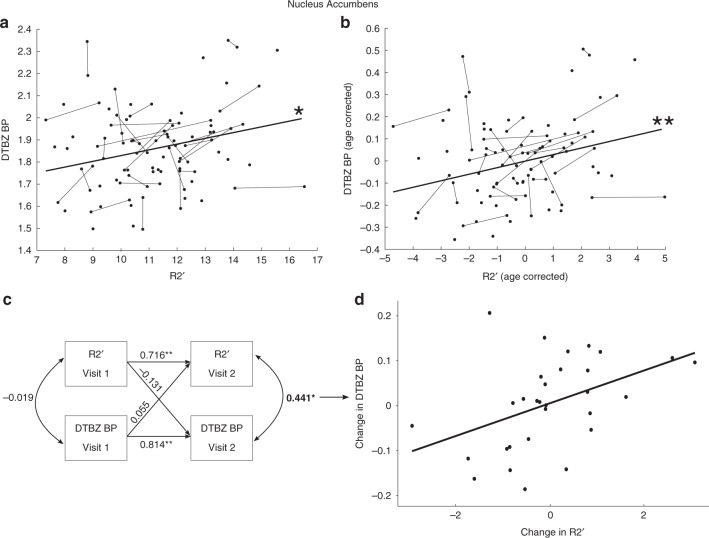
The development of the striatum dopamine (DA) system through human adolescence, a time of increased sensation seeking and vulnerability to the emergence of psychopathology, has been difficult to study due to pediatric restrictions on direct in vivo assessments of DA. Here, we applied neuroimaging in a longitudinal sample of n = 146 participants aged 12–30. R2′, an MR measure of tissue iron which co-localizes with DA vesicles and is necessary for DA synthesis, was assessed across the sample. In the 18–30 year-olds (n = 79) we also performed PET using [11C]dihydrotetrabenazine (DTBZ), a measure of presynaptic vesicular DA storage, and [11C]raclopride (RAC), an indicator of D2/D3 receptor availability. We observed decreases in D2/D3 receptor availability with age, while presynaptic vesicular DA storage (as measured by DTBZ), which was significantly associated with R2′ (standardized coefficient = 0.29, 95% CI = [0.11, 0.48]), was developmentally stable by age 18. Our results provide new evidence for maturational specialization of the striatal DA system through adolescence.
Overview
The claims and statistics of this paper haven’t been checked yet.
Run the check — free, no account needed. It reads the open-access text and checks the sources it cites.
Integrity signals
Registry
Cites
Statistics
Figures
Verification
Claims the paper makes
The claims and statistics of this paper haven’t been checked yet.
Run the check — free, no account needed. It reads the open-access text and checks the sources it cites.
Claims other papers make about this one
Each is checked against this paper’s own text: does it say what the citing paper says it does?
The cited source says what the claim attributes to it.
In the citing paper
“Functional imaging studies further indicate that significant changes occur in the function of subcortical structures and in their relationship with cortical regions, notably in nuclei associated with reward and motivation [26–29].”
From Adolescent nicotine exposure and persistent neurocircuitry changes: unveiling lifelong psychiatric risks (2025) · cites this paper as [29]
Self-check
Percentages, sums and reported differences re-computed from the paper’s own numbers.
The self-check needs the paper’s full text.
Citations
Cites
(1998) · Cited by 36,049
AFNI: Software for analysis and visualization of functional magnetic resonance neuroimages
Cox, R. W. (1996) · Computers and Biomedical Research · doi:10.1006/cbmr.1996.0014 · Cited by 10,913
Fischl, B. (2012) · NeuroImage · doi:10.1016/j.neuroimage.2012.01.021 · Cited by 9,526
The adolescent brain and age-related behavioral manifestations
Spear, L. P. (2000) · Neuroscience & Biobehavioral Reviews · doi:10.1016/s0149-7634(00)00014-2 · Cited by 5,433
The reward circuit: linking primate anatomy and human imaging
Haber, S. N., & Knutson, B. (2010) · Neuropsychopharmacology · doi:10.1038/npp.2009.129 · Cited by 3,718
Why do many psychiatric disorders emerge during adolescence?
Paus, T., Keshavan, M., & Giedd, J. N. (2008) · Nature Reviews Neuroscience · doi:10.1038/nrn2513 · Cited by 3,123
Simplified Reference Tissue Model for PET Receptor Studies
Lammertsma, A. A., & Hume, S. P. (1996) · NeuroImage · doi:10.1006/nimg.1996.0066 · Cited by 2,025
The role of iron in brain ageing and neurodegenerative disorders
Ward, R. J., Zucca, F. A., Duyn, J. H., Crichton, R. R., & Zecca, L. (2014) · The Lancet Neurology · doi:10.1016/s1474-4422(14)70117-6 · Cited by 1,819
Schizophrenia: Caused by a fault in programmed synaptic elimination during adolescence?
Feinberg, I. (1982) · Journal of Psychiatric Research · doi:10.1016/0022-3956(82)90038-3 · Cited by 1,395
Importance of investing in adolescence from a developmental science perspective
Dahl, R. E., Allen, N. B., Wilbrecht, L., & Suleiman, A. B. (2018) · Nature · doi:10.1038/nature25770 · Cited by 1,196
A dual systems model of adolescent risk‐taking
Steinberg, L. (2010) · Developmental Psychobiology · doi:10.1002/dev.20445 · Cited by 1,187
Imaging iron stores in the brain using magnetic resonance imaging
Haacke, E. M., Cheng, N. Y. C., House, M. J., Liu, Q., Neelavalli, J., Ogg, R. J., Khan, A., Ayaz, M., Kirsch, W., & Obenaus, A. (2005) · Magnetic Resonance Imaging · doi:10.1016/j.mri.2004.10.001 · Cited by 995
The dual systems model: Review, reappraisal, and reaffirmation
Shulman, E. P., Smith, A. R., Silva, K., Icenogle, G., Duell, N., Chein, J., & Steinberg, L. (2016) · Developmental Cognitive Neuroscience · doi:10.1016/j.dcn.2015.12.010 · Cited by 867
Adolescence as a neurobiological critical period for the development of higher-order cognition
Larsen, B., & Luna, B. (2018) · Neuroscience & Biobehavioral Reviews · doi:10.1016/j.neubiorev.2018.09.005 · Cited by 853
Self-citationTamnes, C. K., Østby, Y., Fjell, A. M., Westlye, L. T., Due-Tønnessen, P., & Walhovd, K. B. (2010) · Cerebral Cortex · doi:10.1093/cercor/bhp118 · Cited by 779
Interactions of iron, dopamine and neuromelanin pathways in brain aging and Parkinson's disease
Zucca, F. A., Segura-Aguilar, J., Ferrari, E., Muñoz, P., Paris, I., Sulzer, D., Sarna, T., Casella, L., & Zecca, L. (2017) · Progress in Neurobiology · doi:10.1016/j.pneurobio.2015.09.012 · Cited by 659
Ichise, M., Liow, J., Lu, J., Takano, A., Modell, K. J., Toyama, H., Suhara, T., Suzuki, K., Innis, R. B., & Carson, R. E. (2003) · Journal of Cerebral Blood Flow & Metabolism · doi:10.1097/01.wcb.0000085441.37552.ca · Cited by 651
Martinez, D., Slifstein, M., Broft, A., Mawlawi, O., Hwang, D.-R., Huang, Y., Cooper, T., Kegeles, L., Zarahn, E., Abi-Dargham, A., et al. (2003) · Journal of Cerebral Blood Flow & Metabolism · doi:10.1097/01.wcb.0000048520.34839.1a · Cited by 569
An Integrative Model of the Maturation of Cognitive Control
Luna, B., Marek, S., Larsen, B., Tervo-Clemmens, B., & Chahal, R. (2015) · Annual Review of Neuroscience · doi:10.1146/annurev-neuro-071714-034054 · Cited by 550
Self-citationTeicher, M. H., Andersen, S. L., & Hostetter, J. C. (1995) · Developmental Brain Research · doi:10.1016/0165-3806(95)00109-q · Cited by 505
Imaging dopamine receptors in humans with [11C]-(+)-PHNO: Dissection of D3 signal and anatomy
Tziortzi, A. C., Searle, G. E., Tzimopoulou, S., Salinas, C. A., Beaver, J. D., Jenkinson, M., Laruelle, M. A., Rabiner, E. A., & Gunn, R. N. (2010) · NeuroImage · doi:10.1016/j.neuroimage.2010.06.044 · Cited by 442
Eshel, N., Nelson, E. E., Blair, R. J., Pine, D. S., & Ernst, M. (2007) · Neuropsychologia · doi:10.1016/j.neuropsychologia.2006.10.004 · Cited by 355
Synapse elimination and plasticity in developing human cerebral cortex.
Huttenlocher, P. R. (1984) · PubMed · Cited by 351
Harden, K. P., & Tucker-Drob, E. M. (2011) · Developmental Psychology · doi:10.1037/a0023279 · Cited by 342
Sex differences in dopamine receptor overproduction and elimination
Andersen, S. L., Rutstein, M. D., Benzo, J., Hostetter, J. C., & Teicher, M. H. (1997) · Neuroreport · doi:10.1097/00001756-199704140-00034 · Cited by 316
Longitudinal four-dimensional mapping of subcortical anatomy in human development
Raznahan, A., Shaw, P. W., Lerch, J. P., Clasen, L. S., Greenstein, D., Berman, R., Pipitone, J., Chakravarty, M. M., & Giedd, J. N. (2014) · Proceedings of the National Academy of Sciences · doi:10.1073/pnas.1316911111 · Cited by 312
Age-related Iron Deposition in the Basal Ganglia: Quantitative Analysis in Healthy Subjects
Aquino, D., Bizzi, A., Grisoli, M., Garavaglia, B., Bruzzone, M. G., Nardocci, N., Savoiardo, M., & Chiapparini, L. (2009) · Radiology · doi:10.1148/radiol.2522081399 · Cited by 296
Karrer, T. M., Josef, A. K., Mata, R., Morris, E. D., & Samanez-Larkin, G. R. (2017) · Neurobiology of Aging · doi:10.1016/j.neurobiolaging.2017.05.006 · Cited by 264
Ladefoged, C. N., Law, I. G., Anazodo, U., Lawrence, K. S., Izquierdo‐Garcia, D., Catana, C., Burgos, N., Cardoso, M. J., Sebastien Ourselin, A., Hutton, B. F., et al. (2016) · NeuroImage · doi:10.1016/j.neuroimage.2016.12.010 · Cited by 229
Brain iron deposition in Parkinson's disease imaged using the PRIME magnetic resonance sequence
Graham, J. (2000) · Brain · doi:10.1093/brain/123.12.2423 · Cited by 223
Izquierdo‐Garcia, D., Hansen, A. E., Förster, S., Benoit, D., Schachoff, S., Fürst, S., Chen, K. T., Chonde, D. B., & Catana, C. (2014) · Journal of Nuclear Medicine · doi:10.2967/jnumed.113.136341 · Cited by 218
Moll, G. H., Mehnert, C., Wicker, M., Bock, N., Rothenberger, A., Rüther, E., & Huether, G. (2000) · Developmental Brain Research · doi:10.1016/s0165-3806(99)00182-0 · Cited by 204
Iron Storage within Dopamine Neurovesicles Revealed by Chemical Nano-Imaging
Ortega, R., Cloetens, P., Devès, G., Carmona, A., & Bohic, S. (2007) · PLoS ONE · doi:10.1371/journal.pone.0000925 · Cited by 183
Tarazi, F. I., Tomasini, E. C., & Baldessarini, R. J. (1998) · Neuroscience Letters · doi:10.1016/s0304-3940(98)00644-2 · Cited by 179
Giorgi, O., Montis, M. G. D., Porceddu, M. L., Mele, S., Calderini, G., Toffano, G., & Biggio, G. (1987) · Developmental Brain Research · doi:10.1016/0165-3806(87)90053-8 · Cited by 174
Savalia, N. K., Agres, P. F., Chan, M. Y., Feczko, E., Kennedy, K. M., & Wig, G. S. (2016) · Human Brain Mapping · doi:10.1002/hbm.23397 · Cited by 167
Rao, P. A., Molinoff, P. B., & Joyce, J. N. (1991) · Developmental Brain Research · doi:10.1016/0165-3806(91)90045-k · Cited by 165
Nyberg, L. H., Karalija, N., Salami, A., Andersson, M., Wåhlin, A., Kaboodvand, N., Köhncke, Y., Axelsson, J., Rieckmann, A., Papenberg, G., et al. (2016) · Proceedings of the National Academy of Sciences · doi:10.1073/pnas.1606309113 · Cited by 164
Functional Imaging by I0‐ and T2* ‐parameter mapping using multi‐image EPI
Speck, O., & Hennig, J. (1998) · Magnetic Resonance in Medicine · doi:10.1002/mrm.1910400210 · Cited by 147
Dennison, M., Whittle, S., Yücel, M., Vijayakumar, N., Kline, A., Simmons, J., & Allen, N. B. (2013) · Developmental Science · doi:10.1111/desc.12057 · Cited by 146
Tarazi, F. I., Tomasini, E. C., & Baldessarini, R. J. (1998) · Developmental Brain Research · doi:10.1016/s0165-3806(98)00111-4 · Cited by 141
van Duijvenvoorde, A. C. K., Achterberg, M., Braams, B. R., Peters, S., & Crone, E. A. (2015) · NeuroImage · doi:10.1016/j.neuroimage.2015.04.069 · Cited by 138
Wu, Q., Reith, M. E. A., Kuhar, M. J., Carroll, F. I., & Garris, P. A. (2001) · Journal of Neuroscience · doi:10.1523/jneurosci.21-16-06338.2001 · Cited by 130
Alakurtti, K., Johansson, J. J., Joutsa, J., Laine, M., Bäckman, L., Nyberg, L. H., & Rinne, J. O. (2015) · Journal of Cerebral Blood Flow & Metabolism · doi:10.1038/jcbfm.2015.53 · Cited by 125
Haacke, E. M., Miao, Y., Liu, M., Habib, C. A., Katkuri, Y., Liu, T., Yang, Z., Lang, Z., Hu, J., & Wu, J. (2010) · Journal of Magnetic Resonance Imaging · doi:10.1002/jmri.22293 · Cited by 124
Regularized Field Map Estimation in MRI
Funai, A. K., Fessler, J. A., Yeo, D. T. B., Olafsson, V. T., & Noll, D. C. (2008) · IEEE Transactions on Medical Imaging · doi:10.1109/tmi.2008.923956 · Cited by 121
Self-citationThe Limited Memory Conjugate Gradient Method
Hager, W. W., & Zhang, H. (2013) · SIAM Journal on Optimization · doi:10.1137/120898097 · Cited by 116
Iron, dopamine, genetics, and hormones in the pathophysiology of restless legs syndrome
Khan, F. H., Ahlberg, C. D., Chow, C. A., Shah, D. R., & Koo, B. B. (2017) · Journal of Neurology · doi:10.1007/s00415-017-8431-1 · Cited by 114
A linear model for estimation of neurotransmitter response profiles from dynamic PET data
Normandin, M. D., Schiffer, W. K., & Morris, E. D. (2011) · NeuroImage · doi:10.1016/j.neuroimage.2011.07.002 · Cited by 94
Reversible, irreversible and effective transverse relaxation rates in normal aging brain at 3T
Sedlacik, J., Boelmans, K., Löbel, U., Holst, B., Siemonsen, S., & Fiehler, J. (2013) · NeuroImage · doi:10.1016/j.neuroimage.2013.08.051 · Cited by 84
Adisetiyo, V., Jensen, J. H., Tabesh, A., Deardorff, R. L., Fieremans, E., Martino, A. D., Gray, K. M., Castellanos, F. X., & Helpern, J. A. (2014) · Radiology · doi:10.1148/radiol.14140047 · Cited by 80
In vivo evidence of neurophysiological maturation of the human adolescent striatum
Larsen, B., & Luna, B. (2015) · Developmental Cognitive Neuroscience · doi:10.1016/j.dcn.2014.12.003 · Cited by 78
Self-citationCalipari, E. S., Huggins, K. N., Mathews, T. A., & Jones, S. R. (2012) · Neurochemistry International · doi:10.1016/j.neuint.2012.07.008 · Cited by 73
Seaman, K. L., Smith, C. T., Juarez, E. J., Dang, L. C., Castrellon, J. J., Burgess, L. L., Juan, M. D. S., Kundzicz, P. M., Cowan, R. L., Zald, D. H., et al. (2019) · Human Brain Mapping · doi:10.1002/hbm.24585 · Cited by 70
Cragg, S. J., Hille, C. J., & Greenfield, S. A. (2002) · Journal of Neuroscience · doi:10.1523/jneurosci.22-13-05705.2002 · Cited by 66
Disrupted iron regulation in the brain and periphery in cocaine addiction
Ersche, K. D., Acosta‐Cabronero, J., Jones, P. S., Ziauddeen, H., van Swelm, R. P. L., Laarakkers, C. M., Raha‐Chowdhury, R., & Williams, G. B. (2017) · Translational Psychiatry · doi:10.1038/tp.2016.271 · Cited by 66
Salvatore, M. F., & Pruett, B. S. (2012) · PLoS ONE · doi:10.1371/journal.pone.0029867 · Cited by 62
Chan, G. L.-Y., Holden, J. E., Stoessl, A. J., Samii, A., Doudet, D. J., Dobko, T., Morrison, K., Adam, M. J., Schulzer, M., Calne, D. B., et al. (1999) · PubMed · Cited by 56
Dopamine Synthesis Capacity is Associated with D2/3 Receptor Binding but Not Dopamine Release
Berry, A. S., Shah, V. D., Furman, D. J., White, R. L., Baker, S., O'Neil, J., Janabi, M., D'Esposito, M., & Jagust, W. (2017) · Neuropsychopharmacology · doi:10.1038/npp.2017.180 · Cited by 55
Peterson, E., Kwon, D., Luna, B., Larsen, B., Prouty, D., Bellis, M. D. D., Voyvodic, J. T., Liu, C., Li, W., Pohl, K. M., et al. (2018) · Human Brain Mapping · doi:10.1002/hbm.24461 · Cited by 47
Self-citationShape variability of the human striatum—Effects of age and gender
Koikkalainen, J. R., Hirvonen, J., Nyman, M. J., Lötjönen, J. M. P., Hietala, J. A., & Ruotsalainen, U. (2006) · NeuroImage · doi:10.1016/j.neuroimage.2006.08.039 · Cited by 46
Developmental variation in regional brain iron and its relation to cognitive functions in childhood
Hect, J. L., Daugherty, A. M., Hermez, K., & Thomason, M. E. (2018) · Developmental Cognitive Neuroscience · doi:10.1016/j.dcn.2018.05.004 · Cited by 46
Garris, P. A., Carroll, F. I., Kuhar, M. J., Reith, M., & Wu, Q. (2000) · Journal of Neuroscience Nursing · Cited by 46
Teslovich, T., Mulder, M., Franklin, N. T., Ruberry, E. J., Millner, A., Somerville, L. H., Simen, P., Durston, S., & Casey, B. J. (2014) · Developmental Science · doi:10.1111/desc.12092 · Cited by 45
Salvatore, M. F., Fisher, B., Surgener, S. P., Gerhardt, G. A., & Rouault, T. A. (2005) · Molecular Brain Research · doi:10.1016/j.molbrainres.2005.06.002 · Cited by 38
Walhovd, K. B., Tamnes, C. K., Bjørnerud, A., Due‐Tønnessen, P., Holland, D., Dale, A. M., & Fjell, A. M. (2014) · Cerebral Cortex · doi:10.1093/cercor/bht424 · Cited by 36
Larsen, B., Verstynen, T., Yeh, F.-C. F., & Luna, B. (2017) · Cerebral Cortex · doi:10.1093/cercor/bhx162 · Cited by 32
Self-citationYoder, K. K., Kareken, D. A., & Morris, E. D. (2007) · Neuroscience Letters · doi:10.1016/j.neulet.2007.10.017 · Cited by 31
Test–retest variability of [11C]raclopride‐binding potential in nontreatment‐seeking alcoholics
Yoder, K. K., Albrecht, D., Kareken, D. A., Federici, L. M., Perry, K. M., Patton, E. A., Zheng, Q., Mock, B. H., O’Connor, S., & Herring, C. M. (2010) · Synapse · doi:10.1002/syn.20874 · Cited by 25
Levodopa effects on [11C]raclopride binding in the resting human brain
BLACK, K. J., Piccirillo, M. L., Koller, J. M., Hseih, T., Wang, L., & Mintun, M. A. (2015) · F1000Research · doi:10.12688/f1000research.5672.1 · Cited by 18
Maturational and Aging Effects on Human Brain Apparent Transverse Relaxation
Wang, J., Shaffer, M. L., Eslinger, P. J., Sun, X., Weitekamp, C. W., Patel, M. M., Dossick, D., Gill, D. J., Connor, J. R., & Yang, Q. (2012) · PLoS ONE · doi:10.1371/journal.pone.0031907 · Cited by 17
Leroux‐Nicollet, I., & Costentin, J. (1994) · Journal of Neural Transmission · doi:10.1007/bf01277946 · Cited by 15
MODEL-BASED MR IMAGE RECONSTRUCTION WITH COMPENSATION FOR THROUGH-PLANE FIELD INHOMOGENEITY
Fessler, J. A., & Noll, D. C. (2007) · IEEE International Symposium on Biomedical Imaging: From Nano to Macro (ISBI) · doi:10.1109/isbi.2007.357003 · Cited by 13
Radioligands for Imaging Vesicular Monoamine Transporters
Kilbourn, M. R. (2014) · Springer eBooks · doi:10.1007/978-3-642-42014-6_27 · Cited by 12
Seaman, K. L., Smith, C. T., Juarez, E. J., et al. (2018) · bioRxiv (Cold Spring Harbor Laboratory) · doi:10.1101/358200 · Cited by 8
Research works printed but not linked: 1
- Gunn, R., Coello, C. & Searle, G. Molecular Imaging And Kinetic Analysis Toolbox (MIAKAT)—a quantitative software package for the analysis of PET neuroimaging data. J. Nucl. Med 57, 1928–1928 (2016).
Cited by
Sorted by citations
Mesocorticolimbic Dopamine Pathways Across Adolescence: Diversity in Development
Reynolds, L. M., & Flores, C. (2021) · Frontiers in Neural Circuits · doi:10.3389/fncir.2021.735625 · Cited by 137
Adolescent brain development and contextual influences: a decade in review
Galván, A. (2021) · Journal of Research on Adolescence · doi:10.1111/jora.12687 · Cited by 77
Koirala, S., Grimsrud, G. J., Mooney, M. A., Larsen, B., Feczko, E., Elison, J. T., Nelson, S. M., Nigg, J. T., Tervo‐Clemmens, B., & Fair, D. A. (2024) · Nature reviews. Neuroscience · doi:10.1038/s41583-024-00869-z · Cited by 78
Self-citationChinese Color Nest Project : An accelerated longitudinal brain-mind cohort
Liu, S., Wang, Y., Zhang, Q., Zhou, Q., Cao, L., Jiang, C., Zhang, Z., Yang, N., Dong, Q., & Zuo, X. (2021) · Developmental Cognitive Neuroscience · doi:10.1016/j.dcn.2021.101020 · Cited by 67
Parr, A. C., Calabro, F., Larsen, B., Tervo-Clemmens, B., Elliot, S., Foran, W., Olafsson, V., & Luna, B. (2021) · Progress in Neurobiology · doi:10.1016/j.pneurobio.2021.101997 · Cited by 60
Self-citation
Metadata
- Authors
- Larsen, B., Olafsson, V., Calabro, F., Laymon, C., Tervo-Clemmens, B., Campbell, E., Minhas, D., Montez, D., Price, J., & Luna, B.
- Affiliations
- University of Pittsburgh (US)
- Northeastern University (US)
- Harvard University (US)
- Corresponding author
- Larsen, Bart
- ORCID
- Venue
- Nature Communications
- Published
- 2020-02-12
- Volume
- 11
- Issue
- 1
- Pages
- 846-846
- ISSN
- 2041-1723 (ISSN-L)
- PMC
- PMC7015913
- OpenAlex
- W3005739685
- Cited by
- 107
- References
- 76
- Citations by year
- 2026: 16 · 2025: 20 · 2024: 16 · 2023: 17 · 2022: 17 · 2021: 16 · 2020: 5
- FWCI
- 6.26
- Type
- journal article
- Language
- en
- Open access
- gold
- doi.org
- Locations
- Nature Communications (publishedVersion, cc-by)
- PubMed Central (publishedVersion, cc-by)
- PubMed (publishedVersion)
- RePEc: Research Papers in Economics (submittedVersion)
- DOAJ (DOAJ: Directory of Open Access Journals) (submittedVersion, cc-by-sa)
- Funders
- U.S. Department of Health & Human Services | NIH | U.S. National Library of Medicine (5R01MH080243-07)
- Topics
- Life Sciences › Neuroscience › Cellular and Molecular Neuroscience › Neurotransmitter Receptor Influence on Behavior
- Life Sciences › Neuroscience › Cognitive Neuroscience › Neural and Behavioral Psychology Studies
- Health Sciences › Medicine › Radiology, Nuclear Medicine and Imaging › Advanced MRI Techniques and Applications
- Keywords
- adolescence, dihydrotetrabenazine, raclopride, R2* MRI, tissue iron, brain maturation, longitudinal neuroimaging
- MeSH terms
- Cognitive Neuroscience, Adolescent, Adult, Age Factors, Child, Corpus Striatum, Dopamine, Female, Humans, Kinetics, Magnetic Resonance Imaging, Male, Models, Biological, Tetrabenazine, Receptors, Dopamine D2, Raclopride, Positron-Emission Tomography, Receptors, Dopamine D3, Young Adult, Neuroimaging
About these checks
Registry, retraction, Cites and statistics results are computed, not judged, with one exception: a printed reference that no index lists is read once by a model before it counts as a citation error. Claims are extracted by a model and kept only when their quote appears verbatim in the text; each cited source is then checked, with a model’s help, for whether it says what the claim relies on. Those verdicts can be wrong — read the claim’s quote and the cited source before relying on one.
Statistics and arithmetic are recomputed, not judged.
Confidence bands are shown only for fields calibrated on our annotated test set. None is calibrated yet, so no band is shown.
What counts as a claim ›How we measure confidence ›How we check this ›
Are you an author, or do you see a problem with this page? Request a review
