

Mesenchymal stem cell perspective: cell biology to clinical progress
Open accessThe DOI matches its Crossref record; 162 of the 163 references it cites were found in a registry or an index.
Abstract
The terms MSC and MSCs have become the preferred acronym to describe a cell and a cell population of multipotential stem/progenitor cells commonly referred to as mesenchymal stem cells, multipotential stromal cells, mesenchymal stromal cells, and mesenchymal progenitor cells. The MSCs can differentiate to important lineages under defined conditions in vitro and in limited situations after implantation in vivo. MSCs were isolated and described about 30 years ago and now there are over 55,000 publications on MSCs readily available. Here, we have focused on human MSCs whenever possible. The MSCs have broad anti-inflammatory and immune-modulatory properties. At present, these provide the greatest focus of human MSCs in clinical testing; however, the properties of cultured MSCs in vitro suggest they can have broader applications. The medical utility of MSCs continues to be investigated in over 950 clinical trials. There has been much progress in understanding MSCs over the years, and there is a strong foundation for future scientific research and clinical applications, but also some important questions remain to be answered. Developing further methods to understand and unlock MSC potential through intracellular and intercellular signaling, biomedical engineering, delivery methods and patient selection should all provide substantial advancements in the coming years and greater clinical opportunities. The expansive and growing field of MSC research is teaching us basic human cell biology as well as how to use this type of cell for cellular therapy in a variety of clinical settings, and while much promise is evident, careful new work is still needed.
Overview
The claims and statistics of this paper haven’t been checked yet.
Free: it reads the open-access text and checks the first 5 claims that cite a source we can check. Create a free account to check every claim.
Integrity signals
Registry
Cites
Statistics
Figures



Verification
Claims the paper makes
The claims and statistics of this paper haven’t been checked yet.
Free: it reads the open-access text and checks the first 5 claims that cite a source we can check. Create a free account to check every claim.
Claims other papers make about this one
Each is checked against this paper’s own text: does it say what the citing paper says it does?
The source backs part of the claim, not all of it — or backs it for a different population, condition or comparison.
In the citing paper
“In many clinical trials, MSCs have shown little benefit [1, 5, 9].”
From Harmonised culture procedures minimise but do not eliminate mesenchymal stromal cell donor and tissue variability in a decentralised multicentre manufacturing approach (2023) · cites this paper as [1]
Experimental — accuracy not yet measuredThe source backs part of the claim, not all of it — or backs it for a different population, condition or comparison.
In the citing paper
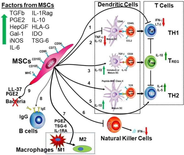
“factors segregated by MSCs such as IL-1RA [73], [74] inhibit the product of TNF-α segregated by IL-1α-activated macrophages [73], on the other hand, inflammatory factors segregated by macrophages, such as TNF-α [4], [63] and LPS [63] activate MSCs to segregate anti-inflammatory mediators, including TSG-6 [4], [63], IL-10 [4], [63] and PGE2 [4], [63]”
From Mesenchymal stem cell secretome for regenerative medicine: Where do we stand? (2025) · cites this paper as [4]
Experimental — accuracy not yet measuredThe source backs part of the claim, not all of it — or backs it for a different population, condition or comparison.
In the citing paper
“MSC therapies demonstrated cartilage regeneration and early osteoarthritis improvement, but high costs and ethical concerns remain barriers to widespread adoption.”
From Advancements in Regenerative Therapies for Orthopedics: A Comprehensive Review of Platelet-Rich Plasma, Mesenchymal Stem Cells, Peptide Therapies, and Biomimetic Applications (2025) · cites this paper as [19]
Experimental — accuracy not yet measuredThe cited source says what the claim attributes to it.
In the citing paper
“MSCs have shown their therapeutic effects in numerous disease models as well as in some clinical trials [4].”
From Mesenchymal stem cell secretome for regenerative medicine: Where do we stand? (2025) · cites this paper as [4]
The cited source says what the claim attributes to it.
In the citing paper
“MSCs were shown to polarize macrophages from an inflammatory M1 phenotype to an anti-inflammatory M2 phenotype [4].”
From Mesenchymal stem cell secretome for regenerative medicine: Where do we stand? (2025) · cites this paper as [4]
Self-check
Percentages, sums and reported differences re-computed from the paper’s own numbers.
The self-check needs the paper’s full text.
Citations
Cites
Multilineage potential of adult human mesenchymal stem cells
Pittenger, M. F., Mackay, A. M., Beck, S. C., Jaiswal, R. K., Douglas, R., Mosca, J. D., Moorman, M. A., Simonetti, D. W., Craig, S., & Marshak, D. R. (1999) · Science · doi:10.1126/science.284.5411.143 · Cited by 21,052 · cited 20×
“MSCs are still largely defined by their in vitro expression of a restricted subset of cell surface proteins, and their capacity for stimulus-induced tri-lineage differentiation.(6,31) While this minimalistic definition has sufficed for almost two decades and is still widely used today, the field should benefit from th…”
BackgroundInfluentialSelf-citationDominici, M., Le Blanc, K., Mueller, I., Slaper-Cortenbach, I., Marini, F. C., Krause, D. S., Deans, R. J., Keating, A., Prockop, D. J., & Horwitz, E. M. (2006) · Cytotherapy · doi:10.1080/14653240600855905 · Cited by 17,766 · cited 1×
“MSCs are still largely defined by their in vitro expression of a restricted subset of cell surface proteins, and their capacity for stimulus-induced tri-lineage differentiation.(6,31) While this minimalistic definition has sufficed for almost two decades and is still widely used today, the field should benefit from th…”
BackgroundMatrix elasticity directs stem cell lineage specification
Engler, A. J., Sen, S., Sweeney, H. L., & Discher, D. E. (2006) · Cell · doi:10.1016/j.cell.2006.06.044 · Cited by 13,915 · cited 4×
“Growing MSCs for a longer time on either a soft or a stiff substrate progressively commits the cells to the corresponding lineages, making them refractory to rapid lineage switching by both soluble factors and substrate changes.(96,99) The molecular basis for this commitment— be it DNA methylation, microRNA, soluble f…”
BackgroundResultInfluentialSelf-citationMultilineage Cells from Human Adipose Tissue: Implications for Cell-Based Therapies
Zuk, P. A., Zhu, M., Mizuno, H., Huang, J., Futrell, J. W., Katz, A. J., Benhaim, P., Lorenz, H. P., & Hedrick, M. H. (2001) · Tissue Engineering · doi:10.1089/107632701300062859 · Cited by 7,956
Role of YAP/TAZ in mechanotransduction
Dupont, S., Morsut, L., Aragona, M., Enzo, E., Giulitti, S., Cordenonsi, M., Zanconato, F., Le Digabel, J., Forcato, M., Bicciato, S., et al. (2011) · Nature · doi:10.1038/nature10137 · Cited by 5,979 · cited 1×
“2b) into the nucleus in cells on stiff substrates to promote expression of differentiation genes for “stiff lineages”.(98,106) Further, MSCs respond to the surface curvature of their substrate.”
BackgroundHuman mesenchymal stem cells modulate allogeneic immune cell responses
Aggarwal, S., & Pittenger, M. F. (2005) · Blood · doi:10.1182/blood-2004-04-1559 · Cited by 4,511
Self-citationA perivascular origin for mesenchymal stem cells in multiple human organs
Crisan, M., Yap, S., Casteilla, L., Chen, C.-W., Corselli, M., Park, T. S., Andriolo, G., Sun, B., Zheng, B., Zhang, L., et al. (2008) · Cell Stem Cell · doi:10.1016/j.stem.2008.07.003 · Cited by 3,991 · cited 2×
“Further, although bone marrow was first used for MSC isolation and considered a renewable source, MSC-like cells have been isolated from many tissue sources including harvested adipose tissue,7,8 umbilical Wharton’s jelly,9 placenta,10,11 skin73 and the roots of shed teeth.74 The studies of microvascular pericytes soo…”
Self-citationMesenchymal and haematopoietic stem cells form a unique bone marrow niche
Méndez-Ferrer, S., Michurina, T. V., Ferraro, F., Mazloom, A. R., MacArthur, B. D., Lira, S. A., Scadden, D. T., Ma’ayan, A., Enikolopov, G. N., & Frenette, P. S. (2010) · Nature · doi:10.1038/nature09262 · Cited by 3,374 · cited 1×
“MSCs in bone marrow can exhibit dendritic shapes and express some neuro-typical markers such as CD271, TRK A, B and C mRNAs, and a moderate fraction of MSCs cultured on soft gels that mimic the softness of marrow or neural tissue favor expression of nestin, β3 tubulin, and other neuro-typic traits.(96,97) However, adi…”
ResultDi Nicola, M., Carlo-Stella, C., Magni, M., Milanesi, M., Longoni, P. D., Matteucci, P., Grisanti, S., & Gianni, A. M. (2002) · Blood · doi:10.1182/blood.v99.10.3838 · Cited by 3,226 · cited 1×
“Contemporaneous with early MSC/hematopoietic stem cell transplant clinical studies, in vitro studies tested allo-MSCs in mixed donor lymphocyte reactions and revealed the MSCs prevented lymphocyte proliferation, and do not cause apoptosis of T cells, rather the T cells will respond to subsequent lymphocyte challenge w…”
ResultEvidence for Cardiomyocyte Renewal in Humans
Bergmann, O., Bhardwaj, R. D., Bernard, S., Zdunek, S., Barnabé-Heider, F., Walsh, S., Zupicich, J., Alkass, K., Buchholz, B. A., Druid, H., et al. (2009) · Science · doi:10.1126/science.1164680 · Cited by 3,130 · cited 1×
“brain neurons, chondrocytes, and cardiomyocytes where the cell half-life is on the order of ~50 years.(132) When a tissue is damaged by trauma or disease, these regenerative repair processes may be accelerated, but this capacity diminishes with age, and each tissue ages somewhat differently.”
Background
Research works printed but not linked: 1
- Pittenger, M. F., Mbalaviele, G., Black, M., Mosca, J. D. & Marshak D. R. (2001). Adult mesenchymal stem cells from bone marrow. Primary Mesenchymal Cells, Ch. 10, Human Cell Culture, Vol. 5 (eds Koller, M. et al.) pp 190-207 (Kluwer Academic Publishers, Dordrecht, Netherlands, 2001).
Cited by
Fibroblasts: Origins, definitions, and functions in health and disease
Plikus, M. V., Wang, X., Sinha, S., Forte, E., Thompson, S. M., Herzog, E. L., Driskell, R. R., Rosenthal, N., Biernaskie, J., & Horsley, V. (2021) · Cell · doi:10.1016/j.cell.2021.06.024 · Cited by 1,141
Recent Updates of Diagnosis, Pathophysiology, and Treatment on Osteoarthritis of the Knee
Jang, S., Lee, K., & Ju, J. H. (2021) · International Journal of Molecular Sciences · doi:10.3390/ijms22052619 · Cited by 803
“Additionally, MSCs have unique immunomodulatory properties and can reduce inflammation and support other cells, enhancing angiogenesis, cell survival, and differentiation [82,83].”
BackgroundFibroblast and myofibroblast activation in normal tissue repair and fibrosis
Younesi, F. S., Miller, A. E., Barker, T. H., Rossi, F. M. V., & Hinz, B. (2024) · Nature Reviews Molecular Cell Biology · doi:10.1038/s41580-024-00716-0 · Cited by 681
“The most common mesenchymal myofibroblast precursors include adipocytes, pericytes, smooth muscle cells (SMCs), multipotent mesenchymal stromal or stem cells (MSCs) 29–31 , and bona fide fibroblasts 19,29,32,33 .”
InfluentialPrimorac, D., Molnar, V., Rod, E., Jeleč, Ž., Čukelj, F., Matišić, V., Vrdoljak, T., Hudetz, D., Hajsok, H., & Borić, I. (2020) · Genes · doi:10.3390/genes11080854 · Cited by 542
Engineering the next generation of cell-based therapeutics
Bashor, C. J., Hilton, I. B., Bandukwala, H., Smith, D. M., & Veiseh, O. (2022) · Nature Reviews Drug Discovery · doi:10.1038/s41573-022-00476-6 · Cited by 439
Metadata
- Authors
- Pittenger, M. F., Discher, D. E., Péault, B. M., Phinney, D. G., Hare, J. M., & Caplan, A. I.
- Affiliations
- University of Maryland, Baltimore (US)
- University of Pennsylvania (US)
- University of California, Los Angeles (US)
- Scripps Research Institute (US)
- University of Miami (US)
- Case Western Reserve University (US)
- Corresponding author
- Pittenger, Mark F.
- Péault, Bruno M.
- Venue
- npj Regenerative Medicine
- Published
- 2019-12-02
- Volume
- 4
- Issue
- 1
- Pages
- 22-22
- ISSN
- 2057-3995 (ISSN-L)
- PMC
- PMC6889290
- OpenAlex
- W2991416247
- Cited by
- 2,008
- References
- 163
- Citations by year
- 2026: 187 · 2025: 272 · 2024: 348 · 2023: 318 · 2022: 346 · 2021: 385 · 2020: 148
- FWCI
- 27.07
- Type
- journal article
- Language
- en
- Open access
- gold
- doi.org
- Licence
- cc-by
- creativecommons.org/licenses/by/4.0 (tdm)
- creativecommons.org/licenses/by/4.0 (vor)
- Locations
- npj Regenerative Medicine (publishedVersion, cc-by)
- PubMed Central (publishedVersion, cc-by)
- PubMed (publishedVersion)
- DOAJ (DOAJ: Directory of Open Access Journals) (submittedVersion)
- Funders
- National Heart, Lung, and Blood Institute (R01HL124106)
- National Cancer Institute (U01CA254886)
- National Institute of Biomedical Imaging and Bioengineering (EB021911, P41EB021911)
- Topics
- Life Sciences › Biochemistry, Genetics and Molecular Biology › Cell Biology › Mesenchymal stem cell research
- Health Sciences › Dentistry › Periodontics › Periodontal Regeneration and Treatments
- Health Sciences › Medicine › Hematology › Hematopoietic Stem Cell Transplantation
- Keywords
- mesenchymal stem cells, cell differentiation, immunomodulation, anti-inflammatory activity, clinical trials, cell therapy, intercellular communication, cell delivery, patient selection, biomedical engineering
About these checks
Registry, retraction, Cites and statistics results are computed, not judged, with one exception: a printed reference that no index lists is read once by a model before it counts as a citation error. Claims are extracted by a model and kept only when their quote appears verbatim in the text; each cited source is then checked, with a model’s help, for whether it says what the claim relies on. Those verdicts can be wrong — read the claim’s quote and the cited source before relying on one.
Statistics and arithmetic are recomputed, not judged.
Confidence bands are shown only for fields calibrated on our annotated test set. None is calibrated yet, so no band is shown.
What counts as a claim ›How we measure confidence ›How we check this ›
Are you an author, or do you see a problem with this page? Request a review
How to cite
Cite CoChat for the checks on this page, and the paper for its own findings.
- This page
- CoChat. (2026). Citation check: Mesenchymal stem cell perspective: cell biology to clinical progress. https://app.cochat.ai/papers/10.1038/s41536-019-0083-6
- The paper
- Pittenger, M. F., Discher, D. E., Péault, B. M., Phinney, D. G., Hare, J. M., & Caplan, A. I. (2019). Mesenchymal stem cell perspective: cell biology to clinical progress. npj Regenerative Medicine. https://doi.org/10.1038/s41536-019-0083-6
Data sources
Bibliographic data: Semantic Scholar, OpenAlex, Crossref.
Contains information from the Semantic Scholar Academic Graph Datasets, made available under the ODC Attribution License (ODC-BY 1.0).
Semantic Scholar’s data is described in: Kinney, R., et al. (2023). The Semantic Scholar Open Data Platform. arXiv:2301.10140.
