

The DOI matches its Crossref record; 36 of the 37 references it cites were found in a registry or an index, and 1 are not checked yet.
Understanding metric-related pitfalls in image analysis validation
Open accessAbstract
Validation metrics are key for tracking scientific progress and bridging the current chasm between artificial intelligence research and its translation into practice. However, increasing evidence shows that, particularly in image analysis, metrics are often chosen inadequately. Although taking into account the individual strengths, weaknesses and limitations of validation metrics is a critical prerequisite to making educated choices, the relevant knowledge is currently scattered and poorly accessible to individual researchers. Based on a multistage Delphi process conducted by a multidisciplinary expert consortium as well as extensive community feedback, the present work provides a reliable and comprehensive common point of access to information on pitfalls related to validation metrics in image analysis. Although focused on biomedical image analysis, the addressed pitfalls generalize across application domains and are categorized according to a newly created, domain-agnostic taxonomy. The work serves to enhance global comprehension of a key topic in image analysis validation.
Overview
The claims and statistics of this paper haven’t been checked yet.
Run the check — free, no account needed. It reads the open-access text and checks the sources it cites.
Integrity signals
Registry
Cites
Statistics
Figures
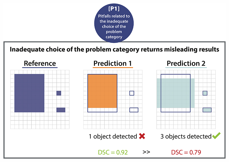
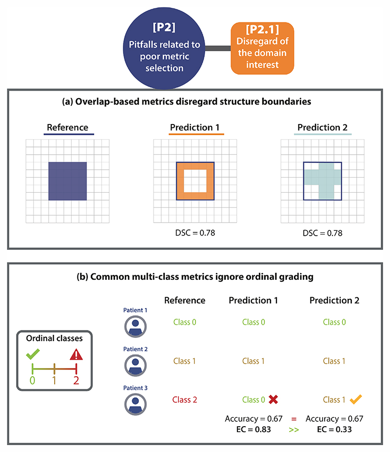
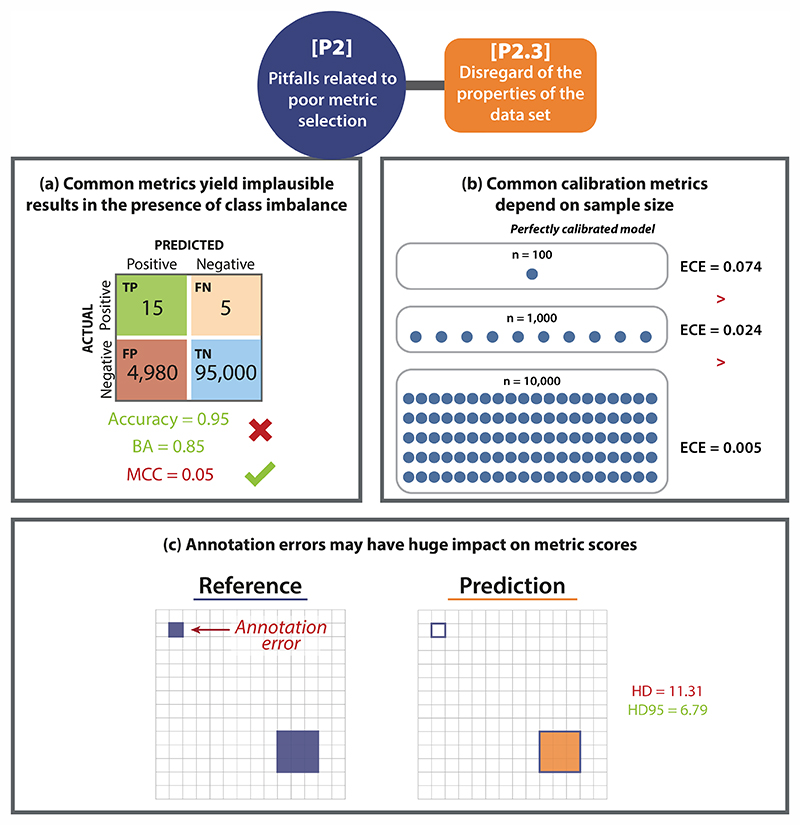
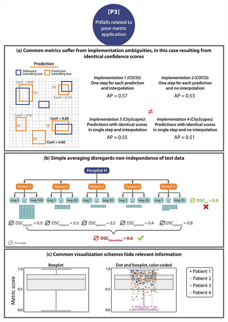
Verification
Claims the paper makes
The claims and statistics of this paper haven’t been checked yet.
Run the check — free, no account needed. It reads the open-access text and checks the sources it cites.
Claims other papers make about this one
Each is checked against this paper’s own text: does it say what the citing paper says it does?
The source backs part of the claim, not all of it — or backs it for a different population, condition or comparison.
In the citing paper
“Acceptance testing of AI auto contouring algorithms typically involves both area‐based metrics (e.g., Dice Similarity Coefficient, Intersection over Union (IoU)) and distance‐based metrics (e.g., Hausdorff Distance) evaluated against reference standard contours. 11 , 12 , 13 , 14 These metrics are well established for assessing contour agreement, but they say nothing about where the model is confi”
From Uncertainty quantification of U‐Net based segmentation tool using conformal prediction (2026) · cites this paper as [13]
Experimental — accuracy not yet measured
Self-check
Percentages, sums and reported differences re-computed from the paper’s own numbers.
The self-check needs the paper’s full text.
Citations
Cites
Microsoft COCO: Common Objects in Context
Lin, T.-Y., Maire, M., Belongie, S., Hays, J., Perona, P., Ramanan, D., Dollár, P., & Zitnick, C. L. (2014) · Lecture Notes in Computer Science · doi:10.1007/978-3-319-10602-1_48 · Cited by 45,460
The pascal visual object classes (VOC) challenge
Everingham, M., Van Gool, L., Williams, C. K. I., Winn, J., & Zisserman, A. (2010) · International Journal of Computer Vision · doi:10.1007/s11263-009-0275-4 · Cited by 19,599
Yang (2021) · doi:10.1038/s41598-020-79139-8 · Cited by 5,035
Cellpose: a generalist algorithm for cellular segmentation
Stringer, C., Wang, T., Michaelos, M., & Pachitariu, M. (2021) · Nature Methods · doi:10.1038/s41592-020-01018-x · Cited by 4,369
Metrics for evaluating 3D medical image segmentation: analysis, selection, and tool
Taha, A. A., & Hanbury, A. (2015) · BMC Medical Imaging · doi:10.1186/s12880-015-0068-x · Cited by 3,247
Self-citationCell Detection with Star-Convex Polygons
Schmidt, U., Weigert, M., Broaddus, C., & Myers, G. (2018) · Lecture Notes in Computer Science · doi:10.1007/978-3-030-00934-2_30 · Cited by 1,826
Delphi methodology in healthcare research: How to decide its appropriateness
Nasa, P., Jain, R., & Juneja, D. (2021) · World Journal of Methodology · doi:10.5662/wjm.v11.i4.116 · Cited by 1,821
The Liver Tumor Segmentation Benchmark (LiTS)
Bilic, P., Christ, P., Li, H. B., Vorontsov, E., Ben-Cohen, A., Kaissis, G., Szeskin, A., Jacobs, C., Mamani, G. E. H., Chartrand, G., et al. (2023) · Medical Image Analysis · doi:10.1016/j.media.2022.102680 · Cited by 1,412
Self-citationHow to Evaluate Foreground Maps
Margolin, R., Zelnik‐Manor, L., & Tal, A. (2014) · IEEE Computer Society Conference on Computer Vision and Pattern Recognition/Proceedings - IEEE Computer Society Conference on Computer Vision and Pattern Recognition/Proceedings · doi:10.1109/cvpr.2014.39 · Cited by 1,143
Metrics for Multi-Class Classification: an OverviewPreprint
Grandini, M., Bagli, E., & Visani, G. (2020) · arXiv (Cornell University) · doi:10.48550/arxiv.2008.05756 · Cited by 892
Research works printed but not linked: 1
- Lennerz, J. K., Green, U., Williamson, D. F. K. & Mahmood, F. A unifying force for the realization of medical ai. NPJ Digit. Med. 5, 172 (2022).
Cited by
Sorted by citations
Items 101–200 of 230
Machine learning y deep learning en medicina interna: desmitificando conceptos
Ramos-Ruperto, L., Mora-Delgado, J., Rodríguez‐González, A., Sicilia, M., Pardilla, M. J., Sempere, J. M., & Puchades, R. (2025) · Revista Clínica Española · doi:10.1016/j.rce.2025.502412 · Cited by 3
Risks and rewards of AI democratization
van der Sommen, F., & de Groof, J. (2024) · United European Gastroenterology Journal · doi:10.1002/ueg2.12560 · Cited by 3
Detection of Local Prostate Cancer Recurrence from PET/CT Scans Using Deep Learning
Korb, M., Efetürk, H., Jedamzik, T., Hartrampf, P. E., Kosmala, A., Serfling, S. E., Dirk, R., Michalski, K., Buck, A. K., Werner, R. A., et al. (2025) · Cancers · doi:10.3390/cancers17091575 · Cited by 2
Automatic contour quality assurance using deep-learning based contours
Marquez, B., Fuentes, D., Peterson, C. B., Rhee, D. J., Douglas, R. J., Mumme, R. P., Jhingran, A., Pollard‐Larkin, J., Prajapati, S., Whitaker, T. J., et al. (2025) · Physics in Medicine and Biology · doi:10.1088/1361-6560/ade5e6 · Cited by 2
Anatomy‐Guided, Modality‐Agnostic Segmentation of Neuroimaging Abnormalities
Lteif, D., Appapogu, D., Bargal, S. A., Plummer, B. A., & Kolachalama, V. B. (2025) · Human Brain Mapping · doi:10.1002/hbm.70329 · Cited by 2
Li, F., Wang, J., Hong, Y., Yang, X., Jiang, J., Wang, Z., Zeng, S., Fan, Y., Lyu, G., Li, P., et al. (2025) · Biomedical Signal Processing and Control · doi:10.1016/j.bspc.2025.109110 · Cited by 2
SEM-based analysis of pore uniformity: effects of pore positions and sizes
Papia, E.-M., Kondi, A., Singh, V., Tiwari, M. K., Gogolides, Ε., & Constantoudis, V. (2026) · Materials Today Communications · doi:10.1016/j.mtcomm.2026.115342 · Cited by 2
Preempting the Trough of Disillusionment in Surgical AI
Marwaha, J. S., & Hashimoto, D. A. (2026) · JAMA Surgery · doi:10.1001/jamasurg.2025.6035 · Cited by 2
Self-citationEicke, M., Geissler, K., Heepe, J., Krueger, M. B., Winther, H. B., Heymann, A.-K., Knörr, F., Bucher, A. M., Herrmann, J., Benoit, C., et al. (2026) · European Journal of Radiology Artificial Intelligence · doi:10.1016/j.ejrai.2026.100096 · Cited by 2
Ramani, R. S., Jones, B., Chaurasia, A., Khazaei, Y., Schwärzler, J., Uribe, S. E., Schwendicke, F., & Tichý, A. (2026) · International Dental Journal · doi:10.1016/j.identj.2026.109566 · Cited by 2
Dwyer, M. G., Bergsland, N., Bartnik, A., Jakimovski, D., Noteboom, S., Schoonheim, M. M., Steenwijk, M. D., Pei, J., Clayton, D., & Zivadinov, R. (2026) · Communications Medicine · doi:10.1038/s43856-026-01683-7 · Cited by 7
Subcutaneous edema segmentation on abdominal CT using multi-class labels and iterative annotation
Bhadra, S., Liu, J., & Summers, R. M. (2024) · International Journal of Computer Assisted Radiology and Surgery · doi:10.1007/s11548-024-03262-4 · Cited by 2
Self-citationAssessing MedSAM's Generalization to White Matter Hyperintensities in Magnetic Resonance Imaging
Rodrigues, J., do Carmo, D. S., & Rittner, L. (2024) · International Symposium on Medical Information Processing and Analysis (SIPAIM) · doi:10.1109/sipaim62974.2024.10783572 · Cited by 2
Model-Based Neural Networks for Thermographic Image Reconstruction
Galiger, G., Azadi, N., Lehner, B., Huemer, M., & Kovács, P. (2024) · IEEE Conference on Information Technology and Data Science (CITDS) · doi:10.1109/citds62610.2024.10791362 · Cited by 2
Evaluating Medical Image Segmentation Models Using Augmentation
Sayed, M., Saba-Sadiya, S., Wichtlhuber, B., Dietz, J., Neitzel, M., Keller, L., Roig, G., & Bucher, A. M. (2024) · Tomography · doi:10.3390/tomography10120150 · Cited by 2
Application-driven validation of posteriors in inverse problems
Adler, T. J., Nölke, J.-H., Reinke, A., Tizabi, M. D., Gruber, S., Trofimova, D., Ardizzone, L., Jäger, P. F., Buettner, F., Koethe, U., et al. (2025) · Medical Image Analysis · doi:10.1016/j.media.2025.103474 · Cited by 2
Self-citationPhitidis, J., O’Neil, A. Q., Whiteley, W. N., Alex, B., Wardlaw, J. M., Bernabéu, M. O., & Hernández, M. D. C. V. (2025) · Computer Methods and Programs in Biomedicine · doi:10.1016/j.cmpb.2025.108715 · Cited by 2
Tuning vision foundation models for rectal cancer segmentation from CT scans
Zhang, H., Guo, W., Wan, S., Zou, B., Wang, W., Qiu, C., Liu, K., Jin, P., & Yang, J. (2025) · Communications Medicine · doi:10.1038/s43856-025-00953-0 · Cited by 2
Galba, T., Livada, Č., & Baumgartner, A. (2025) · Applied Sciences · doi:10.3390/app16010433 · Cited by 3
Connectivity of the Piriform Cortex and its Implications in Temporal Lobe EpilepsyPreprint
Lucas, A., Jaskir, M., Sinha, N., Pattnaik, A. R., Mouchtaris, S., Josyula, M., Petillo, N., Roth, R. W., Dikeçligil, G. N., Bonilha, L., et al. (2024) · medRxiv · doi:10.1101/2024.07.21.24310778 · Cited by 2
Cao, T., Durkee, M. S., Ai, J., Hara, S., Andrade, M. S., Chang, A., Casella, G., Chong, A. S., Giger, M. L., & Clark, M. R. (2026) · bioRxiv (Cold Spring Harbor Laboratory) · doi:10.64898/2026.01.14.699505 · Cited by 2
Kundu, S. S., Kujawa, A., Ivory, M., Barfoot, T., Shapey, J., & Vercauteren, T. K. (2025) · SPIE Medical Imaging: Computer-Aided Diagnosis · doi:10.1117/12.3047296 · Cited by 1
Benchmarking Anomaly Detection Methods for Extracardiac Findings in Cardiac MRI
Pinto, E., Costa, P., Silva, C., Pereira, V., Fonseca, J. C., & Queirós, S. (2025) · Applied Sciences · doi:10.3390/app15074027 · Cited by 1
Ba, V. T., Hübner, M., Qasim, A. B., Rees, M., Sellner, J., Seidlitz, S., Christodoulou, E., Özdemir, B., Studier‐Fischer, A., Nickel, F., et al. (2025) · International Journal of Computer Assisted Radiology and Surgery · doi:10.1007/s11548-025-03364-7 · Cited by 1
Self-citationMultimodale Datenverarbeitung mittels KI: der Operationssaal der Zukunft
Eckhoff, J. A., Krauss, D. T., Brunner, S., Bruns, C. J., & Fuchs, H. F. (2025) · Die Chirurgie · doi:10.1007/s00104-025-02377-x · Cited by 1
GOUHFI: A novel contrast- and resolution-agnostic segmentation tool for ultra-high-field MRI
Fortin, M.-A., Kristoffersen, A. L., Larsen, M. S., Lamalle, L., Stirnberg, R., & Goa, P. E. (2025) · Imaging Neuroscience · doi:10.1162/imag.a.960 · Cited by 1
Future perspectives of artificial intelligence in vascular surgery
Lareyre, F., Weaver, M. L., Afifi, R. O., Kiang, S. C., & Raffort, J. (2025) · The Journal of Cardiovascular Surgery · doi:10.23736/s0021-9509.25.13473-3 · Cited by 1
Segmenting the non-enhancing compartment of brain tumor MRIs
Tobias, S., Alexander, B., Simon, W., Maria, T. A., & Lang Elmar, W. (2025) · Annual International Conference of the IEEE Engineering in Medicine and Biology Society (EMBC) · doi:10.1109/embc58623.2025.11254161 · Cited by 1
System under test: Challenges for algorithm benchmarking
Tizabi, M. D., & Reinke, A. (2025) · Advances in magnetic resonance technology and applications · doi:10.1016/b978-0-443-14109-6.00025-0 · Cited by 1
Self-citationAuditing Significance, Metric Choice, and Demographic Fairness in Medical AI Challenges
Lubonja, A., Bassi, P. R. A. S., Li, W., Qiao, H., Burns, R. C., Yuille, A., & Zhou, Z. Z. (2026) · Lecture notes in computer science · doi:10.1007/978-3-032-09513-8_13 · Cited by 3
Erenstein, H., Krijnen, W. P., van der Heij-Meijer, A., & van Ooijen, P. M. A. (2026) · European Journal of Radiology Artificial Intelligence · doi:10.1016/j.ejrai.2026.100071 · Cited by 1
Ethical and Legal Concerns of Deepfake Technology in Biomedical Imaging: A Comprehensive Survey
Rajpar, S. A., Chen, H., & Ashiru, A. (2026) · Annals of Biomedical Engineering · doi:10.1007/s10439-026-04128-3 · Cited by 1
TOPOS: target organ prediction on scout views for automated CT scan planning
Ziegelmayer, S., Lemke, T., Graf, M., Kim, S. H., Lemke, L., Mertens, C. J., Busch, F., Weller, D., Makowski, M. R., Adams, L. C., et al. (2026) · European Radiology · doi:10.1007/s00330-026-12623-3 · Cited by 1
Regulating digital surgery to balance safety and innovation: a SAGES white paper
Guzman, N. M., Marwaha, J. S., Keller, D. S., Feldman, L. S., Hashimoto, D. A., Madani, A., & Telem, D. A. (2026) · Surgical Endoscopy · doi:10.1007/s00464-026-12875-6 · Cited by 2
Self-citationJi, X., Feng, Y., Lu, H., Chu, D., & Han, Q. (2026) · Nutrients · doi:10.3390/nu18132119 · Cited by 1
Perret, S., Albe, L., Edet‐Sanson, A., Tonnelet, D., Carlier, T., Kohi, C., Barbaud, M., Modzelewski, R., Véra, P., Augusto, L., et al. (2026) · EJNMMI Research · doi:10.1186/s13550-026-01468-y · Cited by 1
Korsten, T., Kuiper, G. M., Su, R., Ruurda, J. P. H., Verhagen, A. F. T. M., Mariani, M., Khalil, Y. A., & Sadeghi, A. H. (2026) · npj Digital Surgery · doi:10.1038/s44484-026-00013-7 · Cited by 1
Language-Guided Segmentation of Medical Images: A Review of Foundation Models
Qamar, S. (2026) · Bioengineering · doi:10.3390/bioengineering13070803 · Cited by 1
Şahin, M. E., Ulutaş, H., Coşar, H. İ., Bicer, T., Günay, R. B., & Büyük, S. K. (2026) · Bioengineering · doi:10.3390/bioengineering13070840 · Cited by 1
Deng, K., Hao, X., Peng, B., Chen, P., Ren, Y., Zuo, C., & Yi, X. (2026) · Cold Regions Science and Technology · doi:10.1016/j.coldregions.2026.105086 · Cited by 1
Zhou, Y., Kanesan, J., Chow, C., & Xu, C. (2026) · Computerized Medical Imaging and Graphics · doi:10.1016/j.compmedimag.2026.102807 · Cited by 1
Validation of Simulation-Based Evaluation by Inclusion of Unlabeled Data and Meta-ablation
Ploner, S. B., Schneider, L.-S., Hwang, Y., Hassan, A. R., Waheed, N. K., Fujimoto, J. G., & Maier, A. (2026) · Lecture notes in computer science · doi:10.1007/978-3-032-38072-2_61 · Cited by 1
Zhou, Y., Zhao, S., Sonneck, J., & Chen, J. (2024) · IEEE International Symposium on Biomedical Imaging (ISBI) · doi:10.1109/isbi56570.2024.10635298 · Cited by 1
Self-citationTrainee Essay Prize Winner: AI-powered fracture detection: transforming orthopaedic practices
Thomson, F. (2025) · Bone and Joint 360 · doi:10.1302/2048-0105.143.360236 · Cited by 1
Bouzid, A. I. H., Benlala, I., de Senneville, B. D., Baldacci, F., Macey, J., Hassen, W. B., Su, P., Bui, S., Zysman, M., Bustin, A., et al. (2025) · Journal of Magnetic Resonance Imaging · doi:10.1002/jmri.70196 · Cited by 1
Advancing standards in biomedical image analysis validation: A perspective on Metrics Reloaded
Reinke, A., Tizabi, M. D., & Maier‐Hein, L. (2025) · Clinical and Translational Medicine · doi:10.1002/ctm2.70237 · Cited by 1
Self-citationSlice, Subtract, Segment: Efficient 2D and Physiologically Informed 3D Brain Tumor SegmentationPreprint
Chhetri, L., & Anand, A. (2026) · bioRxiv (Cold Spring Harbor Laboratory) · doi:10.64898/2026.10.04.756528 · Cited by 1
Uncertainty quantification of U‐Net based segmentation tool using conformal prediction
Borden, B. J., Graham‐Knight, J. B., Lasserre, P., Lucas, S., & Rajapakshe, R. D. (2026) · Journal of Applied Clinical Medical Physics · doi:10.1002/acm2.70663
Chierici, A. P., Lareyre, F., Iannelli, A., Poggi, E., Goffart, S., Guzzi, L., Delingette, H., & Raffort, J. (2024) · doi:10.2196/preprints.57617
Glomeruli Segmentation in Whole-Slide Images: Is Better Local Performance Always Better?
Sánchez, M., Sánchez, H., de Arenaza, C. P., Ribalta, D., Terreros, N. A., Cámara, Ó., & Galdrán, A. (2025) · Lecture notes in computer science · doi:10.1007/978-3-031-77786-8_21
Calibrated Models for DME Progression Prediction from Ultra-Wide Field Retinal Images
Galdrán, A. (2025) · Lecture notes in computer science · doi:10.1007/978-3-031-86651-7_5
How to Evaluate Generated skVCT from MVCT? Optimal Parameters and Clinical Validation
Boily, C., Mazellier, J.-P., & Meyer, P. (2025) · IEEE International Symposium on Biomedical Imaging (ISBI) · doi:10.1109/isbi60581.2025.10980660
Validierung von künstliche Intelligenz-Algorithmen für die chirurgische Praxis
Reinke, A. (2025) · Die Chirurgie · doi:10.1007/s00104-025-02348-2
Self-citationA quantitative comparison between human experts and AI at estimating tumor-stroma ratioPreprint
Pai, A., Brunekreef, J., Marcus, E., Sanders, J., Guimaraes, M. D. S., de Menezes, R. X., Sánchez, C. I., Horlings, H. M., & Teuwen, J. (2025) · medRxiv · doi:10.1101/2025.08.26.25334451
Self-citationHierarchical clustering algorithm based on an adaptive purified neighborhood graph
Qin, S., Feng, J., Yang, D., & Chen, Z. (2025) · Cluster Computing · doi:10.1007/s10586-025-05350-9
Gunawan, I., Dey, M., Neumann, D., Kohane, F. V., He, Y., Meijering, E. H. W., & Lock, J. G. (2025) · bioRxiv (Cold Spring Harbor Laboratory) · doi:10.1101/2025.09.15.676186
Self-citationCuocolo, R., Stanzione, A., Ponsiglione, A., Ugga, L., & Koçak, B. (2025) · Elsevier eBooks · doi:10.1016/b978-0-443-29242-2.00012-4
Enhancing Lesion Detection in Rat CT Images: A Deep Learning-Based Super-Resolution Study
Ham, S., Jeong, S. H., Lee, H., Nam, Y. J., Lee, H., Choi, J. Y., Lee, Y.-S., Park, Y. H., Park, S. A., Kim, W., et al. (2025) · Biomedicines · doi:10.3390/biomedicines13102421
From Clever Composites to Credible Technologies
Gao, Q., Zheng, L., Xu, D., & der Bruggen, B. V. (2025) · Membranes · doi:10.3390/membranes15110342
Machine learning and deep learning in internal medicine: demystifying concepts
Ramos-Ruperto, L., Mora-Delgado, J., Rodríguez‐González, A., Sicilia, M., Pardilla, M. J., Sempere, J. M., & Puchades, R. (2025) · Revista Clínica Española (English Edition) · doi:10.1016/j.rceng.2025.502412
Metrics Reloaded: A framework for trustworthy image analysis validation
Kavur, A. E., Reinke, A., Jäger, P. F., Isensee, F., Rädsch, T., Dietlinde Tizabi, M., & Maier‐Hein, L. (2024) · Zenodo (CERN European Organization for Nuclear Research) · doi:10.5281/zenodo.13830075
Self-citationHolzschuh, J. C., Mix, M., Freitag, M. T., Hölscher, T., Braune, A., Kotzerke, J., Vrachimis, A., Doolan, P., Ilhan, H., Marinescu, I. M., et al. (2024) · Figshare · doi:10.6084/m9.figshare.26758547
Senbi, A., Huang, T., Lyu, F., Li, Q., Tao, Y., Shao, W., Chen, Q., Wang, C., Wang, S., Zhou, T., et al. (2026) · Lecture notes in computer science · doi:10.1007/978-981-95-5634-2_17
A Novel Distance‐Based Metric for Quality Assessment in Image Segmentation
Rottmayer, N., & Redenbach, C. (2026) · IET Image Processing · doi:10.1049/ipr2.70296
Cortez, R. C., & Krishnamoorthy, B. (2026) · SSRN Electronic Journal · doi:10.2139/ssrn.6318904
Erenstein, H., den Broeck, J. V., van der Heij-Meijer, A., Krijnen, W. P., Scafoglieri, A., Jager-Wittenaar, H., Sealy, M. J., & van Ooijen, P. M. A. (2026) · Journal of Imaging · doi:10.3390/jimaging12030135
Measuring Neural Network Similarity
Kist, A. M. (2026) · F1000Research · doi:10.12688/f1000research.178206.1
Güzel, M., & Baykan, Ö. K. (2026) · Applied Sciences · doi:10.3390/app16094164
How do lower-limb landmarking errors impact morphometrics in total knee arthroplasty planning?
Roch, Y., Lozano, D., Fougeron, N., Gaulin, B., Horteur, C., Yohan Payan, A., Bucki, M., & Perrier, A. (2026) · European Journal of Radiology · doi:10.1016/j.ejrad.2026.112894
Overgaard, A., Molnar, K., Jurtz, V. I., Lie, M. E. K., & Nyengaard, J. R. (2026) · Computers in Biology and Medicine · doi:10.1016/j.compbiomed.2026.111738
Zhong, Q. (2026) · International Conference on Vision, Advanced Imaging and Computer Technology (VAICT) · doi:10.1109/vaict69205.2026.11519335
CC-DiT: A conditional cold diffusion transformer for retinal vessel segmentation
Dai, X., Bing, L., Cao, W., Yu, Z., & Chen, X. (2026) · Information Sciences · doi:10.1016/j.ins.2026.123668
Toward trustworthy healthcare AI: designing academic research for translation readiness
Hekler, A., & Buettner, F. (2026) · Molecular Systems Biology · doi:10.1038/s44320-026-00219-4
Self-citationTurginbekov, A., Alzhanov, A., Tolegenov, M., & Hasegawa, H. (2026) · SSRN Electronic Journal · doi:10.2139/ssrn.6926199
Seghier, M. L. (2026) · International Journal of Imaging Systems and Technology · doi:10.1002/ima.70393
Visconti-Lopez, F. J., & Lozada-Martínez, I. D. (2026) · Medical Education & Clinical Practice · doi:10.21142/mecp.2026.1.2.e017
Super-resolution of periapical and bitewing digital radiographs using convolutional neural network
Pourahmadiyan-Nadiki, H., Alemohammad, N., Mohammadi-Bassir, M., & Hamze, F. (2026) · BMC Medical Imaging · doi:10.1186/s12880-026-02504-y
Garrido-Martínez, J. F., Garrido-López, J. M., Zapata, J., & Martínez-Alajarín, J. (2026) · Sensors · doi:10.3390/s26134109
Chirurgie vasculaire et intelligence artificielle
Lareyre, F., Raffort, J., Ducasse, É., & Caradu, C. (2026) · EMC - Techniques chirurgicales - Chirurgie vasculaire · doi:10.1016/s0246-0459(26)50237-5
Sun, S. Y., Long, C., Dong, X., Ming, Z., Tan, Y., & Ruan, P. (2026) · Scientific Reports · doi:10.1038/s41598-026-62118-w
Organ Segmentation with Machine Learning Models
Barberis, A., Menegaki, O., Panagiotakopoulou, A., Vezakis, A., Vezakis, I., Kakkos, I., & Matsopoulos, G. K. (2026) · Journal of Imaging · doi:10.3390/jimaging12080335
Montero, A. M. B., Huet‐Dastarac, M., Perri, D. D., Hofstede, D., Birimac, N. E., Geerts, M., Roberfroid, B., Quéré, E., Lee, J. A., Roelofs, E., et al. (2026) · Physics and Imaging in Radiation Oncology · doi:10.1016/j.phro.2026.101051
Generative Inpainting as a Diagnostic Tool for Marine Wildlife Detection RobustnessPreprint
Zhou, J., Aghakishiyeva, G., Johnston, D. W., & Bent, B. (2026) · SSRN Electronic Journal · doi:10.2139/ssrn.7206689
Khazem, S. (2026) · Lecture notes in computer science · doi:10.1007/978-3-032-31397-3_45
Two-loop validation of neural network-based pneumothorax segmentation under class imbalance
Kosinov, A. V., Krupchatnikov, R. A., Серебровский, В. В., Efremov, M. A., & Kondrashov, D. S. (2026) · Proceedings of the Southwest State University Series IT Management Computer Science Computer Engineering Medical Equipment Engineering · doi:10.21869/2223-1536-2026-16-4-122-149
Adhikary, S., Chabi, N., & Mastmeyer, A. (2026) · Research Square · doi:10.21203/rs.3.rs-10551828/v1
Ho, Q. T., Bui, N. H., Tran, T. D., Khuat, Q. H., Tran, N. T., Le, X. C., Nguyen, H. Q., Nguyen, T. T., Nguyen, V. T., Mai, D. T., et al. (2026) · Journal of Imaging · doi:10.3390/jimaging12080364
Gulsen, A. (2026) · Journal of Imaging Informatics in Medicine · doi:10.1007/s10278-026-02186-6
Cosma, C., Molnár, D., Botoncea, M., Nicolescu, C. L., Molnar, C., & Butiurcă, V. O. (2026) · Medicina · doi:10.3390/medicina62091705
Firdaus, M. N. (2026) · SSRN Electronic Journal · doi:10.2139/ssrn.7331360
Mu, F., Qin, C., Zhang, J., Sun, M., Han, Y., & Chi, C. (2026) · Frontiers in Forests and Global Change · doi:10.3389/ffgc.2026.1886196
MMCA-SAM: Multimodal Cross-Attention SAM for Landslide Segmentation with Topographic Guidance
Shi, J., Pan, S., & Meng, Q. (2026) · International Conference on Geoinformatics · doi:10.1109/geoinformatics72247.2026.11665377
Can Dental AI Really Beat Dentists? DentalPair-Cert for Rigorous AI–Dentist InferencePreprint
Alve, S. R., Rahman, S., & Meem, S. M. A. C. (2026) · medRxiv · doi:10.64898/2026.09.01.26361874
Reference Workflows Shape Earth Observation Objects and Model ComparisonsPreprint
Yang, F., Zhang, W., Tang, J., Wang, L., & Tong, B. (2026) · SSRN Electronic Journal · doi:10.2139/ssrn.7398381
Wang, J., Hong, Y., & Chen, X. (2026) · Cross Strait Radio Science and Wireless Technology Conference (CSRSWTC) · doi:10.1109/csrswtc71154.2026.11669767
Recent advances in deep learning for biological microscopy image analysis beyond segmentation
Cao, K., Gao, C., Zhang, X., Pan, X., Chen, J., & Wang, H. (2026) · BioTechniques · doi:10.1080/07366205.2026.2730974
Self-citationHu, J., Zhang, Z., Lei, P., Zhang, J., Zhu, X., & Li, B. (2026) · Frontiers in Aging Neuroscience · doi:10.3389/fnagi.2026.1930675
Nouman, M., & Haraguchi, R. (2026) · BioMedInformatics · doi:10.3390/biomedinformatics6050074
Lorenzen, U. S., Bracco, M. I., Avril, S., Rouet, L., Jensen, A. K., & Eiberg, J. (2026) · European Journal of Vascular and Endovascular Surgery · doi:10.1016/j.ejvs.2026.09.041
Metadata
- Authors
- Reinke, A., Tizabi, M. D., Baumgartner, M., Eisenmann, M., Heckmann-Nötzel, D., Kavur, A. E., Rädsch, T., Sudre, C. H., Acion, L., Antonelli, M., Arbel, T., Bakas, S., Benis, A., Buettner, F., Cardoso, M. J., Cheplygina, V., Chen, J., Christodoulou, E., Cimini, B. A., Farahani, K., Ferrer, L., Galdran, A., van Ginneken, B., Glocker, B., Godau, P., Hashimoto, D. A., Hoffman, M. M., Huisman, M., Isensee, F., Jannin, P., Kahn, C. E., Kainmueller, D., Kainz, B., Karargyris, A., Kleesiek, J., Kofler, F., Kooi, T., Kopp-Schneider, A., Kozubek, M., Kreshuk, A., Kurc, T., Landman, B. A., Litjens, G., Madani, A., Maier-Hein, K., Martel, A. L., Meijering, E., Menze, B., Moons, K. G. M., Müller, H., Nichyporuk, B., Nickel, F., Petersen, J., Rafelski, S. M., Rajpoot, N., Reyes, M., Riegler, M. A., Rieke, N., Saez-Rodriguez, J., Sánchez, C. I., Shetty, S., Summers, R. M., Taha, A. A., Tiulpin, A., Tsaftaris, S. A., Van Calster, B., Varoquaux, G., Yaniv, Z. R., Jäger, P. F., & Maier-Hein, L.
- Affiliations
- German Cancer Research Center (DE)
- King's College London (GB)
- Mila - Quebec Artificial Intelligence Institute (CA)
- Indiana University Indianapolis (US)
- Holon Institute of Technology (IL)
- Goethe University Frankfurt (DE)
- IT University of Copenhagen (DK)
- Leibniz Institute for Analytical Sciences - ISAS (DE)
- Broad Institute (US)
- National Cancer Institute (US)
- Consejo Nacional de Investigaciones Científicas y Técnicas (AR)
- Radboud University Medical Center (NL)
- Imperial College London (GB)
- University of Pennsylvania (US)
- University Health Network (CA)
- Inserm (FR)
- University of Potsdam (DE)
- Friedrich-Alexander-Universität Erlangen-Nürnberg (DE)
- Institut de Chirurgie Guidée par l'Image (FR)
- Essen University Hospital (DE)
- Lunit (South Korea) (KR)
- Masaryk University (CZ)
- European Molecular Biology Laboratory (DE)
- Vanderbilt University (US)
- University of Toronto (CA)
- UNSW Sydney (AU)
- University of Zurich (CH)
- Utrecht University (NL)
- University of Geneva (CH)
- University Medical Center Hamburg-Eppendorf (DE)
- Allen Institute for Cell Science (US)
- University of Warwick (GB)
- University of Bern (CH)
- Simula Metropolitan Center for Digital Engineering (NO)
- Google (United States) (US)
- National Institutes of Health (US)
- TU Wien (AT)
- Oulu University Hospital (FI)
- University of Edinburgh (GB)
- National Institute of Allergy and Infectious Diseases (US)
- Corresponding author
- Reinke, Annika
- Tizabi, Minu D.
- Maier-Hein, Klaus
- Jäger, Paul F.
- ORCID
- Reinke, Annika
- Tizabi, Minu D.
- Baumgartner, Michael
- Eisenmann, Matthias
- Kavur, A. Emre
- Rädsch, Tim
- Sudre, Carole H.
- Acion, Laura
- Antonelli, Michela
- Arbel, Tal
- Bakas, Spyridon
- Benis, Arriel
- Buettner, Florian
- Cardoso, M. Jorge
- Cheplygina, Veronika
- Chen, Jianxu
- Christodoulou, Evangelia
- Cimini, Beth A.
- Farahani, Keyvan
- Ferrer, Luciana
- van Ginneken, Bram
- Glocker, Ben
- Godau, Patrick
- Hashimoto, Daniel A.
- Hoffman, Michael M.
- Huisman, Merel
- Isensee, Fabian
- Jannin, Pierre
- Kahn, Charles E.
- Kainmueller, Dagmar
- Kainz, Bernhard
- Karargyris, Alexandros
- Kleesiek, Jens
- Kofler, Florian
- Kooi, Thijs
- Kopp-Schneider, Annette
- Kozubek, Michal
- Kreshuk, Anna
- Landman, Bennett A.
- Litjens, Geert
- Madani, Amin
- Maier-Hein, Klaus
- Martel, Anne L.
- Meijering, Erik
- Menze, Bjoern
- Moons, Karel G. M.
- Müller, Henning
- Nichyporuk, Brennan
- Nickel, Felix
- Petersen, Jens
- Rafelski, Susanne M.
- Rajpoot, Nasir
- Reyes, Mauricio
- Riegler, Michael A.
- Rieke, Nicola
- Saez-Rodriguez, Julio
- Shetty, Shravya
- Summers, Ronald M.
- Taha, Abdel A.
- Tiulpin, Aleksei
- Tsaftaris, Sotirios A.
- Varoquaux, Gaël
- Yaniv, Ziv R.
- Jäger, Paul F.
- Maier-Hein, Lena
- Venue
- Nature Methods
- Published
- 2024-02
- Volume
- 21
- Issue
- 2
- Pages
- 182-194
- ISSN
- 1548-7091 (ISSN-L)
- 1548-7105
- PMC
- PMC11181963
- OpenAlex
- W4353015179
- Cited by
- 241
- References
- 36
- Citations by year
- 2026: 108 · 2025: 82 · 2024: 48
- FWCI
- 12.21
- Type
- journal article
- Language
- en
- Open access
- bronze
- doi.org
- Licence
- Locations
- Nature Methods (publishedVersion)
- PubMed (publishedVersion)
- UvA-DARE (University of Amsterdam) (acceptedVersion, other-oa)
- PubMed Central (acceptedVersion)
- Warwick Research Archive Portal (University of Warwick) (acceptedVersion)
- Zurich Open Repository and Archive (University of Zurich) (acceptedVersion, cc-by-nc)
- Radboud Repository (Radboud University) (submittedVersion)
- PubMed Central (submittedVersion, cc-by)
- HAL (submittedVersion)
- Universitätsbibliographie, Universität Duisburg-Essen (submittedVersion)
- TECNALIA Publications (Fundación TECNALIA Research & Innovation) (submittedVersion, public-domain)
- ArODES (HES-SO (https://www.hes-so.ch/)) (submittedVersion)
- IRIS eCampus Telematic University (Università degli Studi eCampus) (submittedVersion)
- Lirias (KU Leuven) (submittedVersion)
- Fraunhofer-Publica (Fraunhofer-Gesellschaft) (submittedVersion)
- Pure Amsterdam UMC (submittedVersion, cc-by)
- Amsterdam UMC (VU Amsterdam) - Institutional Repository (submittedVersion, other-oa)
- University of Birmingham Research Portal (University of Birmingham) (submittedVersion)
- Edinburgh Research Explorer (University of Edinburgh) (submittedVersion, cc-by)
- PuSH - Publication Server of Helmholtz Zentrum München (submittedVersion)
- Zurich Open Repository and Archive (University of Zurich) (submittedVersion, cc-by-nc)
- Zurich Open Repository and Archive (University of Zurich)
- Funders
- National Science Foundation (GM135019)
- Silicon Valley Community Foundation (2020-225720)
- Foundation for Cardiovascular Research
- Canadian Institute for Advanced Research
- Wellcome Trust (WT 203148/Z/16/Z), 203148, WT213038/Z/18/Z, 213038)
- Alzheimer's Society (AS‐JF‐17‐011)
- Cancer Research UK (C49297/A27294)
- Royal Academy of Engineering (RCSRF1819\8\25)
- European Commission (101002198, HORIZON2020, 101088594)
- Agence Nationale de la Recherche (ANR-10-IAHU-0002, ANR-10–IAHU-02, ANR-10)
- Schweizerischer Nationalfonds zur Förderung der Wissenschaftlichen Forschung (31274, 27294, 225720, 212939, 205320)
- Bundesministerium für Bildung und Forschung (161L0272)
- Nederlandse Organisatie voor Wetenschappelijk Onderzoek
- Sydäntutkimussäätiö
- Oulun Yliopisto (336449, Profi6 336449)
- Novo Nordisk Fonden (NNF21OC0068816)
- Innosuisse - Schweizerische Agentur für Innovationsförderung
- National Institutes of Health (LM2018129, 5u24ca215109-05, 2020-225720, GM135019)
- Natural Sciences and Engineering Research Council of Canada (rgpin-2022-05134)
- Engineering and Physical Sciences Research Council ([WT203148/Z/16/Z], WT213038/Z/18/Z)
- Centre For Medical Engineering, King’s College London (WT 203148/Z/16/Z)
- National Cancer Institute (U24CA180924, UH3CA225021, UG3CA225021, U24CA215109,, 3U24CA215109-02, U01CA242871, 4UH3-CA225021-03, U24CA279629)
- National Institute of General Medical Sciences (P41GM135019)
- National Institute of Neurological Disorders and Stroke (R01NS042645)
- NIH Clinical Center
- Topics
- Life Sciences › Biochemistry, Genetics and Molecular Biology › Biophysics › Cell Image Analysis Techniques
- Physical Sciences › Computer Science › Computer Vision and Pattern Recognition › Medical Image Segmentation Techniques
- Health Sciences › Medicine › Health Informatics › Artificial Intelligence in Healthcare and Education
- Keywords
- validation metrics, medical image analysis, Delphi consensus, expert consensus, reproducibility
About these checks
Registry, retraction, Cites and statistics results are computed, not judged, with one exception: a printed reference that no index lists is read once by a model before it counts as a citation error. Claims are extracted by a model and kept only when their quote appears verbatim in the text; each cited source is then checked, with a model’s help, for whether it says what the claim relies on. Those verdicts can be wrong — read the claim’s quote and the cited source before relying on one.
Statistics and arithmetic are recomputed, not judged.
Confidence bands are shown only for fields calibrated on our annotated test set. None is calibrated yet, so no band is shown.
What counts as a claim ›How we measure confidence ›How we check this ›
Are you an author, or do you see a problem with this page? Request a review
