

The DOI matches its Crossref record; 75 of the 75 references it cites were found in a registry or an index.
Reduced orbitofrontal-thalamic functional connectivity related to suicidal ideation in patients with major depressive disorder
Open accessAbstract
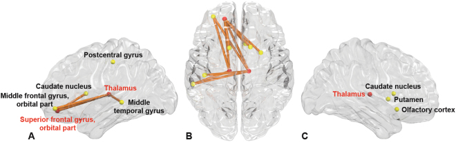
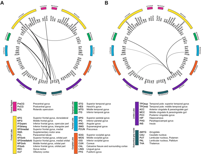
Despite recent developments in neuroimaging, alterations of brain functional connectivity in major depressive disorder (MDD) patients with suicidal ideation are poorly understood. This study investigated specific changes of suicidal ideation in functional connectivity of MDD patients. Whole brain functional connectivity in 46 patients with MDD (23 with suicidal ideation and 23 without) and 36 age- and gender- matched healthy controls were compared using resting-state functional Magnetic Resonance Imaging (fMRI) analyzed with network-based statistics (NBS) and graph-theoretical methods. Decreased functional connectivity in a characterized sub-network was observed in patients with MDD and suicidal ideation (FDR-adjusted p < 0.05). The sub-network included the regions of the fronto-thalamic circuits in the left hemisphere. The network measures of the left superior frontal gyrus, pars orbitalis (r = −0.40, p = 0.009), left thalamus (r = −0.41, p = 0.009), and right thalamus (r = −0.51, p = −0.002) were shown, through graph theoretical analysis, to be significantly negatively correlated with severity of suicidal ideation. The reduced functional connectivity in left orbitofrontal-both thalamic regions with suicidal ideation in MDD were inversely proportional to the severity of suicidality independent from depression severity. These findings suggest problems with decision-making and information integration in MDD patients with suicidal ideation.
Overview
The claims and statistics of this paper haven’t been checked yet.
Run the check — free, no account needed. It reads the open-access text and checks the sources it cites.
Integrity signals
Registry
Cites
Statistics
Figures
Verification
Claims the paper makes
The claims and statistics of this paper haven’t been checked yet.
Run the check — free, no account needed. It reads the open-access text and checks the sources it cites.
Claims other papers make about this one
Each is checked against this paper’s own text: does it say what the citing paper says it does?
In the citing paper
“However, other studies have reported lower rs-fMRI activity and functional connectivity (85, 86, 92, 93), probably due to use of different outcome measures of rs-fMRI activity, for example, regional homogeneity, fractional amplitude of low-frequency fluctuations among studies, and heterogeneity of suicidal ideation.”
From A brain-centric model of suicidal behavior (2020) · cites this paper as [92]
Self-check
Percentages, sums and reported differences re-computed from the paper’s own numbers.
The self-check needs the paper’s full text.
Citations
Cites
Tzourio-Mazoyer, N., Landeau, B., Papathanassiou, D., Crivello, F., Etard, O., Delcroix, N., Mazoyer, B., & Joliot, M. (2002) · NeuroImage · doi:10.1006/nimg.2001.0978 · Cited by 16,319
Jenkinson, M., Bannister, P., Brady, M., & Smith, S. (2002) · NeuroImage · doi:10.1006/nimg.2002.1132 · Cited by 11,888
Complex network measures of brain connectivity: Uses and interpretations
Rubinov, M., & Sporns, O. (2010) · NeuroImage · doi:10.1016/j.neuroimage.2009.10.003 · Cited by 11,522
Development of a Rating Scale for Primary Depressive Illness
Hamilton, M. (1967) · British Journal of Social and Clinical Psychology · doi:10.1111/j.2044-8260.1967.tb00530.x · Cited by 8,410
Assessment of suicidal intention: The Scale for Suicide Ideation.
Beck, A. T., Kovacs, M., & Weissman, A. (1979) · Journal of Consulting and Clinical Psychology · doi:10.1037//0022-006x.47.2.343 · Cited by 3,419
Predicting human resting-state functional connectivity from structural connectivity
Honey, C. J., Sporns, O., Cammoun, L., Gigandet, X., Thiran, J. P., Meuli, R., & Hagmann, P. (2009) · Proceedings of the National Academy of Sciences · doi:10.1073/pnas.0811168106 · Cited by 3,307
Network-based statistic: identifying differences in brain networks
Zalesky, A., Fornito, A., & Bullmore, E. T. (2010) · NeuroImage · doi:10.1016/j.neuroimage.2010.06.041 · Cited by 2,750
Achard, S., Salvador, R., Whitcher, B., Suckling, J., & Bullmore, E. (2006) · The Journal of Neuroscience · doi:10.1523/jneurosci.3874-05.2006 · Cited by 2,393
Resting-state connectivity biomarkers define neurophysiological subtypes of depression
Drysdale, A. T., Grosenick, L., Downar, J., Dunlop, K., Mansouri, F., Meng, Y., Fetcho, R. N., Zebley, B., Oathes, D. J., Etkin, A., et al. (2017) · Nature Medicine · doi:10.1038/nm.4246 · Cited by 2,170
Functional brain networks develop from a local to distributed organization
Fair, D. A., Cohen, A. L., Power, J. D., Dosenbach, N. U. F., Church, J. A., Miezin, F. M., Schlaggar, B. L., & Petersen, S. E. (2009) · PLoS Computational Biology · doi:10.1371/journal.pcbi.1000381 · Cited by 1,530
Cited by
Sorted by citations
Imaging suicidal thoughts and behaviors: a comprehensive review of 2 decades of neuroimaging studies
Schmaal, L., van Harmelen, A.-L., Chatzi, V., Lippard, E. T. C., Toenders, Y. J., Averill, L. A., Mazure, C. M., & Blumberg, H. P. (2020) · Molecular Psychiatry · doi:10.1038/s41380-019-0587-x · Cited by 373
Adolescent Suicide as a Failure of Acute Stress-Response Systems.
Miller, A. B., & Prinstein, M. J. (2019) · Annual Review of Clinical Psychology · doi:10.1146/annurev-clinpsy-050718-095625 · Cited by 235
“In adult samples, similar findings have emerged, suggesting decreased frontolimbic functional connectivity in individuals with suicidal ideation (Du et al. 2017, Kim et al. 2017, Minzenberg et al. 2015).”
BackgroundA brain-centric model of suicidal behavior
Mann, J. J., & Rizk, M. M. (2020) · American Journal of Psychiatry · doi:10.1176/appi.ajp.2020.20081224 · Cited by 214
Li, J., Duan, X., Cui, Q., Chen, H., & Liao, W. (2019) · Psychological Medicine · doi:10.1017/s0033291718001502 · Cited by 208
Liao, W., Li, J., Duan, X., Cui, Q., Chen, H., & Chen, H. (2018) · Human Brain Mapping · doi:10.1002/hbm.24235 · Cited by 132
Graph theory approach for the structural-functional brain connectome of depression
Yun, J., & Kim, Y. (2021) · Progress in Neuro-Psychopharmacology and Biological Psychiatry · doi:10.1016/j.pnpbp.2021.110401 · Cited by 111
Kang, L., Zhang, A., Sun, N., Liu, P., Yang, C., Li, G., Liu, Z., Wang, Y., & Zhang, K. (2018) · BMC Psychiatry · doi:10.1186/s12888-018-1913-6 · Cited by 94
Biological bases of suicidal behaviours: a narrative review
Lengvenyte, A., Conejero, I., Courtet, P., & Olié, E. (2021) · European Journal of Neuroscience · doi:10.1111/ejn.14635 · Cited by 87
Byun, S., Kim, A. Y., Jang, E. H., Kim, S., Choi, K. W., Yu, H. Y., & Jeon, H. J. (2019) · Technology and Health Care · doi:10.3233/thc-199037 · Cited by 84
Self-citationHong, S., Liu, Y. S., Cao, B., Cao, J., Ai, M., Chen, J., Greenshaw, A. J., & Kuang, L. (2020) · Journal of Affective Disorders · doi:10.1016/j.jad.2020.10.077 · Cited by 71
Wagner, G., Li, M., Sacchet, M. D., Richard‐Devantoy, S., Turecki, G., Bär, K., Gotlib, I. H., Walter, M., & Jollant, F. (2021) · Translational Psychiatry · doi:10.1038/s41398-021-01232-x · Cited by 60
Resting-state amplitude of low-frequency fluctuation is associated with suicidal ideation
Lan, M. J., Rizk, M. M., Pantazatos, S. P., Rubin‐Falcone, H., Miller, J. M., Sublette, M. E., Oquendo, M. A., Keilp, J. G., & Mann, J. J. (2019) · Depression and Anxiety · doi:10.1002/da.22888 · Cited by 48
Placebo aiTBS attenuates suicidal ideation and frontopolar cortical perfusion in major depression
Baeken, C., Wu, G., & van Heeringen, K. (2019) · Translational Psychiatry · doi:10.1038/s41398-019-0377-x · Cited by 46
Glăvan, D., Gheorman, V., Greșiță, A., Hermann, D. M., Udriștoiu, I., & Popa‐Wagner, A. (2021) · Scientific Reports · doi:10.1038/s41598-021-98210-6 · Cited by 48
Alterations in functional brain networks in depressed patients with a suicide attempt history
Jung, J., Choi, S.-Y., Han, K., Kim, A., Kang, W., Paik, J., Lee, H. W., & Ham, B. (2019) · Neuropsychopharmacology · doi:10.1038/s41386-019-0560-z · Cited by 47
Using resting-state intrinsic network connectivity to identify suicide risk in mood disorders
Stange, J. P., Jenkins, L. M., Pocius, S. L., Kreutzer, K. A., Bessette, K. L., DelDonno, S. R., Kling, L. R., Bhaumik, R., Welsh, R. C., Keilp, J. G., et al. (2019) · Psychological Medicine · doi:10.1017/s0033291719002356 · Cited by 42
Qiao, D., Zhang, A., Sun, N., Yang, C., Jian-ying, L., Zhao, T., Wang, Y., Xu, Y., Wen, Y., Zhang, K., et al. (2020) · Frontiers in Psychiatry · doi:10.3389/fpsyt.2020.608197 · Cited by 41
Liu, X., He, C., Fan, D., Zang, F., Zhu, Y., Zhang, H., Zhang, Z., Zhang, H., & Xie, C. (2021) · Translational Psychiatry · doi:10.1038/s41398-021-01353-3 · Cited by 40
Zhang, R., Zhang, L., Wei, S., Wang, P., Jiang, X., Tang, Y., & Wang, F. (2021) · Frontiers in Human Neuroscience · doi:10.3389/fnhum.2020.585664 · Cited by 42
Hao, H., Chen, C., Mao, W., Zhong, J., & Dai, Z. (2018) · Journal of Affective Disorders · doi:10.1016/j.jad.2018.10.113 · Cited by 39
Piani, M. C., Maggioni, E., Delvecchio, G., & Brambilla, P. (2022) · Journal of Affective Disorders · doi:10.1016/j.jad.2022.02.003 · Cited by 39
Nugent, A. C., Ballard, E. D., Gilbert, J. R., Tewarie, P. K., Brookes, M. J., & Zarate, C. A. (2020) · NeuroImage: Clinical · doi:10.1016/j.nicl.2020.102378 · Cited by 34
Aberrant brain dynamics in major depressive disorder with suicidal ideation
Yang, J., Liu, Z., Tao, H., Cheng, Y., Fan, Z., Sun, F., Ouyang, X., & Yang, J. (2022) · Journal of Affective Disorders · doi:10.1016/j.jad.2022.07.043 · Cited by 36
Chen, V. C., Chou, Y.-S., Tsai, Y., Huang, Y.-C., McIntyre, R. S., & Weng, J. (2021) · Brain Topography · doi:10.1007/s10548-020-00817-x · Cited by 33
Tang, N., Sun, C., Wang, Y., Li, X., Liu, J., Chen, Y., Sun, L., Rao, Y., Li, S., Qi, S., et al. (2021) · Frontiers in Psychiatry · doi:10.3389/fpsyt.2021.768819 · Cited by 33
Dobbertin, M. D., Blair, K. S., Carollo, E., Blair, J., Dominguez, A. J., & Bajaj, S. (2023) · Frontiers in Psychiatry · doi:10.3389/fpsyt.2023.1083244 · Cited by 34
Yang, J., Palaniyappan, L. K., Xi, C., Cheng, Y., Fan, Z., Chen, C., Zhang, M., Pan, Y., Tao, H., Ouyang, X., et al. (2022) · Journal of Psychiatric Research · doi:10.1016/j.jpsychires.2022.02.003 · Cited by 30
Flexible and specific contributions of thalamic subdivisions to human cognition
Antonucci, L. A., Penzel, N., Pigoni, A., Dominke, C., Kambeitz, J. P., & Pergola, G. (2021) · Neuroscience & Biobehavioral Reviews · doi:10.1016/j.neubiorev.2021.01.014 · Cited by 31
Kim, K., Shin, J., Myung, W., D, M. F. M., Mischoulon, D., Papakostas, G. I., Choi, K. W., Na, E. J., Seo, S. W., Seong, J., et al. (2019) · Scientific Reports · doi:10.1038/s41598-019-43882-4 · Cited by 30
Self-citationParsaei, M., Taghavizanjani, F., Cattarinussi, G., Moghaddam, H. S., Camillo, F. D., Akhondzadeh, S., Sambataro, F., Brambilla, P., & Delvecchio, G. (2023) · Journal of Affective Disorders · doi:10.1016/j.jad.2023.08.034 · Cited by 29
Wáng, Q., He, C., Wang, Z., Fan, D., Zhang, Z., Xie, C., Yan, C., Chen, X., Li, L., Castellanos, F. X., et al. (2023) · Translational Psychiatry · doi:10.1038/s41398-023-02655-4 · Cited by 26
Fu, Z., Jing Sui (Beijing Normal University), my correct affiliation is beijing normal university, N. Q. U. O. S. A. T., Espinoza, R. T., Narr, K. L., Qi, S., Sendi, M. S. E., Abbott, C., & Calhoun, V. D. (2020) · Biological Psychiatry Cognitive Neuroscience and Neuroimaging · doi:10.1016/j.bpsc.2021.07.004 · Cited by 24
Decreased Connection Between Reward Systems and Paralimbic Cortex in Depressive Patients
Bai, T., Zu, M., Chen, Y., Xie, W., Cai, C., Wei, Q., Ji, G., Tian, Y., & Wang, K. (2018) · Frontiers in Neuroscience · doi:10.3389/fnins.2018.00462 · Cited by 23
Fan, D., He, C., Liu, X., Zang, F., Zhu, Y., Zhang, H., Zhang, H., Zhang, Z., & Xie, C. (2022) · Journal of Cerebral Blood Flow & Metabolism · doi:10.1177/0271678x221090998 · Cited by 22
Microglia as a Hub for Suicide Neuropathology: Future Investigation and Prevention Targets
de Andrade, E. G., Ibáñez, F. G., & Tremblay, M. (2022) · Frontiers in Cellular Neuroscience · doi:10.3389/fncel.2022.839396 · Cited by 22
Wang, W., Kang, L., Zhang, N., Guo, X., Wang, P., Zong, X., Yao, L., Bai, H., Cheng, J., Tu, N., et al. (2021) · Neural Plasticity · doi:10.1155/2021/7088856 · Cited by 22
Lucido, M. J., & Dunlop, B. W. (2025) · Brain Sciences · doi:10.3390/brainsci15020161 · Cited by 20
Sun, F., Yan, J., Pang, J., Song, M., Wang, M., Huang, T., & Zhao, Z. (2024) · Journal of Affective Disorders · doi:10.1016/j.jad.2024.01.213 · Cited by 18
Kim, H., Kim, Y., Myung, W., D, M. F. M., Mischoulon, D., Lee, U., Lee, H., Na, E. J., Choi, K. W., Shin, M., et al. (2019) · SLEEP · doi:10.1093/sleep/zsz235 · Cited by 17
Self-citationWang, P., Zhang, R., Jiang, X., Wei, S., Wang, F., & Tang, Y. (2020) · Annals of General Psychiatry · doi:10.1186/s12991-020-00318-y · Cited by 16
Tu, P., Chang, W.-C., Su, T., Lin, W.-C., Li, C., Bai, Y., Tsai, S., & Chen, M. (2024) · Molecular Psychiatry · doi:10.1038/s41380-024-02640-3 · Cited by 17
Na, E. J., Choi, K. W., Hong, J. P., Cho, M. J., D, M. F. M., Mischoulon, D., & Jeon, H. J. (2019) · The Journal of Nervous and Mental Disease · doi:10.1097/nmd.0000000000001050 · Cited by 15
Self-citationReis, J. V., Vieira, R., Portugal‐Nunes, C., Coelho, A., Magalhães, R. J. D. S., Moreira, P. S., Ferreira, S., Picó‐Pérez, M., Sousa, N., Dias, N. S., et al. (2022) · Frontiers in Psychiatry · doi:10.3389/fpsyt.2022.838111 · Cited by 17
Wen, X., Zhang, J., Wei, G., Wu, M., Zhang, Y., Zhang, Q., & Hou, G. (2024) · Journal of Affective Disorders · doi:10.1016/j.jad.2024.10.009 · Cited by 13
Kirshenbaum, J. S., Chahal, R., Ho, T. C., King, L. S., Gifuni, A. J., Mastrovito, D., Coury, S. M., Weisenburger, R. L., & Gotlib, I. H. (2021) · Journal of Child Psychology and Psychiatry · doi:10.1111/jcpp.13512 · Cited by 14
Xue, K., Chen, J., Wei, Y., Chen, Y., Han, S., Wang, C., Zhang, Y., Song, X., & Cheng, J. (2023) · Frontiers in Psychiatry · doi:10.3389/fpsyt.2023.1078779 · Cited by 13
Kim, G.-W., Park, K., Kim, Y., & Jeong, G.-W. (2024) · Aging · doi:10.18632/aging.205662 · Cited by 12
Luo, L., Langley, C., Moreno-López, L., Kendrick, K. M., Menon, D. K., Stamatakis, E. A., & Sahakian, B. J. (2021) · Psychological Medicine · doi:10.1017/s0033291721004724 · Cited by 12
EEG source functional connectivity in patients after a recent suicide attempt
Bankwitz, A., Rüesch, A., Adank, A., Hörmann, C., de Araujo, T. V., Schoretsanitis, G., Kleim, B., & Olbrich, S. (2023) · Clinical Neurophysiology · doi:10.1016/j.clinph.2023.06.025 · Cited by 12
Miller, A. B., & Eisenlohr-Moul, T. A. (2019) · Current Behavioral Neuroscience Reports · doi:10.1007/s40473-019-00185-2 · Cited by 12
Chi, S., Mok, Y. E., Lee, J., Suh, S., Han, C., & Lee, M. (2023) · Journal of Affective Disorders · doi:10.1016/j.jad.2023.09.027 · Cited by 10
Self-citationZhang, Y., Mu, N., Qi, S., Sun, C., Rao, Y., Yang, X., Guo, J., & Mu, Y. (2025) · Frontiers in Psychiatry · doi:10.3389/fpsyt.2025.1486835 · Cited by 9
Yang, C., Duan, Y., Lei, L., Liu, P., Zhang, A., Li, G., Sun, N., Wang, Y., Liu, Z., & Zhang, K. (2022) · Frontiers in Neuroscience · doi:10.3389/fnins.2022.849158 · Cited by 9
Kim, H., Kim, Y., Shin, M., Park, Y., Park, H.-E., D, M. F. M., Mischoulon, D., Park, M. J., Kim, E., & Jeon, H. J. (2022) · Frontiers in Psychiatry · doi:10.3389/fpsyt.2022.607892 · Cited by 10
Self-citationKim, S., Jang, K., Lee, H. S., Shim, S.-H., & Kim, J. S. (2024) · Progress in Neuro-Psychopharmacology and Biological Psychiatry · doi:10.1016/j.pnpbp.2024.110965 · Cited by 9
Self-citationLi, H., Zhang, H., Qin, K., Yin, L., Chen, Z., Zhang, F., Wu, B., Chen, T., Sweeney, J. A., Gong, Q., et al. (2024) · Brain Imaging and Behavior · doi:10.1007/s11682-024-00870-1 · Cited by 9
Zheng, Y., Wu, Y., Liu, Y., Li, D., Liang, X., Chen, Y., Zhang, H., Guo, Y. H., Lu, R., Wang, J., et al. (2023) · Frontiers in Psychiatry · doi:10.3389/fpsyt.2023.1152332 · Cited by 9
Dan, R., Whitton, A. E., Treadway, M. T., Rutherford, A. V., Kumar, P., Ironside, M. L., Kaiser, R. H., Ren, B., & Pizzagalli, D. A. (2024) · Neuropsychopharmacology · doi:10.1038/s41386-024-01842-1 · Cited by 8
Hoptman, M. J., Evans, K. T., Parincu, Z., Sparpana, A., Sullivan, E. F., Ahmed, A. O., & Iosifescu, D. V. (2024) · Frontiers in Psychiatry · doi:10.3389/fpsyt.2024.1408083 · Cited by 8
Risk of Suicide Attempt after Thyroidectomy: A Nationwide Population Study in South Korea
Kim, H., Kim, Y., Shin, M., Choi, K. W., Chung, M. K., & Jeon, H. J. (2020) · Psychiatry Investigation · doi:10.30773/pi.2020.0174 · Cited by 7
Self-citationAaronson, A. L., Smith, B., Krese, K., Barnhart, M., Adamson, M., de Wit, H., Philip, N. S., Brenner, L. A., Bender-Pape, T., & Herrold, A. A. (2024) · The Journal of Neuropsychiatry and Clinical Neurosciences · doi:10.1176/appi.neuropsych.20230044 · Cited by 6
Zhong, H., Zhang, J., Mo, D., Zheng, H., Li, M., Liu, W., Shen, X., Cao, X., & Jia, Y. (2024) · Frontiers in Psychiatry · doi:10.3389/fpsyt.2024.1491042 · Cited by 5
Can optical coherence tomography findings be related with suicide attempt in bipolar disorder?
Onur, Ö. Ş., Yeşilkaya, Ü. H., Subaş, E., Açar, Ö. P. A., Onur, İ. U., Karamustafalıoğlu, N., & Eriş, E. (2023) · Photodiagnosis and Photodynamic Therapy · doi:10.1016/j.pdpdt.2022.103268 · Cited by 5
Dynamic brain activity and thyroid dysregulation in suicidal ideation of MDD
Zhao, S., Wang, A., Han, Y., Liu, Y., Fang, W., Cheng, X., Guan, J., Chen, J., Song, C., Yan, Y., et al. (2025) · Journal of Affective Disorders · doi:10.1016/j.jad.2025.120195 · Cited by 4
An Overview of Machine Learning Applications in Mood Disorders
Topolski, N., Jeong, S. H., & Mwangi, B. (2021) · Cambridge University Press eBooks · doi:10.1017/9781108623018.017 · Cited by 4
(2021) · Cambridge University Press eBooks · doi:10.1017/9781108623018 · Cited by 3
Structural covariance network of inhibition and shifting in major depressive disorder
Liu, X., Li, Y., Li, Q., Hu, Y., Fu, C. H. Y., & Gong, Q. (2025) · Cerebral Cortex · doi:10.1093/cercor/bhaf312 · Cited by 2
Fateh, A. A., Cui, Q., Smahi, A., Yang, Y., Aburas, A., & Chen, H. (2021) · International Conference on Biomedical Engineering and Technology (ICBET) · doi:10.1145/3460238.3460242 · Cited by 2
Unified framework for brain connectivity-based biomarkers in neurodegenerative disorders
Kim, S.-W., Song, Y., Kim, H. J., Noh, Y., Seo, S. W., Na, D. L., & Seong, J. (2022) · Frontiers in Neuroscience · doi:10.3389/fnins.2022.975299 · Cited by 2
Self-citationNeuroimaging Studies of Effects of Psychotherapy in Depression
Bauer, I. E., & Meyer, T. D. (2021) · Cambridge University Press eBooks · doi:10.1017/9781108623018.021 · Cited by 2
Neural biomarkers of suicidal behavior: from cognition and circuits to cells (and back)
Jollant, F., & Mechawar, N. (2022) · Elsevier eBooks · doi:10.1016/b978-0-12-821398-8.00007-2 · Cited by 2
Electrophysiological Biomarkers for Mood Disorders
Ramakrishnan, N., Murphy, N., Selvaraj, S., & Cho, R. Y. (2021) · Cambridge University Press eBooks · doi:10.1017/9781108623018.015 · Cited by 2
Kelsall, N. C., Wang, Y., Gameroff, M. J., Cha, J., Posner, J. E., Talati, A., Weissman, M. M., & van Dijk, M. T. (2025) · Journal of Affective Disorders · doi:10.1016/j.jad.2025.120386 · Cited by 1
Tang, E.-S., Ren, W., & Zou, H. (2025) · World Journal of Psychiatry · doi:10.5498/wjp.v16.i1.108761 · Cited by 1
Fattahi, M., Esmaeil-Zadeh, M., Hamid Soltanian-zadeh, A., Rostami, R., Mansouri, J., & Hossein‐Zadeh, G. (2025) · Frontiers in Human Neuroscience · doi:10.3389/fnhum.2024.1427532 · Cited by 2
Individual differences in policy precision: Links to suicidal risk and network dynamics
Yoon, D., Kim, J., Kim, D. H., Shin, D. W., Bong, S. H., Kim, J., Park, H., Jeon, H. J., & Jeong, B. (2025) · NeuroImage · doi:10.1016/j.neuroimage.2025.121479 · Cited by 1
Self-citationYamashita, A., Itahashi, T., Sakai, Y., Takamura, M., Togo, H., Yoshihara, Y., Okada, T., Yamagata, H., Harada, K., Takagishi, H., et al. (2026) · Imaging Neuroscience · doi:10.1162/imag.a.1121 · Cited by 1
Brain Imaging and the Mechanisms of Antidepressant Action
Godlewska, B. R., Selvaraj, S., & Cowen, P. J. (2021) · Cambridge University Press eBooks · doi:10.1017/9781108623018.020 · Cited by 1
Effects of Lithium on Brain Structure in Bipolar Disorder
Kaur, J., Kafantaris, V., & Szeszko, P. R. (2021) · Cambridge University Press eBooks · doi:10.1017/9781108623018.018 · Cited by 1
Functional Connectome in Bipolar Disorder
Cha, J., & Anand, A. (2021) · Cambridge University Press eBooks · doi:10.1017/9781108623018.008 · Cited by 1
He, Z., & Lu, F. (2022) · Frontiers in Neuroscience · doi:10.3389/fnins.2022.1012050 · Cited by 1
Imaging Genetic and Epigenetic Markers in Mood Disorders
Poletti, S., Mazza, E. A., Vai, B., & Benedetti, F. (2021) · Cambridge University Press eBooks · doi:10.1017/9781108623018.012
Brain Imaging of Reward Dysfunction in Unipolar and Bipolar Disorders
Kumar, P., Jiang, Y., & Whitton, A. E. (2021) · Cambridge University Press eBooks · doi:10.1017/9781108623018.006
Resting-State Functional Connectivity in Unipolar Depression
Chen, Z., & Gong, Q. (2021) · Cambridge University Press eBooks · doi:10.1017/9781108623018.007
Brain Imaging Methods in Mood Disorders
Selvaraj, S., Brambilla, P., & Soares, J. C. (2021) · Cambridge University Press eBooks · doi:10.1017/9781108623018.002
Kious, B. M., & Renshaw, P. F. (2021) · Cambridge University Press eBooks · doi:10.1017/9781108623018.009
Neuroimaging Brain Inflammation in Mood Disorders
Meyer, J. H. (2021) · Cambridge University Press eBooks · doi:10.1017/9781108623018.011
Functional Near-Infrared Spectroscopy Studies in Mood Disorders
Matsuo, K., & Matsubara, T. (2021) · Cambridge University Press eBooks · doi:10.1017/9781108623018.014
Neuroanatomical Findings in Unipolar Depression and the Role of the Hippocampus
Arnone, D. (2021) · Cambridge University Press eBooks · doi:10.1017/9781108623018.003
Neuroimaging Biomarkers in Pediatric Mood Disorders
West, M. P., Tang, W. J., & Singh, M. K. (2021) · Cambridge University Press eBooks · doi:10.1017/9781108623018.005
fMRI Neurofeedback as Treatment for Depression
Skottnik, L., & Linden, D. E. J. (2021) · Cambridge University Press eBooks · doi:10.1017/9781108623018.013
Neuroanatomical Findings in Bipolar Disorder
Tronchin, G., & McDonald, C. (2021) · Cambridge University Press eBooks · doi:10.1017/9781108623018.004
Imaging Glutamatergic and GABAergic Abnormalities in Mood Disorders
Scotti-Muzzi, E., Otaduy, M. C. G., Soeiro‐de‐Souza, M. G., & Machado‐Vieira, R. (2021) · Cambridge University Press eBooks · doi:10.1017/9781108623018.010
Molecular Imaging of Dopamine and Antipsychotics in Bipolar Disorder
Jauhar, S. (2021) · Cambridge University Press eBooks · doi:10.1017/9781108623018.019
Magnetoencephalography Studies in Mood Disorders
Nugent, A. C. (2021) · Cambridge University Press eBooks · doi:10.1017/9781108623018.016
Selvaraj, S., Brambilla, P., & Soares, J. C. (2021) · Cambridge University Press eBooks · doi:10.1017/9781108623018.022
Selvaraj, S., Brambilla, P., & Soares, J. C. (2021) · Cambridge University Press eBooks · doi:10.1017/9781108623018.001
Namaky, N., Swearingen, H. R., Winter, J., Bozzay, M. L., Primack, J. M., Philip, N. S., & Barredo, J. (2023) · BMJ Open · doi:10.1136/bmjopen-2022-070654
Dong, M. N. T. K. T. (2018) · theses.fr (ABES) · doi:10.70675/a7dbfb58z251cz4ff1z929ez333caaa06eb8
Metadata
- Authors
- Kim, K., Kim, S.-W., Myung, W., Han, C. E., Fava, M., Mischoulon, D., Papakostas, G. I., Seo, S. W., Cho, H., Seong, J.-K., & Jeon, H. J.
- Affiliations
- Seoul National University (KR)
- Korea University (KR)
- CHA University Bundang Medical Center (KR)
- Harvard University (US)
- Samsung Medical Center (KR)
- Samsung Biomedical Research Institute (KR)
- Corresponding author
- Seong, Joon-Kyung
- Jeon, Hong Jin
- ORCID
- Venue
- Scientific Reports
- Published
- 2017-11-17
- Volume
- 7
- Issue
- 1
- Pages
- 15772-15772
- ISSN
- 2045-2322 (ISSN-L)
- PMC
- PMC5693996
- OpenAlex
- W2768253102
- Cited by
- 107
- References
- 75
- Citations by year
- 2026: 3 · 2025: 10 · 2024: 12 · 2023: 10 · 2022: 11 · 2021: 36 · 2020: 7 · 2019: 12 · 2018: 6
- FWCI
- 3.93
- Type
- journal article
- Language
- en
- Open access
- gold
- doi.org
- Locations
- Scientific Reports (publishedVersion, cc-by)
- PubMed Central (publishedVersion, cc-by)
- PubMed (publishedVersion)
- Digital Access to Scholarship at Harvard (DASH) (Harvard University) (submittedVersion, cc-by)
- DOAJ (DOAJ: Directory of Open Access Journals) (submittedVersion)
- Funders
- National Research Foundation
- Ministry of Science, ICT and Future Planning (2017M3C7A1048092, 2016M3C7A1947307, NRF-2017M3A9F1027323)
- National Research Foundation of Korea (NRF-2017M3A9F1027323, 2017M3A9F1027323, 2016M3C7A1947307, NRF-2016M3C7A1947307, No. NRF-2017M3A9F1027323; PI HJJ)
- Ministry of Science ICT and Future Planning
- Ministry of Education, Science and Technology (NRF-2017M3A9F1027323, 2011-0013064, 2016M3C7A1947307, NRF-2016M3C7A1947307)
- Ministry of Science and ICT, South Korea
- Samsung (SMO1161491)
- Topics
- Life Sciences › Neuroscience › Cognitive Neuroscience › Functional Brain Connectivity Studies
- Health Sciences › Medicine › Pharmacology (medical) › Treatment of Major Depression
- Social Sciences › Psychology › Experimental and Cognitive Psychology › Mental Health Research Topics
- Keywords
- major depressive disorder, suicidal ideation, orbitofrontal cortex, thalamus, functional connectivity, resting-state fMRI, network-based statistics, graph theory, decision-making, information integration, suicide risk
- MeSH terms
- Adult, Major Depressive Disorder, Female, Humans, Male, Middle Aged, Nerve Net, Thalamus, Prefrontal Cortex, Suicidal Ideation
About these checks
Registry, retraction, Cites and statistics results are computed, not judged, with one exception: a printed reference that no index lists is read once by a model before it counts as a citation error. Claims are extracted by a model and kept only when their quote appears verbatim in the text; each cited source is then checked, with a model’s help, for whether it says what the claim relies on. Those verdicts can be wrong — read the claim’s quote and the cited source before relying on one.
Statistics and arithmetic are recomputed, not judged.
Confidence bands are shown only for fields calibrated on our annotated test set. None is calibrated yet, so no band is shown.
What counts as a claim ›How we measure confidence ›How we check this ›
Are you an author, or do you see a problem with this page? Request a review
