

The DOI matches its Crossref record; 61 of the 61 references it cites were found in a registry or an index.
Comparative profiling of cortical gene expression in Alzheimer’s disease patients and mouse models demonstrates a link between amyloidosis and neuroinflammation
Open accessAbstract
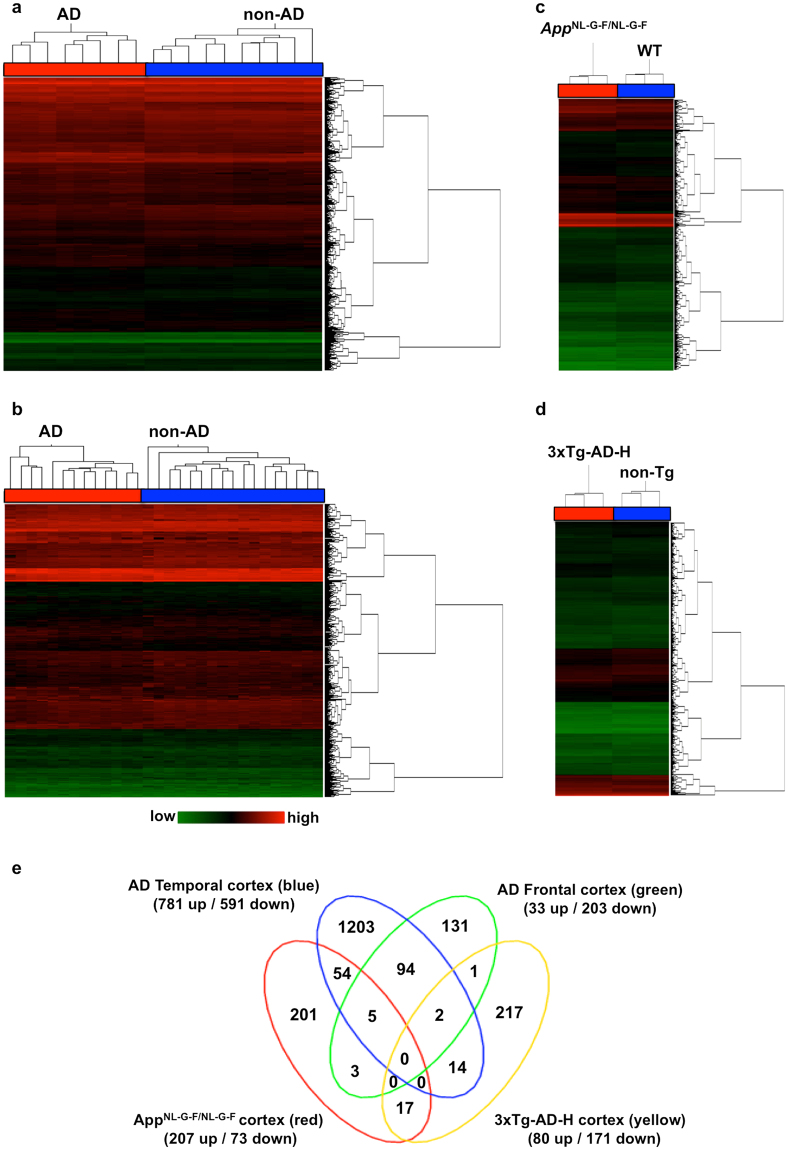
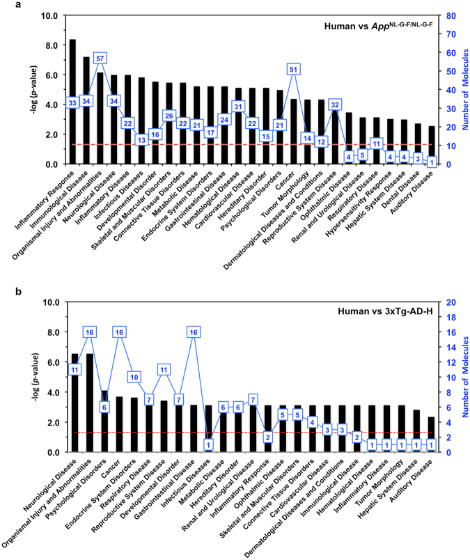
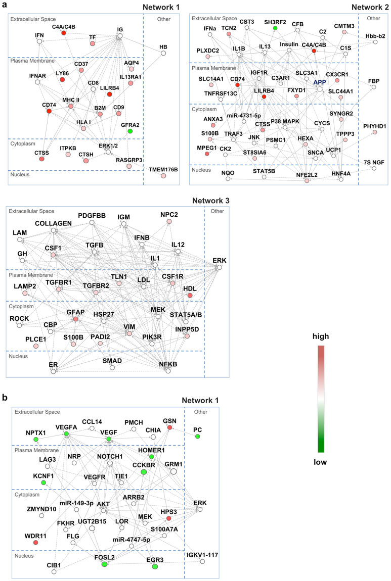
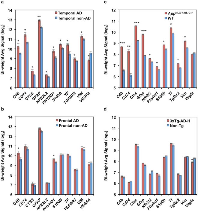
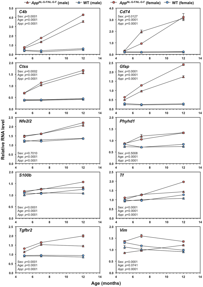
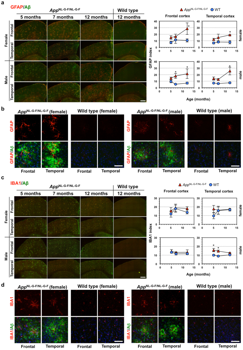
Abstract Alzheimer’s disease (AD) is the most common form of dementia, characterized by accumulation of amyloid β (Aβ) and neurofibrillary tangles. Oxidative stress and inflammation are considered to play an important role in the development and progression of AD. However, the extent to which these events contribute to the Aβ pathologies remains unclear. We performed inter-species comparative gene expression profiling between AD patient brains and theAppNL-G-F/NL-G-Fand 3xTg-AD-H mouse models. Genes commonly altered inAppNL-G-F/NL-G-Fand human AD cortices correlated with the inflammatory response or immunological disease. Among them, expression of AD-related genes (C4a/C4b,Cd74,Ctss,Gfap,Nfe2l2,Phyhd1,S100b,Tf,Tgfbr2, andVim) was increased in theAppNL-G-F/NL-G-Fcortex as Aβ amyloidosis progressed with exacerbated gliosis, while genes commonly altered in the 3xTg-AD-H and human AD cortices correlated with neurological disease. TheAppNL-G-F/NL-G-Fcortex also had altered expression of genes (Abi3,Apoe,Bin2,Cd33,Ctsc,Dock2,Fcer1g,Frmd6,Hck,Inpp5D,Ly86,Plcg2,Trem2,Tyrobp) defined as risk factors for AD by genome-wide association study or identified as genetic nodes in late-onset AD. These results suggest a strong correlation between cortical Aβ amyloidosis and the neuroinflammatory response and provide a better understanding of the involvement of gender effects in the development of AD.
Overview
The claims and statistics of this paper haven’t been checked yet.
Run the check — free, no account needed. It reads the open-access text and checks the sources it cites.
Integrity signals
Registry
Cites
Statistics
Figures
Verification
Claims the paper makes
The claims and statistics of this paper haven’t been checked yet.
Run the check — free, no account needed. It reads the open-access text and checks the sources it cites.
Claims other papers make about this one
Each is checked against this paper’s own text: does it say what the citing paper says it does?
The source backs part of the claim, not all of it — or backs it for a different population, condition or comparison.
In the citing paper
“and in transgenic mice carrying mutations associated with early AD (APP KM670/671 NL, PNSEN1 M146V) or overexpressing the human tau-4R/2N isoform (P301L) [34, 35].”
From Alzheimer’s disease phospholipase C-gamma-2 (PLCG2) protective variant is a functional hypermorph (2019) · cites this paper as [34]
Experimental — accuracy not yet measuredThe cited source says what the claim attributes to it.
In the citing paper
“Although some studies have identified selected overlaps in expression changes between AD mouse models and human brains (Bennett et al., 2018; Castillo et al., 2017; Mostafavi et al., 2018; Neuner et al., 2019; Rojo et al., 2017), other studies have questioned the overall degree of conservation (Burns et al., 2015; Galatro et al., 2017; Hargis and Blalock, 2017).”
From Meta-Analysis of the Alzheimer’s Disease Human Brain Transcriptome and Functional Dissection in Mouse Models (2020) · cites this paper as Castillo et al., 2017
The cited source says what the claim attributes to it.
In the citing paper
“Another study similarly found an upregulation of INPP5D RNA expression in both human AD patients and an App knock-in mouse model [170].”
From Microglial function, INPP5D/SHIP1 signaling, and NLRP3 inflammasome activation: implications for Alzheimer’s disease (2023) · cites this paper as [170]
The cited source says what the claim attributes to it.
In the citing paper
“Comparative gene expression profiling of known genetic risk factors in AD revealed an increase of PLCG2 gene expression in the cortex of AppNL-G-F/NL-G-F mice (165).”
From The role of PLCγ2 in immunological disorders, cancer, and neurodegeneration (2021) · cites this paper as [165]
In the citing paper
“LILRB4 (leukocyte immunoglobulin-like receptor, subfamily B, member 4), orthologues have also been shown to be increased with amyloid deposition and specifically associated with amyloid plaques (Wirz et al., 2013; Kamphuis et al., 2016; Castillo et al., 2017).”
From Genetic variability in response to amyloid beta deposition influences Alzheimer’s disease risk (2019) · cites this paper as Castillo et al., 2017
Self-check
Percentages, sums and reported differences re-computed from the paper’s own numbers.
The self-check needs the paper’s full text.
Citations
Cites
1 retracted
Bialas, A. R., & Stevens, B. (2013) · Nature Neuroscience · doi:10.1038/nn.3560 · Cited by 519
RetractedQuerfurth, H. W., & LaFerla, F. M. (2010) · New England Journal of Medicine · doi:10.1056/nejmra0909142 · Cited by 4,569
Self-citationMeta-Analysis of 74,046 Individuals Identifies 11 New Susceptibility Loci for Alzheimer’s Disease
(EADI), E. A. D. I., Lambert, J.-C., (GERAD), G. A. E. R. I. A. D., Alzheimer's Disease Genetic Consortium (ADGC), (CHARGE), C. F. H. A. A. R. I. G. E., Ibrahim-Verbaas, C. A., Harold, D., Naj, A. C., Sims, R., Bellenguez, C., et al. (2013) · Nature Genetics · doi:10.1038/ng.2802 · Cited by 4,336
Triple-Transgenic Model of Alzheimer’s Disease with Plaques and Tangles
Oddo, S., Caccamo, A., Shepherd, J. D., Murphy, M. P., Golde, T. E., Kayed, R., Metherate, R., Mattson, M. P., Akbari, Y., & LaFerla, F. M. (2003) · Neuron · doi:10.1016/s0896-6273(03)00434-3 · Cited by 4,126
Self-citationComplement and microglia mediate early synapse loss in Alzheimer mouse models
Hong, S., Beja-Glasser, V. F., Nfonoyim, B. M., Frouin, A., Li, S., Ramakrishnan, S., Merry, K. M., Shi, Q., Rosenthal, A., Barres, B. A., et al. (2016) · Science · doi:10.1126/science.aad8373 · Cited by 3,343
Immune attack: the role of inflammation in Alzheimer disease
Heppner, F. L., Ransohoff, R. M., & Becher, B. (2015) · Nature Reviews Neuroscience · doi:10.1038/nrn3880 · Cited by 2,134
Neuroinflammation in Alzheimer's disease: current evidence and future directions
Calsolaro, V., & Edison, P. (2016) · Alzheimer's & Dementia · doi:10.1016/j.jalz.2016.02.010 · Cited by 1,726
Integrated Systems Approach Identifies Genetic Nodes and Networks in Late-Onset Alzheimer’s Disease
Zhang, B., Gaiteri, C., Bodea, L.-G., Wang, Z., McElwee, J., Podtelezhnikov, A. A., Zhang, C., Xie, T., Tran, L., Dobrin, R., et al. (2013) · Cell · doi:10.1016/j.cell.2013.03.030 · Cited by 1,657
Single App knock-in mouse models of Alzheimer’s disease
Saito, T., Matsuba, Y., Mihira, N., Takano, J., Nilsson, P., Itohara, S., Iwata, N., & Saido, T. C. (2014) · Nature Neuroscience · doi:10.1038/nn.3697 · Cited by 1,366
Self-citationBillings, L. M., Oddo, S., Green, K. N., McGaugh, J. L., & LaFerla, F. M. (2005) · Neuron · doi:10.1016/j.neuron.2005.01.040 · Cited by 1,294
Self-citation
Research works printed but not linked: 1
- Pfaffi, M. W. Relative quantification, in real-time PCR (ed. Dorak M. T), Taylor & Francis Group, NY, pp63–82, 2006.
Cited by
Sorted by citations
Items 101–165 of 165
Papazoglou, A., Henseler, C., Weickhardt, S., Teipelke, J., Παπάζογλου, Π., Daubner, J., Schiffer, T., Krings, D., Broich, K., Hescheler, J., et al. (2024) · PLoS ONE · doi:10.1371/journal.pone.0296959 · Cited by 9
Dystonin/BPAG1 modulates diabetes and Alzheimer’s disease cross-talk: a meta-analysis
Cheng, J., Liu, H., Hwang, S., Hsu, L., Lin, W., & Tsai, F. (2019) · Neurological Sciences · doi:10.1007/s10072-019-03879-3 · Cited by 10
A Human Embryonic Stem Cell Model of Aβ-Dependent Chronic Progressive Neurodegeneration
Ubina, T., Magallanes, M., Srivastava, S., Warden, C., Yee, J., & Salvaterra, P. M. (2019) · Frontiers in Neuroscience · doi:10.3389/fnins.2019.01007 · Cited by 9
Godfrey, R. K., Alsop, E. B., Bjork, R. T., Chauhan, B. S., Ruvalcaba, H. C., Antone, J., Gittings, L. M., Michael, A. F., Williams, C., Hala’ufia, G., et al. (2023) · Acta Neuropathologica Communications · doi:10.1186/s40478-023-01656-0 · Cited by 13
Diverse signaling mechanisms and heterogeneity of astrocyte reactivity in Alzheimer's disease
Qin, H., Zhou, L., Haque, F. T., Martín-Jiménez, C., Trang, A., Benveniste, E. N., & Wang, Q. (2023) · Journal of Neurochemistry · doi:10.1111/jnc.16002 · Cited by 10
Lim, S., Tran, D. N., Kieu, Z., Chen, C., Villanueva, E., Ghiaar, S., Gallup, V., Zumkehr, J., Cribbs, D. H., Rodríguez‐Ortiz, C. J., et al. (2020) · Molecular Neurobiology · doi:10.1007/s12035-020-01894-6 · Cited by 8
PLCG2 as a Risk Factor for Alzheimer's Disease.
Tsai, A. P., Dong, C., Preuss, C., Moutinho, M., Lin, P. B.-C., Hajicek, N., Sondek, J., Bissel, S. J., Oblak, A. L., Carter, G. W., et al. (2020) · doi:10.21203/rs.3.rs-38216/v1 · Cited by 7
A Standardized Brain Molecular Atlas: A Resource for Systems Modeling and Simulation
Shichkova, P., Coggan, J. S., Markram, H., & Keller, D. X. (2021) · Frontiers in Molecular Neuroscience · doi:10.3389/fnmol.2021.604559 · Cited by 7
Changes in Expression of Key Genes in Alzheimer’s Disease: A Specific Brain Tissue Change
Rasmussen, L. T., de Lábio, R. W., dos Santos, M. P., Fredi, B. M., Chagas, E. F. B., Chen, E., Turecki, G., Smith, M. D. A. C., & Payão, S. L. M. (2024) · The Journals of Gerontology Series A · doi:10.1093/gerona/glae023 · Cited by 7
A Set of Dysregulated Target Genes to Reduce Neuroinflammation at Molecular Level
Massimini, M., Bachetti, B., Vedove, E. D., Benvenga, A., Pierro, F. D., & Bernabò, N. (2022) · International Journal of Molecular Sciences · doi:10.3390/ijms23137175 · Cited by 7
Willin/FRMD6 Mediates Mitochondrial Dysfunction Relevant to Neuronal Aβ Toxicity
Chen, D., Yu, W., Aitken, L., & Gunn‐Moore, F. J. (2022) · Cells · doi:10.3390/cells11193140 · Cited by 7
Ala‐Nisula, T., Sah‐Teli, S. K., Ronkainen, V., Dimova, E. Y., & Koivunen, P. (2023) · FEBS Letters · doi:10.1002/1873-3468.14666 · Cited by 5
Guo, X., Qin, C., Zhu, M., Wang, J., Sun, J. F., & Dai, L. (2018) · Dalton Transactions · doi:10.1039/c8dt02246a · Cited by 6
Wan, Y., Al-Ouran, R., Mangleburg, C. G., Lee, T. V., Allison, K., Neuner, S., Kaczorowski, C. C., Phillip, V., Howell, G. R., Martini-Stoica, H., et al. (2019) · bioRxiv (Cold Spring Harbor Laboratory) · doi:10.1101/506873 · Cited by 5
Reactive Astrocytes with Reduced Function of Glutamate Transporters in the App NL-G-F Knock-in Mice
Srivastava, I., Goikolea, J., Kaya, T. A., Latorre‐Leal, M., Eroli, F., Iglesias, M. P., Álvarez-Jiménez, L., Arroyo‐García, L. E., Shimozawa, M., Nilsson, P., et al. (2025) · ACS Chemical Neuroscience · doi:10.1021/acschemneuro.4c00714 · Cited by 8
Yin, W., Cerda-Hernández, N., Castillo-Morales, A., Ruiz-Tejada-Segura, M. L., Monzón-Sandoval, J., Moreno-Castilla, P., Pérez-Ortega, R., Bermudez-Rattoni, F., Urrutia, A. O., & Gutiérrez, H. (2020) · Frontiers in Neuroscience · doi:10.3389/fnins.2020.602642 · Cited by 5
Widjaya, M. A., Liu, C.-H., Lee, S., & Cheng, W. (2023) · Journal of Molecular Neuroscience · doi:10.1007/s12031-023-02152-9 · Cited by 5
Tianma Formula Alleviates Dementia via ACER2-Mediated Sphingolipid Signaling Pathway Involving Aβ
Lin, H., Wu, S., Weng, Z., Wang, H., Shi, R., Tian, M., Wang, Y., He, H., Wang, Y., Liu, X., et al. (2021) · Evidence-based Complementary and Alternative Medicine · doi:10.1155/2021/6029237 · Cited by 5
The KEAP1-NRF2 system and neurodegenerative diseases
Uruno, A., & Yamamoto, M. P. (2023) · Antioxidants and Redox Signaling · doi:10.1089/ars.2023.0005 · Cited by 8
TREM2 R47H exacerbates immune response in Alzheimer’s disease brainPreprint
Korvatska, O., Kiianitsa, K., Ratushny, A., Matsushita, M., Beeman, N., Chien, W.-M., Satoh, J.-I., Dorschner, M., Keene, C., Bammler, T., et al. (2018) · bioRxiv · doi:10.1101/499319 · Cited by 4
(2020) · doi:10.1101/2020.05.19.104216 · Cited by 5
Combined multi-omics and brain pathology reveal novel biomarkers for alzheimer’s disease
Zhao, Q., Gou, C., Luo, G., Dai, S., Wang, D., Zhang, S., Wang, F., Xu, H. M., Han, Y., & Wang, S. (2025) · Scientific Reports · doi:10.1038/s41598-025-21983-7 · Cited by 4
Multifactorial glial responses and their contributions to Alzheimer's disease continuum
Hijioka, M., Manabe, T., & Saito, T. (2023) · Clinical and Experimental Neuroimmunology · doi:10.1111/cen3.12745 · Cited by 4
Self-citationNiu, L., Yin, R., Li, J., Cao, X., Yu, J., Tan, L., & Initiative, A. D. N. (2019) · Journal of Alzheimer s Disease · doi:10.3233/jad-190153 · Cited by 4
Interactions of Cellular Energetic Gene Clusters in the Alzheimer’s Mouse Brain
Raju, R. P., Cai, L., Tyagi, A., & Pugazhenthi, S. (2023) · Molecular Neurobiology · doi:10.1007/s12035-023-03551-0 · Cited by 3
Pathological Roles of INPP5D in Alzheimer’s Disease
Chu, Y. N., Akahori, A., Takatori, S., & Tomita, T. (2023) · Advances in experimental medicine and biology · doi:10.1007/978-3-031-31978-5_30 · Cited by 3
Bartolo, N. D., Mortimer, N., Manter, M. A., Sanchez, N., Riley, M. M., O’Malley, T. T., & Hooker, J. M. (2022) · ACS Chemical Neuroscience · doi:10.1021/acschemneuro.2c00607 · Cited by 3
Chen, S., Chang, Y., Li, L., Acosta, D. M., Morrison, C. T., Wang, C., Julian, D. L., Hester, M. E., Serrano, G. E., Beach, T. G., et al. (2021) · SSRN Electronic Journal · doi:10.21203/rs.3.rs-855234/v1 · Cited by 3
A new role for PHYHD1 and related dioxygenases: demethylation of 2′- O -methylated nucleosides
Stonkus, J., Rutkienė, R., Meškienė, R., Jasiūnienė, M., Aučynaitė, A., Kalinienė, L., Lazutka, J., Balciunas, D., Vilkaitis, G., & Meškys, R. (2025) · Nucleic Acids Research · doi:10.1093/nar/gkaf1379 · Cited by 2
The role of DAP12 in immune-related inflammatory diseases
Lu, J., Shi, L., Jin, G., Yang, Y., Zhu, F., & Zhou, G. (2026) · Inflammation Research · doi:10.1007/s00011-026-02256-z · Cited by 3
Hernández-Contreras, K. A., Hernández‐Aguilar, M. E., Herrera‐Covarrubias, D., Rojas-Durán, F., & Aranda-Abreu, G. E. (2021) · Revista de Fomento Social · doi:10.24875/rmn.20000087 · Cited by 4
Fergany, A., Zong, C., Ekuban, F. A., Wu, B., Ueha, S., Shichino, S., Matsushima, K., Iwakura, Y., Ichihara, S., & Ichihara, G. (2023) · Archives of Toxicology · doi:10.1007/s00204-023-03627-9 · Cited by 2
Du, Y., Wang, D., Katis, V. L., Zoeller, E. L., Qui, M., Levey, A. I., Gileadi, O., & Fu, H. (2024) · SLAS DISCOVERY · doi:10.1016/j.slasd.2024.100177 · Cited by 2
PLCG2 as a Risk Factor for Alzheimer's DiseasePreprint
Tsai, A. P., Dong, C., Preuß, C., Moutinho, M., Lin, P. B., Hajicek, N., Sondek, J., Bissel, S. J., Oblak, A. L., Carter, G. W., et al. (2020) · doi:10.21203/rs.3.rs-33082/v1 · Cited by 2
Naia, L., Shimozawa, M., Bereczki, E., Li, X., Liu, J., Jiang, R., Leal, N. S., Pinho, C. M., Berger, E., Falk, V. L., et al. (2023) · bioRxiv (Cold Spring Harbor Laboratory) · doi:10.1101/2023.03.07.531542 · Cited by 2
Koivumäki, M., Martiskainen, H., Takalo, M., Lehtisalo, J., Ngandu, T., FinnGen, Snellman, A., Hiltunen, M. O., & Rinne, J. O. (2026) · Alzheimer s Research & Therapy · doi:10.1186/s13195-026-01984-y · Cited by 1
A human embryonic stem cell model of Aβ-dependent chronic progressive neurodegenerationPreprint
Ubina, T., Magallanes, M., Srivastava, S., Warden, C., Yee, J., & Salvaterra, P. M. (2018) · bioRxiv (Cold Spring Harbor Laboratory) · doi:10.1101/419978 · Cited by 1
Barthelson, K., Newman, M., & Lardelli, M. T. (2021) · bioRxiv (Cold Spring Harbor Laboratory) · doi:10.1101/2021.02.16.431539 · Cited by 1
Zhu, K., He, Q., Tsai, S., Maheepala, D. C., Henrion, M., Zaidi, S. S. A., Lau, B., Tang, A., Cadiz, M. P., Hodos-Nkhereanye, R., et al. (2022) · bioRxiv (Cold Spring Harbor Laboratory) · doi:10.1101/2022.10.19.512953 · Cited by 1
Transcriptional signatures of progressive neuropathology in transgenic tau and amyloid mouse modelsPreprint
Castanho, I., Murray, T. K., Hannon, E. J., Jeffries, A. R., Walker, E., Laing, E., Baulf, H., Harvey, J., Randall, A. D., Moore, K., et al. (2019) · bioRxiv (Cold Spring Harbor Laboratory) · doi:10.1101/548578
Castanho, I., Murray, T. K., Hannon, E. J., Jeffries, A. R., Walker, E., Laing, E., Baulf, H., Harvey, J., Randall, A. D., Moore, K., et al. (2019) · SSRN Electronic Journal · doi:10.2139/ssrn.3377366
Honda, K., Saito, Y., Saito, H., Toyoda, M., Abe, R., Saito, T., Saido, T. C., Michikawa, M., Taru, H., Sobu, Y., et al. (2022) · SSRN Electronic Journal · doi:10.1016/j.neurobiolaging.2022.12.003 · Cited by 16
Self-citationNieraad, H. (2022) · Publication Server of Goethe University Frankfurt am Main (Goethe University Frankfurt) · doi:10.21248/gups.68191
Chen, Q., Aguirre, L., Zhao, H., Borrego, F., de Rojas, I., Su, L., Li, P. P., Zhang, B., Kokovay, E., Lechleiter, J. D., et al. (2023) · medRxiv · doi:10.1101/2023.10.30.23297431
Saido, T. C. (2024) · Elsevier eBooks · doi:10.1016/b978-0-443-28979-8.00023-1
Self-citationNeuroinflammation: Microgliosis and astrocytosis in the days of microscopic omics
Saido, T. C. (2024) · Elsevier eBooks · doi:10.1016/b978-0-443-28979-8.00020-6
Self-citationPei, L., Hu, W., Wang, L., & Cai, H. (2025) · Journal of Neuropathology & Experimental Neurology · doi:10.1093/jnen/nlaf048 · Cited by 1
Koivumäki, M., Martiskainen, H., Takalo, M., Lehtisalo, J., Ngandu, T., Snellman, A., Hiltunen, M., & Rinne, J. O. (2025) · doi:10.21203/rs.3.rs-7823342/v1
Guo, L., Gaunt, J. R., Cheah, C. C. H., Chen, A. I., Claudine, S., Dawe, G. S., Goh, E. L. K., Kho, S.-H., Kumar, P., Lim, G. G. Y., et al. (2025) · Frontiers in Neuroscience · doi:10.3389/fnins.2025.1629830
Self-citationWang, Y., Sun, Y., Wang, Y., Jia, S., Qiao, Y., Zhou, Z., Shao, W., 张向飞, Guo, J., Zhang, B., et al. (2024) · Figshare · doi:10.6084/m9.figshare.26635863
Pauls, E., Bayod, S., Mateo, L., Alcalde, V., Juan-Blanco, T., Sánchez‐Soto, M., Saido, T. C., Saito, T., Berrenguer-Llergo, A., Attolini, C. S., et al. (2021) · Figshare · doi:10.6084/m9.figshare.16880438
Self-citationWang, Y., Sun, Y., Wang, Y., Jia, S., Qiao, Y., Zhou, Z., Shao, W., 张向飞, Guo, J., Zhang, B., et al. (2024) · Figshare · doi:10.6084/m9.figshare.26635878
Wang, Y., Sun, Y., Wang, Y., Jia, S., Qiao, Y., Zhou, Z., Shao, W., 张向飞, Guo, J., Zhang, B., et al. (2024) · Figshare · doi:10.6084/m9.figshare.26635890
Wang, Y., Sun, Y., Wang, Y., Jia, S., Qiao, Y., Zhou, Z., Shao, W., 张向飞, Guo, J., Zhang, B., et al. (2024) · Figshare · doi:10.6084/m9.figshare.26635866
Wang, Y., Sun, Y., Wang, Y., Jia, S., Qiao, Y., Zhou, Z., Shao, W., Zhang, X., Guo, J., Zhang, B., et al. (2024) · Figshare · doi:10.6084/m9.figshare.26635875
Swanson, M. E. V., Scotter, E. L., Smyth, L. C. D., Murray, H. C., Ryan, B., Turner, C., Faull, R. L. M., Dragunow, M., & Curtis, M. A. (2020) · Figshare · doi:10.6084/m9.figshare.13120600
Wang, Y., Sun, Y., Wang, Y., Jia, S., Qiao, Y., Zhou, Z., Shao, W., 张向飞, Guo, J., Zhang, B., et al. (2024) · Figshare · doi:10.6084/m9.figshare.26635881
Wang, Y., Sun, Y., Wang, Y., Jia, S., Qiao, Y., Zhou, Z., Shao, W., Zhang, X., Guo, J., Zhang, B., et al. (2024) · Figshare · doi:10.6084/m9.figshare.26635872
Wang, Y., Sun, Y., Wang, Y., Jia, S., Qiao, Y., Zhou, Z., Shao, W., 张向飞, Guo, J., Zhang, B., et al. (2024) · Figshare · doi:10.6084/m9.figshare.26635884
Wang, Y., Sun, Y., Wang, Y., Jia, S., Qiao, Y., Zhou, Z., Shao, W., Zhang, X., Guo, J., Zhang, B., et al. (2024) · Figshare · doi:10.6084/m9.figshare.26635887
Wang, Y., Sun, Y., Wang, Y., Jia, S., Qiao, Y., Zhou, Z., Shao, W., 张向飞, Guo, J., Zhang, B., et al. (2024) · Figshare · doi:10.6084/m9.figshare.26635869
Analysis of GRK2 aggregation in the pathology of Alzheimer disease in animal models
Alla, J. A., Perhal, A. F., Fu, X., Langer, A., el Faramawy, Y., & Quitterer, U. (2026) · Cell Reports Medicine · doi:10.1016/j.xcrm.2026.102707
Shang, X., Maimaiti, N., Tian, F., 廖春波, Sun, H., Zhou, X., Kong, W., Sun, Q., Wang, L., & Ma, X. F. (2026) · Frontiers in Cellular and Infection Microbiology · doi:10.3389/fcimb.2026.1835965
Yang, D., Guo, W., & Wang, B. (2026) · Metabolic Brain Disease · doi:10.1007/s11011-026-01930-9
Metadata
- Authors
- Castillo, E., Leon, J., Mazzei, G., Abolhassani, N., Haruyama, N., Saito, T., Saido, T., Hokama, M., Iwaki, T., Ohara, T., Ninomiya, T., Kiyohara, Y., Sakumi, K., LaFerla, F. M., & Nakabeppu, Y.
- Affiliations
- Kyushu University (JP)
- RIKEN (JP)
- Hisayama Research Institute For Lifestyle Diseases (JP)
- University of California, Irvine (US)
- Corresponding author
- Nakabeppu, Yusaku
- ORCID
- Venue
- Scientific Reports
- Published
- 2017-12-19
- Volume
- 7
- Issue
- 1
- Pages
- 17762-17762
- ISSN
- 2045-2322 (ISSN-L)
- PMC
- PMC5736730
- OpenAlex
- W2771127066
- Cited by
- 158
- References
- 61
- Citations by year
- 2026: 5 · 2025: 12 · 2024: 16 · 2023: 29 · 2022: 21 · 2021: 24 · 2020: 23 · 2019: 18 · 2018: 10
- FWCI
- 10.31
- Type
- journal article
- Language
- en
- Update notices
- correction (2021-09-09)
- Open access
- gold
- doi.org
- Locations
- Scientific Reports (publishedVersion, cc-by)
- Europe PMC (PubMed Central) (publishedVersion, cc-by)
- PubMed Central (publishedVersion, cc-by)
- PubMed (publishedVersion)
- Kyushu University Institutional Repository (QIR) (Kyushu University) (submittedVersion, cc-by)
- DOAJ (DOAJ: Directory of Open Access Journals) (submittedVersion)
- eScholarship (California Digital Library) (submittedVersion, cc-by)
- Funders
- Japan Agency for Medical Research and Development (H25-ninchisho-ippann-004)
- Ministry of Health, Labour and Welfare (H20-ninchisho-ippan-004)
- Fakultet Medicinskih Nauka, Univerziteta U Kragujevcu
- Japan Society for the Promotion of Science (26290017, 22221004, 15K15085, 17H01391)
- Topics
- Life Sciences › Neuroscience › Cellular and Molecular Neuroscience › Alzheimer's disease research and treatments
- Life Sciences › Neuroscience › Neurology › Neuroinflammation and Neurodegeneration Mechanisms
- Health Sciences › Medicine › Psychiatry and Mental health › Dementia and Cognitive Impairment Research
- Keywords
- Alzheimer's disease, neuroinflammation, cortical gene expression, gliosis, oxidative stress, sex differences
- MeSH terms
- Aged, Aged, 80 and over, Alzheimer Disease, Amyloidosis, Animals, Brain, Disease Models, Animal, Female, Gliosis, Humans, Inflammation, Male, Middle Aged, Gene Expression, Amyloid beta-Peptides, Gene Expression Profiling, Mice, Genome-Wide Association Study
About these checks
Registry, retraction, Cites and statistics results are computed, not judged, with one exception: a printed reference that no index lists is read once by a model before it counts as a citation error. Claims are extracted by a model and kept only when their quote appears verbatim in the text; each cited source is then checked, with a model’s help, for whether it says what the claim relies on. Those verdicts can be wrong — read the claim’s quote and the cited source before relying on one.
Statistics and arithmetic are recomputed, not judged.
Confidence bands are shown only for fields calibrated on our annotated test set. None is calibrated yet, so no band is shown.
What counts as a claim ›How we measure confidence ›How we check this ›
Are you an author, or do you see a problem with this page? Request a review
