

The DOI matches its Crossref record; 61 of the 61 references it cites were found in a registry or an index.
Comparative profiling of cortical gene expression in Alzheimer’s disease patients and mouse models demonstrates a link between amyloidosis and neuroinflammation
Open accessAbstract
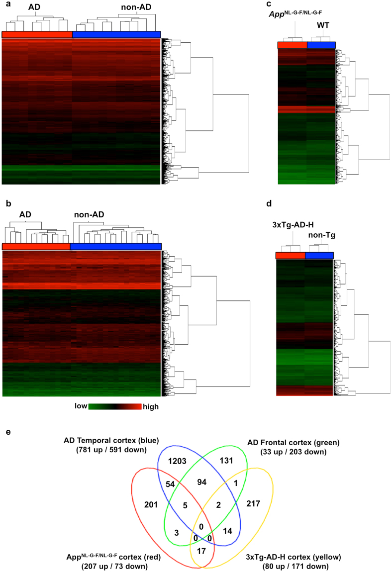
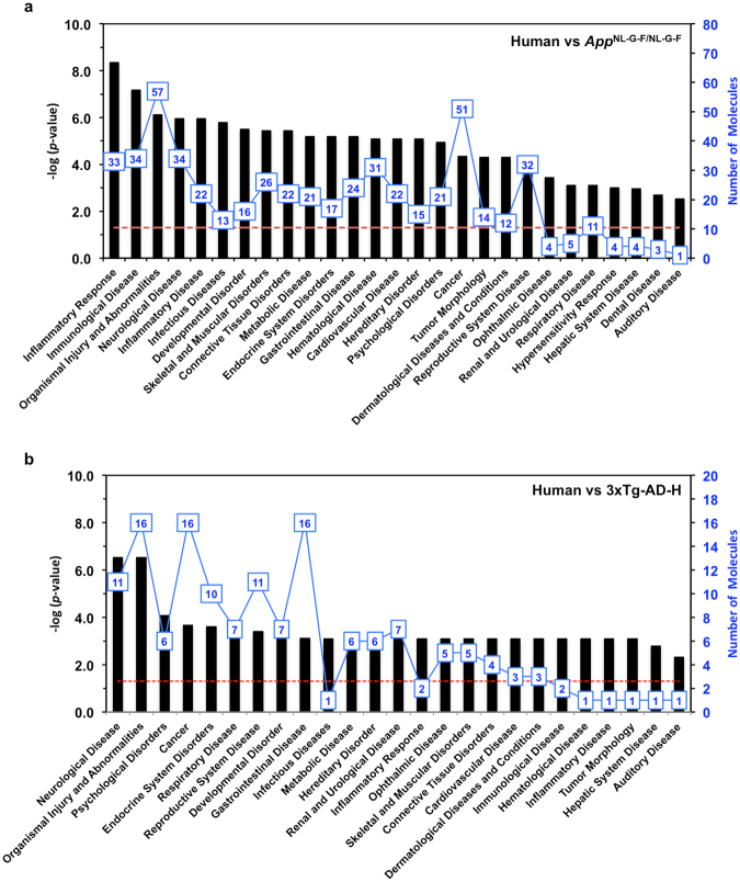
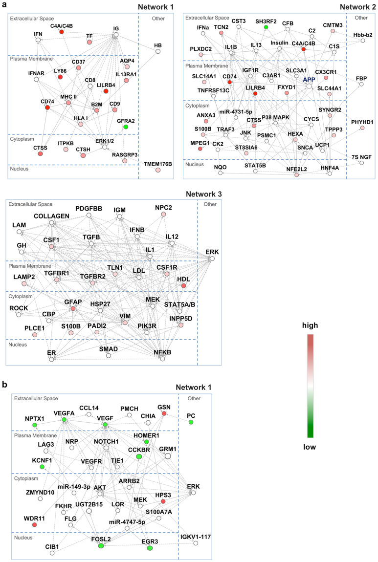
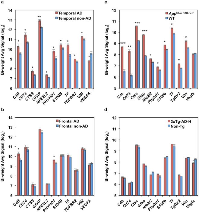
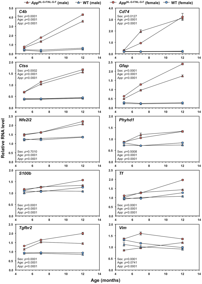
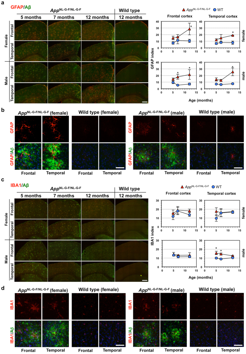
Abstract Alzheimer’s disease (AD) is the most common form of dementia, characterized by accumulation of amyloid β (Aβ) and neurofibrillary tangles. Oxidative stress and inflammation are considered to play an important role in the development and progression of AD. However, the extent to which these events contribute to the Aβ pathologies remains unclear. We performed inter-species comparative gene expression profiling between AD patient brains and theAppNL-G-F/NL-G-Fand 3xTg-AD-H mouse models. Genes commonly altered inAppNL-G-F/NL-G-Fand human AD cortices correlated with the inflammatory response or immunological disease. Among them, expression of AD-related genes (C4a/C4b,Cd74,Ctss,Gfap,Nfe2l2,Phyhd1,S100b,Tf,Tgfbr2, andVim) was increased in theAppNL-G-F/NL-G-Fcortex as Aβ amyloidosis progressed with exacerbated gliosis, while genes commonly altered in the 3xTg-AD-H and human AD cortices correlated with neurological disease. TheAppNL-G-F/NL-G-Fcortex also had altered expression of genes (Abi3,Apoe,Bin2,Cd33,Ctsc,Dock2,Fcer1g,Frmd6,Hck,Inpp5D,Ly86,Plcg2,Trem2,Tyrobp) defined as risk factors for AD by genome-wide association study or identified as genetic nodes in late-onset AD. These results suggest a strong correlation between cortical Aβ amyloidosis and the neuroinflammatory response and provide a better understanding of the involvement of gender effects in the development of AD.
Overview
The claims and statistics of this paper haven’t been checked yet.
Run the check — free, no account needed. It reads the open-access text and checks the sources it cites.
Integrity signals
Registry
Cites
Statistics
Figures
Verification
Claims the paper makes
The claims and statistics of this paper haven’t been checked yet.
Run the check — free, no account needed. It reads the open-access text and checks the sources it cites.
Claims other papers make about this one
Each is checked against this paper’s own text: does it say what the citing paper says it does?
The source backs part of the claim, not all of it — or backs it for a different population, condition or comparison.
In the citing paper
“and in transgenic mice carrying mutations associated with early AD (APP KM670/671 NL, PNSEN1 M146V) or overexpressing the human tau-4R/2N isoform (P301L) [34, 35].”
From Alzheimer’s disease phospholipase C-gamma-2 (PLCG2) protective variant is a functional hypermorph (2019) · cites this paper as [34]
Experimental — accuracy not yet measuredThe cited source says what the claim attributes to it.
In the citing paper
“Although some studies have identified selected overlaps in expression changes between AD mouse models and human brains (Bennett et al., 2018; Castillo et al., 2017; Mostafavi et al., 2018; Neuner et al., 2019; Rojo et al., 2017), other studies have questioned the overall degree of conservation (Burns et al., 2015; Galatro et al., 2017; Hargis and Blalock, 2017).”
From Meta-Analysis of the Alzheimer’s Disease Human Brain Transcriptome and Functional Dissection in Mouse Models (2020) · cites this paper as Castillo et al., 2017
The cited source says what the claim attributes to it.
In the citing paper
“Another study similarly found an upregulation of INPP5D RNA expression in both human AD patients and an App knock-in mouse model [170].”
From Microglial function, INPP5D/SHIP1 signaling, and NLRP3 inflammasome activation: implications for Alzheimer’s disease (2023) · cites this paper as [170]
The cited source says what the claim attributes to it.
In the citing paper
“Comparative gene expression profiling of known genetic risk factors in AD revealed an increase of PLCG2 gene expression in the cortex of AppNL-G-F/NL-G-F mice (165).”
From The role of PLCγ2 in immunological disorders, cancer, and neurodegeneration (2021) · cites this paper as [165]
In the citing paper
“LILRB4 (leukocyte immunoglobulin-like receptor, subfamily B, member 4), orthologues have also been shown to be increased with amyloid deposition and specifically associated with amyloid plaques (Wirz et al., 2013; Kamphuis et al., 2016; Castillo et al., 2017).”
From Genetic variability in response to amyloid beta deposition influences Alzheimer’s disease risk (2019) · cites this paper as Castillo et al., 2017
Self-check
Percentages, sums and reported differences re-computed from the paper’s own numbers.
The self-check needs the paper’s full text.
Citations
Cites
1 retracted
Bialas, A. R., & Stevens, B. (2013) · Nature Neuroscience · doi:10.1038/nn.3560 · Cited by 519
RetractedQuerfurth, H. W., & LaFerla, F. M. (2010) · New England Journal of Medicine · doi:10.1056/nejmra0909142 · Cited by 4,569
Self-citationMeta-Analysis of 74,046 Individuals Identifies 11 New Susceptibility Loci for Alzheimer’s Disease
(EADI), E. A. D. I., Lambert, J.-C., (GERAD), G. A. E. R. I. A. D., Alzheimer's Disease Genetic Consortium (ADGC), (CHARGE), C. F. H. A. A. R. I. G. E., Ibrahim-Verbaas, C. A., Harold, D., Naj, A. C., Sims, R., Bellenguez, C., et al. (2013) · Nature Genetics · doi:10.1038/ng.2802 · Cited by 4,336
Triple-Transgenic Model of Alzheimer’s Disease with Plaques and Tangles
Oddo, S., Caccamo, A., Shepherd, J. D., Murphy, M. P., Golde, T. E., Kayed, R., Metherate, R., Mattson, M. P., Akbari, Y., & LaFerla, F. M. (2003) · Neuron · doi:10.1016/s0896-6273(03)00434-3 · Cited by 4,126
Self-citationComplement and microglia mediate early synapse loss in Alzheimer mouse models
Hong, S., Beja-Glasser, V. F., Nfonoyim, B. M., Frouin, A., Li, S., Ramakrishnan, S., Merry, K. M., Shi, Q., Rosenthal, A., Barres, B. A., et al. (2016) · Science · doi:10.1126/science.aad8373 · Cited by 3,343
Immune attack: the role of inflammation in Alzheimer disease
Heppner, F. L., Ransohoff, R. M., & Becher, B. (2015) · Nature Reviews Neuroscience · doi:10.1038/nrn3880 · Cited by 2,134
Neuroinflammation in Alzheimer's disease: current evidence and future directions
Calsolaro, V., & Edison, P. (2016) · Alzheimer's & Dementia · doi:10.1016/j.jalz.2016.02.010 · Cited by 1,726
Integrated Systems Approach Identifies Genetic Nodes and Networks in Late-Onset Alzheimer’s Disease
Zhang, B., Gaiteri, C., Bodea, L.-G., Wang, Z., McElwee, J., Podtelezhnikov, A. A., Zhang, C., Xie, T., Tran, L., Dobrin, R., et al. (2013) · Cell · doi:10.1016/j.cell.2013.03.030 · Cited by 1,657
Single App knock-in mouse models of Alzheimer’s disease
Saito, T., Matsuba, Y., Mihira, N., Takano, J., Nilsson, P., Itohara, S., Iwata, N., & Saido, T. C. (2014) · Nature Neuroscience · doi:10.1038/nn.3697 · Cited by 1,366
Self-citationBillings, L. M., Oddo, S., Green, K. N., McGaugh, J. L., & LaFerla, F. M. (2005) · Neuron · doi:10.1016/j.neuron.2005.01.040 · Cited by 1,294
Self-citationARUK Consortium, Sims, R., GERAD/PERADES, C., van der Lee, S. J., Naj, A. C., Bellenguez, C., Badarinarayan, N., Jakobsdottir, J., Kunkle, B. W., Boland, A., et al. (2017) · Nature Genetics · doi:10.1038/ng.3916 · Cited by 1,012
(2016) · London School of Economics and Political Science Research Online (London School of Economics and Political Science) · Cited by 874
Multimodal techniques for diagnosis and prognosis of Alzheimer's disease
Perrin, R. J., Fagan, A. M., & Holtzman, D. M. (2009) · Nature · doi:10.1038/nature08538 · Cited by 702
Late onset Alzheimer’s disease genetics implicates microglial pathways in disease risk
Efthymiou, A. G., & Goate, A. M. (2017) · Molecular Neurodegeneration · doi:10.1186/s13024-017-0184-x · Cited by 562
Khan, U. A., Liu, L., Provenzano, F. A., Berman, D. E., Profaci, C. P., Sloan, R., Mayeux, R., Duff, K. E., & Small, S. A. (2014) · Nature Neuroscience · doi:10.1038/nn.3606 · Cited by 561
Riese, R. J., Wolf, P. R., Brömme, D., Natkin, L. W., Villadangos, J. A., Ploegh, H. L., & Chapman, H. A. (1996) · Immunity · doi:10.1016/s1074-7613(00)80249-6 · Cited by 536
Estrogen: A master regulator of bioenergetic systems in the brain and body
Rettberg, J. R., Yao, J., & Brinton, R. D. (2014) · Frontiers in Neuroendocrinology · doi:10.1016/j.yfrne.2013.08.001 · Cited by 534
Cortical Change in Alzheimer's Disease Detected with a Disease-specific Population-based Brain Atlas
Thompson, P. M., Mega, M. S., Woods, R. P., Zoumalan, C. I., Lindshield, C., Blanton, R. E., Moussai, J., Holmes, C. J., Cummings, J. L., & Toga, A. W. (2001) · Cerebral Cortex · doi:10.1093/cercor/11.1.1 · Cited by 447
Carter, S. F., Schöll, M., Almkvist, O., Wall, A., Engler, H., Långström, B., & Nordberg, A. (2012) · Journal of Nuclear Medicine · doi:10.2967/jnumed.110.087031 · Cited by 442
Sex and the development of Alzheimer's disease
Pike, C. J. (2016) · Journal of Neuroscience Research · doi:10.1002/jnr.23827 · Cited by 416
Isolation of glia from Alzheimer’s mice reveals inflammation and dysfunction
Orre, M., Kamphuis, W., Osborn, L. M., Jansen, A. H. P., Kooijman, L., Bossers, K., & Hol, E. M. (2014) · Neurobiology of Aging · doi:10.1016/j.neurobiolaging.2014.06.004 · Cited by 394
Complement — tapping into new sites and effector systems
Kolev, M. V., Friec, G. L., & Kemper, C. (2014) · Nature reviews. Immunology · doi:10.1038/nri3761 · Cited by 367
Complement activation and inhibition: a delicate balance
Sjöberg, A. P., Trouw, L. A., & Blom, A. M. (2009) · Trends in Immunology · doi:10.1016/j.it.2008.11.003 · Cited by 364
Altered Expression of Diabetes-Related Genes in Alzheimer's Disease Brains: The Hisayama Study
Hokama, M., Oka, S., Leon, J., Ninomiya, T., Honda, H., Sasaki, K., Iwaki, T., Ohara, T., Sasaki, T., LaFerla, F. M., et al. (2013) · Cerebral Cortex · doi:10.1093/cercor/bht101 · Cited by 323
Self-citationMatarin, M., Salih, D. A., Yasvoina, M., Cummings, D. M., Guelfi, S., Liu, W., Nahaboo Solim, M. A., Moens, T. G., Paublete, R. M., Ali, S. S., et al. (2015) · Cell Reports · doi:10.1016/j.celrep.2014.12.041 · Cited by 290
Snyder, H. M., Asthana, S., Bain, L., Brinton, R., Craft, S., Dubal, D. B., Espeland, M. A., Gatz, M., Mielke, M. M., Raber, J., et al. (2016) · Alzheimer's & Dementia · doi:10.1016/j.jalz.2016.08.004 · Cited by 262
Cognitive deficits in single App knock-in mouse models
Masuda, A., Kobayashi, Y., Kogo, N., Saito, T., Saido, T. C., & Itohara, S. (2016) · Neurobiology of Learning and Memory · doi:10.1016/j.nlm.2016.07.001 · Cited by 248
Self-citationImaging the progression of Alzheimer pathology through the brain
Smith, A. D. (2002) · Proceedings of the National Academy of Sciences · doi:10.1073/pnas.082107399 · Cited by 202
Sekar, S., McDonald, J., Cuyugan, L., Aldrich, J., Kurdoglu, A., Adkins, J., Serrano, G., Beach, T. G., Craig, D. W., Valla, J., et al. (2015) · Neurobiology of Aging · doi:10.1016/j.neurobiolaging.2014.09.027 · Cited by 199
Assessment of β-amyloid deposits in human brain: a study of the BrainNet Europe Consortium
Alafuzoff, I., Thal, D. R., Arzberger, T., Bogdanovic, N., Al-Sarraj, S., Bodi, I., Boluda, S., Bugiani, O., Duyckaerts, C., Gelpi, E., et al. (2009) · Acta Neuropathologica · doi:10.1007/s00401-009-0485-4 · Cited by 193
Genomic variants, genes, and pathways of Alzheimer's disease: An overview
Naj, A. C., & Schellenberg, G. D. (2016) · American Journal of Medical Genetics Part B Neuropsychiatric Genetics · doi:10.1002/ajmg.b.32499 · Cited by 186
Landel, V., Baranger, K., Virard, I., Loriod, B., Khrestchatisky, M., Rivera, S., Benech, P., & Féron, F. (2014) · Molecular Neurodegeneration · doi:10.1186/1750-1326-9-33 · Cited by 176
Toward more predictive genetic mouse models of Alzheimer’s disease
Onos, K. D., Sukoff Rizzo, S. J., Howell, G. R., & Sasner, M. (2016) · Brain Research Bulletin · doi:10.1016/j.brainresbull.2015.12.003 · Cited by 156
A genome-wide association study of aging
Walter, S., Atzmon, G., Demerath, E. W., Garcia, M. E., Kaplan, R. C., Meena Kumari, B., Lunetta, K. L., Milaneschi, Y., Tanaka, T., Tranah, G. J., et al. (2011) · Neurobiology of Aging · doi:10.1016/j.neurobiolaging.2011.05.026 · Cited by 151
Abolhassani, N., Leon, J., Sheng, Z., Oka, S., Hamasaki, H., Iwaki, T., & Nakabeppu, Y. (2017) · Mechanisms of Ageing and Development · doi:10.1016/j.mad.2016.05.005 · Cited by 140
Self-citationBasal forebrain atrophy correlates with amyloid β burden in Alzheimer's disease
Kerbler, G. M., Fripp, J., Rowe, C. C., Villemagne, V. L., Salvado, O., Rose, S., & Coulson, E. J. (2014) · NeuroImage Clinical · doi:10.1016/j.nicl.2014.11.015 · Cited by 128
Systemic immune system alterations in early stages of Alzheimer's disease
Zhang, R., Miller, R. G., Madison, C. A., Jin, X., Honrada, R., Harris, W., Katz, J., Forshew, D. A., & McGrath, M. S. (2013) · Journal of Neuroimmunology · doi:10.1016/j.jneuroim.2013.01.002 · Cited by 122
Expression of CD74 is increased in neurofibrillary tangles in Alzheimer's disease
Bryan, K. J., Zhu, X., Harris, P. L., Perry, G., Castellani, R. J., Smith, M. A., & Casadesus, G. (2008) · Molecular Neurodegeneration · doi:10.1186/1750-1326-3-13 · Cited by 114
Lysosomal Network Proteins as Potential Novel CSF Biomarkers for Alzheimer’s Disease
Armstrong, A., Mattsson, N., Appelqvist, H., Janefjord, C., Sandin, L., Agholme, L., Olsson, B., Svensson, S., Blennow, K., Zetterberg, H., et al. (2013) · NeuroMolecular Medicine · doi:10.1007/s12017-013-8269-3 · Cited by 113
New Mouse Model of Alzheimer’s
Nilsson, P., Saito, T., & Saido, T. C. (2014) · ACS Chemical Neuroscience · doi:10.1021/cn500105p · Cited by 107
Self-citationDifferential modulation of astrocyte cytokine gene expression by TGF-beta.
Benveniste, E. N., Kwon, J., Chung, W.-C., Sampson, J. B., Pandya, K., & Tang, L. (1994) · The Journal of Immunology · doi:10.4049/jimmunol.153.11.5210 · Cited by 99
Cerebrospinal fluid levels of complement proteins C3, C4 and CR1 in Alzheimer’s disease
Daborg, J., Andréasson, U., Pekna, M., Lautner, R., Hanse, E., Minthon, L., Blennow, K., Hansson, O., & Zetterberg, H. (2012) · Journal of Neural Transmission · doi:10.1007/s00702-012-0797-8 · Cited by 87
Lysosomal processing of amyloid precursor protein to Aβ peptides: a distinct role for cathepsin S
Munger, J. S., Haass, C., Lemere, C. A., Shi, G., Wong, W., Teplow, D. B., Selkoe, D. J., & Chapman, H. A. (1995) · Biochemical Journal · doi:10.1042/bj3110299 · Cited by 85
CD74 interacts with APP and suppresses the production of Aβ
Matsuda, S., Matsuda, Y., & D'adamio, L. (2009) · Molecular Neurodegeneration · doi:10.1186/1750-1326-4-41 · Cited by 79
Matza, D., Kerem, A., Medvedovsky, H., Lantner, F., & Shachar, I. (2002) · Immunity · doi:10.1016/s1074-7613(02)00455-7 · Cited by 78
MIF, CD74 and other partners in kidney disease: Tales of a promiscuous couple
Sánchez-Niño, M. D., Sanz, A. B., Ruiz‐Andrés, O., Poveda, J., Izquierdo, M. C., Selgas, R., Egido, J., & Ortíz, A. (2012) · Cytokine & Growth Factor Reviews · doi:10.1016/j.cytogfr.2012.08.001 · Cited by 76
Piperigkou, Z., Bouris, P., Onisto, M., Franchi, M., Kletsas, D., Theocharis, A. D., & Karamanos, N. K. (2016) · Matrix Biology · doi:10.1016/j.matbio.2016.05.003 · Cited by 73
Presymptomatic cortical thinning in familial Alzheimer disease
Weston, P. S. J., Nicholas, J. M., Lehmann, M., Ryan, N. S., Liang, Y., Macpherson, K., Marc Modat, A., Rossor, M. N., Schott, J. M., Sebastien Ourselin, A., et al. (2016) · Neurology · doi:10.1212/wnl.0000000000003322 · Cited by 71
Genes associated with the progression of neurofibrillary tangles in Alzheimer’s disease
Miyashita, A., Hatsuta, H., Kikuchi, M., Nakaya, A., Saito, Y., Tsukie, T., Hara, N., Ogishima, S., Kitamura, N., Akazawa, K., et al. (2014) · Translational Psychiatry · doi:10.1038/tp.2014.35 · Cited by 65
Wirz, K. T. S., Bossers, K., Stargardt, A., Kamphuis, W., Swaab, D. F., Hol, E. M., & Verhaagen, J. (2013) · Neurobiology of Aging · doi:10.1016/j.neurobiolaging.2012.11.008 · Cited by 65
Complement C4A and C4B Gene Copy Number Study in Alzheimer's Disease Patients
Zorzetto, M., Datturi, F., Divizia, L., Pistono, C., Campo, I., Silvestri, A. D., Cuccia, M., & Ricevuti, G. (2016) · Current Alzheimer Research · doi:10.2174/1567205013666161013091934 · Cited by 45
State of Play in Alzheimer’s Disease Genetics
Zhu, J.-B., Tan, C., Tan, L., & Yu, J. (2017) · Journal of Alzheimer s Disease · doi:10.3233/jad-170062 · Cited by 44
Assessing Candidate Serum Biomarkers for Alzheimer's Disease: A Longitudinal Study
Zabel, M. K., Schrag, M., Mueller, C., Zhou, W., Crofton, A. R., Petersen, F. F., Dickson, A., & Kirsch, W. M. (2012) · Journal of Alzheimer s Disease · doi:10.3233/jad-2012-112012 · Cited by 41
Kiyota, T., Zhang, G., Morrison, C. M., Bosch, M. E., Weir, R. A., Lu, Y., Dong, W., & Gendelman, H. E. (2015) · Molecular Therapy · doi:10.1038/mt.2015.142 · Cited by 41
Chen, Y., Zhang, K.-S., Yang, L., & He, Q. (2016) · Cell Adhesion & Migration · doi:10.1080/19336918.2016.1221567 · Cited by 33
Biomarkers in Alzheimer’s Disease-Recent Update
Sharma, S. K., & Lipincott, W. (2017) · Current Alzheimer Research · doi:10.2174/1567205014666170220141822 · Cited by 28
AβPP Accumulation and/or Intraneuronal Amyloid-β Accumulation? The 3xTg-AD Mouse Model Revisited
Wirths, O., Dins, A., & Bayer, T. A. (2012) · Journal of Alzheimer s Disease · doi:10.3233/jad-2011-111529 · Cited by 26
Loss of TGFβ Receptor Type 2 Expression Impairs Estrogen Response and Confers Tamoxifen Resistance
Busch, S., Sims, A. H., Stål, O., Fernö, M., & Landberg, G. (2015) · Cancer Research · doi:10.1158/0008-5472.can-14-1583 · Cited by 26
Accumulation of carboxy-terminal fragments of APP increases phosphodiesterase 8B.
Kametani, F., & Haga, S. (2015) · Neurobiology of Aging · doi:10.1016/j.neurobiolaging.2014.09.029 · Cited by 23
Ito, H., Shinotoh, H., Shimada, H., Miyoshi, M., Yanai, K., Okamura, N., Takano, H., Takahashi, H., Arakawa, R., Kodaka, F., et al. (2013) · European Journal of Nuclear Medicine and Molecular Imaging · doi:10.1007/s00259-013-2620-7 · Cited by 20
Cuello, A. C. (2016) · Current Alzheimer Research · doi:10.2174/156720501401161130204127 · Cited by 4
Research works printed but not linked: 1
- Pfaffi, M. W. Relative quantification, in real-time PCR (ed. Dorak M. T), Taylor & Francis Group, NY, pp63–82, 2006.
Cited by
Sorted by citations
Spatial Transcriptomics and In Situ Sequencing to Study Alzheimer’s Disease
Chen, W.-T., Lu, A., Craessaerts, K., Pavie, B., Sala Frigerio, C., Corthout, N., Qian, X., Laláková, J., Kühnemund, M., Voytyuk, I., et al. (2020) · Cell · doi:10.1016/j.cell.2020.06.038 · Cited by 949
Self-citationInflammation: the link between comorbidities, genetics, and Alzheimer’s disease
Newcombe, E. A., Camats-Perna, J., Silva, M. L., Valmas, N., Huat, T. J., & Medeiros, R. (2018) · Journal of Neuroinflammation · doi:10.1186/s12974-018-1313-3 · Cited by 532
Metal Toxicity Links to Alzheimer's Disease and Neuroinflammation
Huat, T. J., Camats‐Perna, J., Newcombe, E. A., Valmas, N., Kitazawa, M., & Medeiros, R. (2019) · Journal of Molecular Biology · doi:10.1016/j.jmb.2019.01.018 · Cited by 501
Chan, J. M., Quintanal-Villalonga, À., Gao, V. R., Xie, Y., Allaj, V., Chaudhary, O., Masilionis, I., Egger, J. V., Chow, A. T., Walle, T., et al. (2021) · Cancer Cell · doi:10.1016/j.ccell.2021.09.008 · Cited by 464
Wan, Y.-W., Al-Ouran, R., Mangleburg, C. G., Perumal, T. M., Lee, T. V., Allison, K., Swarup, V., Funk, C. C., Gaiteri, C., Allen, M., et al. (2020) · Cell Reports · doi:10.1016/j.celrep.2020.107908 · Cited by 295
“Although some studies have identified selected overlaps in expression changes between AD mouse models and human brains (Bennett et al., 2018; Castillo et al., 2017; Mostafavi et al., 2018; Neuner et al., 2019; Rojo et al., 2017), other studies have questioned the overall degree of conservation (Burns et al.”
Result
Metadata
- Authors
- Castillo, E., Leon, J., Mazzei, G., Abolhassani, N., Haruyama, N., Saito, T., Saido, T., Hokama, M., Iwaki, T., Ohara, T., Ninomiya, T., Kiyohara, Y., Sakumi, K., LaFerla, F. M., & Nakabeppu, Y.
- Affiliations
- Kyushu University (JP)
- RIKEN (JP)
- Hisayama Research Institute For Lifestyle Diseases (JP)
- University of California, Irvine (US)
- Corresponding author
- Nakabeppu, Yusaku
- ORCID
- Venue
- Scientific Reports
- Published
- 2017-12-19
- Volume
- 7
- Issue
- 1
- Pages
- 17762-17762
- ISSN
- 2045-2322 (ISSN-L)
- PMC
- PMC5736730
- OpenAlex
- W2771127066
- Cited by
- 158
- References
- 61
- Citations by year
- 2026: 5 · 2025: 12 · 2024: 16 · 2023: 29 · 2022: 21 · 2021: 24 · 2020: 23 · 2019: 18 · 2018: 10
- FWCI
- 10.31
- Type
- journal article
- Language
- en
- Update notices
- correction (2021-09-09)
- Open access
- gold
- doi.org
- Locations
- Scientific Reports (publishedVersion, cc-by)
- Europe PMC (PubMed Central) (publishedVersion, cc-by)
- PubMed Central (publishedVersion, cc-by)
- PubMed (publishedVersion)
- Kyushu University Institutional Repository (QIR) (Kyushu University) (submittedVersion, cc-by)
- DOAJ (DOAJ: Directory of Open Access Journals) (submittedVersion)
- eScholarship (California Digital Library) (submittedVersion, cc-by)
- Funders
- Japan Agency for Medical Research and Development (H25-ninchisho-ippann-004)
- Ministry of Health, Labour and Welfare (H20-ninchisho-ippan-004)
- Fakultet Medicinskih Nauka, Univerziteta U Kragujevcu
- Japan Society for the Promotion of Science (26290017, 22221004, 15K15085, 17H01391)
- Topics
- Life Sciences › Neuroscience › Cellular and Molecular Neuroscience › Alzheimer's disease research and treatments
- Life Sciences › Neuroscience › Neurology › Neuroinflammation and Neurodegeneration Mechanisms
- Health Sciences › Medicine › Psychiatry and Mental health › Dementia and Cognitive Impairment Research
- Keywords
- Alzheimer's disease, neuroinflammation, cortical gene expression, gliosis, oxidative stress, sex differences
- MeSH terms
- Aged, Aged, 80 and over, Alzheimer Disease, Amyloidosis, Animals, Brain, Disease Models, Animal, Female, Gliosis, Humans, Inflammation, Male, Middle Aged, Gene Expression, Amyloid beta-Peptides, Gene Expression Profiling, Mice, Genome-Wide Association Study
About these checks
Registry, retraction, Cites and statistics results are computed, not judged, with one exception: a printed reference that no index lists is read once by a model before it counts as a citation error. Claims are extracted by a model and kept only when their quote appears verbatim in the text; each cited source is then checked, with a model’s help, for whether it says what the claim relies on. Those verdicts can be wrong — read the claim’s quote and the cited source before relying on one.
Statistics and arithmetic are recomputed, not judged.
Confidence bands are shown only for fields calibrated on our annotated test set. None is calibrated yet, so no band is shown.
What counts as a claim ›How we measure confidence ›How we check this ›
Are you an author, or do you see a problem with this page? Request a review
