

The DOI matches its Crossref record; 60 of the 60 references it cites were found in a registry or an index.
Insights into antioxidant activities and anti-skin-aging potential of callus extract from Centella asiatica (L.)
Open accessAbstract
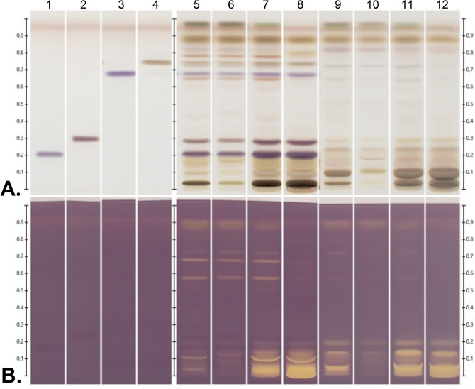
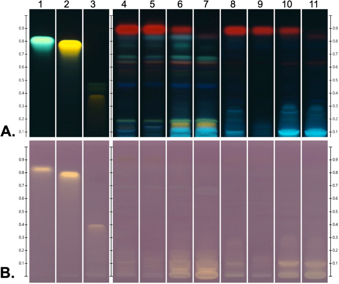
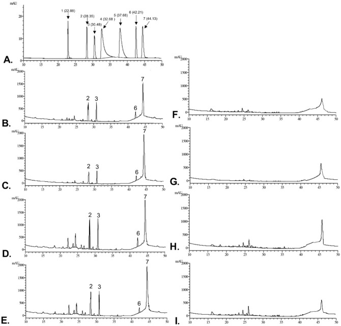
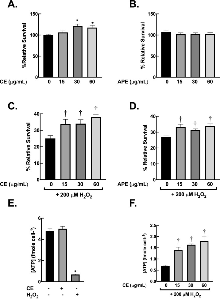
Formation of oxidative stress in dermal fibroblasts plays crucial roles in aging processes of skin. The use of phytochemicals that can promote capacity of fibroblasts to combat oxidative stress is an attractive strategy to prevent skin aging and promote skin beauty. Centella asiatica has been used to treat multitude of diseases for centuries. Previous investigations demonstrated that extracts from C. asiatica have a broad range of beneficial activities through their antioxidant activity. Hence, the extract from this medicinal plant could be a great candidate for anti-skin-aging agent. Callus culture offers a powerful platform for sustainable, rapid and large-scale production of phytochemicals to serve extensive demands of pharmaceutical and cosmeceutical industries. Here, we demonstrated the application of callus culture of Centella asiatica to produce bioactive metabolites. The 50% ethanolic extract of callus culture has distinctive features of chemical compositions and biological profiles. Information from HPTLC-DPPH and HPLC analysis suggested that the callus extract comprises distinctive antioxidant compounds, compared with those isolated from authentic plant. Moreover, results from cell culture experiment demonstrated that callus extract possesses promising antioxidant and anti-skin-aging activities. Pre-treatment with callus extract attenuated H 2 O 2-induced-cytotoxicity on human dermal fibroblasts. The results from RT-qPCR clearly suggested that the upregulation of cellular antioxidant enzymes appeared to be major contributor for the protective effects of callus extract against oxidative stress. Moreover, supplementation with callus extract inhibited induction of matrix metalloprotease-9 following H 2 O 2 exposure, suggesting its potential anti-skin-aging activity. Our results demonstrate the potential utility of C. asiatica callus extract as anti-skin-aging agent in cosmeceutical preparations.
Overview
The claims and statistics of this paper haven’t been checked yet.
Run the check — free, no account needed. It reads the open-access text and checks the sources it cites.
Integrity signals
Registry
Cites
Statistics
Figures
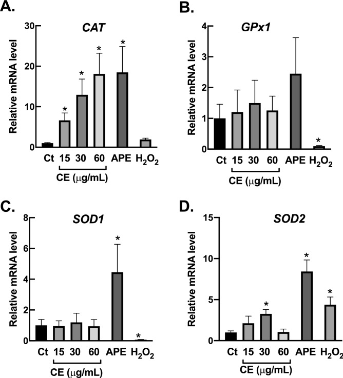
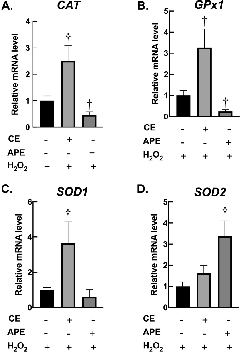
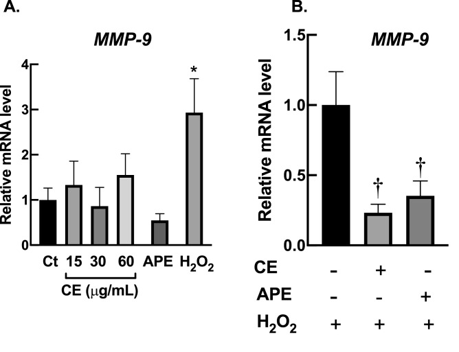
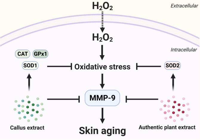
Verification
Claims the paper makes
The claims and statistics of this paper haven’t been checked yet.
Run the check — free, no account needed. It reads the open-access text and checks the sources it cites.
Claims other papers make about this one
Each is checked against this paper’s own text: does it say what the citing paper says it does?
The cited source says what the claim attributes to it.
In the citing paper
“The results showed that the callus extract exhibited strong antioxidant activities and anti‐skin‐aging potential”
From Cosmeceuticals in photoaging: A review. (2024) · cites this paper as [37]
The cited source says what the claim attributes to it.
In the citing paper
“These findings are consistent with earlier reports on the antioxidant properties of C. asiatica [35].”
The check wasn’t confident either way, so it gives no verdict — never disputed.
In the citing paper
“The level of evidence for this study can be categorized as Level IV according to the Oxford Centre for Evidence‐Based Medicine's hierarchical system.”
From Cosmeceuticals in photoaging: A review. (2024) · cites this paper as [37]
Experimental — accuracy not yet measured
Self-check
Percentages, sums and reported differences re-computed from the paper’s own numbers.
The self-check needs the paper’s full text.
Citations
Cites
Superoxide Dismutases: Role in Redox Signaling, Vascular Function, and Diseases
Fukai, T., & Ushio‐Fukai, M. (2011) · Antioxidants and Redox Signaling · doi:10.1089/ars.2011.3999 · Cited by 2,074
The KEAP1-NRF2 system: a thiol-based sensor-effector apparatus for maintaining redox homeostasis
Yamamoto, M., Kensler, T. W., & Motohashi, H. (2018) · Physiological Reviews · doi:10.1152/physrev.00023.2017 · Cited by 1,968
Response of Plant Secondary Metabolites to Environmental Factors
Li, Y., Wen, K., Ruan, X., Zhao, Y., Wei, F., & Wang, Q. (2018) · Molecules · doi:10.3390/molecules23040762 · Cited by 1,744
Role of Matrix Metalloproteinases in Photoaging and Photocarcinogenesis
Pittayapruek, P., Meephansan, J., Prapapan, O., Komine, M., & Ohtsuki, M. (2016) · International Journal of Molecular Sciences · doi:10.3390/ijms17060868 · Cited by 1,099
Oxidative Stress in Aging Human Skin
Rinnerthaler, M., Bischof, J., Streubel, M., Trost, A., & Richter, K. (2015) · Biomolecules · doi:10.3390/biom5020545 · Cited by 1,053
Molecular Mechanisms of Dermal Aging and Antiaging Approaches
Shin, J.-W., Kwon, S.-H., Choi, J.-Y., Na, J.-I., Huh, C.-H., Choi, H.-R., & Park, K.-C. (2019) · International Journal of Molecular Sciences · doi:10.3390/ijms20092126 · Cited by 875
Role of antioxidants in the skin: Anti-aging effects
Masaki, H. (2010) · Journal of Dermatological Science · doi:10.1016/j.jdermsci.2010.03.003 · Cited by 788
Fisher, G. J., Quan, T., Purohit, T., Shao, Y., Cho, M. K., He, T., Varani, J., Kang, S., & Voorhees, J. J. (2009) · The American Journal of Pathology · doi:10.2353/ajpath.2009.080599 · Cited by 487
Biotechnology Applications of Plant Callus Cultures
Efferth, T. (2018) · Engineering · doi:10.1016/j.eng.2018.11.006 · Cited by 444
High-Throughput Relative DPPH Radical Scavenging Capacity Assay
Cheng, Z., Moore, J., & Yu, L. L. (2006) · Journal of Agricultural and Food Chemistry · doi:10.1021/jf0611668 · Cited by 407
Cited by
Sorted by citations
Fernandes, A., Rodrigues, P. M., Pintado, M., & Tavaria, F. K. (2023) · Phytomedicine · doi:10.1016/j.phymed.2023.154824 · Cited by 264
Intradermally delivered mRNA-encapsulating extracellular vesicles for collagen-replacement therapy
You, Y., Tian, Y., Yang, Z., Shi, J., Kwak, K. J., Tong, Y., Estania, A. P., Cao, J., Hsu, W.-H., Liu, Y., et al. (2023) · Nature Biomedical Engineering · doi:10.1038/s41551-022-00989-w · Cited by 258
Liu, S., Zhao, Y., Li, M., Nie, L., Wei, Q., Okoro, O. V., Jafari, H., Wang, S., Deng, J., Chen, J., et al. (2023) · Chemical Engineering Journal · doi:10.1016/j.cej.2023.143016 · Cited by 105
Huang, Y., Liu, J., He, J., Hu, Z., Tan, F., Zhu, X., Yuan, F., & Jiang, Z. (2022) · Cell & Bioscience · doi:10.1186/s13578-022-00776-9 · Cited by 56
Tripathy, S., Verma, D. K., Thakur, M., Chakravorty, N., Sıngh, S., & Srivastav, P. P. (2022) · Food Bioscience · doi:10.1016/j.fbio.2022.101864 · Cited by 57
Chitosan-Coated Niosomes Loaded with Ellagic Acid Present Antiaging Activity in a Skin Cell Line
Abd-Elghany, A. A., & Mohamad, E. A. (2023) · ACS Omega · doi:10.1021/acsomega.2c07254 · Cited by 43
Bouzroud, S., Maaiden, E. E., Sobeh, M., Merghoub, N., Boukcim, H., Kouisni, L., & Kharrassi, Y. E. (2023) · International Journal of Molecular Sciences · doi:10.3390/ijms24021397 · Cited by 48
Aromatic plants as cosmeceuticals: benefits and applications for skin health
Olivero‐Verbel, J., Quintero-Rincón, P., & Caballero-Gallardo, K. (2024) · Planta · doi:10.1007/s00425-024-04550-8 · Cited by 53
Bansal, K., Bhati, H., Vanshita, & Bajpai, M. (2024) · Pharmacological Research - Modern Chinese Medicine · doi:10.1016/j.prmcm.2024.100403 · Cited by 37
Review article vitamin E: a multi-functional ingredient for health enhancement and food preservation
Chu, C. C., Chew, S. C., Liew, W. C., & Nyam, K. L. (2023) · Journal of Food Measurement & Characterization · doi:10.1007/s11694-023-02042-z · Cited by 37
Centella asiatica: Advances in Extraction Technologies, Phytochemistry, and Therapeutic Applications
Hein, Z. M., Gopalakrishna, P. K., Kanuri, A. K., Thomas, W., Hussan, F., Naik, V. R., Shantakumari, N., Che Ramli, M. D., Mohd Moklas, M. A., Che Mohd Nassir, C. M. N., et al. (2025) · Life · doi:10.3390/life15071081 · Cited by 33
Kotb, E. A., El‐Shiekh, R. A., Abd-Elsalam, W. H., Sayed, N. S. E., El-Tanbouly, N. D., & Senousy, A. S. E. (2023) · PLoS ONE · doi:10.1371/journal.pone.0294067 · Cited by 31
Cho, B. S., Lee, H. J., Son, B., Lee, E., Moon, S. Y., Shin, E., Lee, J. J., Hur, J. Y., Park, S. K., Park, C., et al. (2026) · International Journal of Molecular Sciences · doi:10.3390/ijms27114938 · Cited by 27
Can Asiatic Acid from Centella asiatica Be a Potential Remedy in Cancer Therapy?—A Review
Wiciński, M., Fajkiel-Madajczyk, A., Kurant, Z., Gajewska, S., Kurant, D., Kurant, M., & Sousak, M. (2024) · Cancers · doi:10.3390/cancers16071317 · Cited by 29
CORM‐3 Attenuates Oxidative Stress‐Induced Bone Loss via the Nrf2/HO‐1 Pathway
Jin, C., Lin, B., Zheng, G., Tan, K., Liu, G., Yao, Z., Xie, J., Chen, W., Chen, L., Xu, T.-H., et al. (2022) · Oxidative Medicine and Cellular Longevity · doi:10.1155/2022/5098358 · Cited by 24
The Potential Application of Nanocarriers in Delivering Topical Antioxidants
Zazuli, Z., Hartati, R., Rowa, C. R., Asyarie, S., & Satrialdi, S. (2025) · Pharmaceuticals · doi:10.3390/ph18010056 · Cited by 26
Cho, J. H., Jung, H., Kang, D. Y., Sp, N., Shin, W.-S., Lee, J., Park, B. G., Kang, Y. A., Jang, K., & Bae, S. W. (2023) · Current Issues in Molecular Biology · doi:10.3390/cimb45030138 · Cited by 23
Hambali, A., Jusril, N. A., Hashim, N. F. M., Manan, N. A., Adam, S. K., Mehat, M. Z., Adenan, M. I., Stanslas, J., & Hamid, H. A. (2024) · Journal of Alzheimer s Disease · doi:10.3233/jad-230875 · Cited by 23
Cosmeceuticals in photoaging: A review.
Chan, L. K. W., Lee, K. W. A., Lee, C. H., Lam, K. W. P., Lee, K. F. V., Wu, R., Wan, J., Shivananjappa, S., Sky, W. T. H., Choi, H., et al. (2024) · Skin Research and Technology · doi:10.1111/srt.13730 · Cited by 23
Zerbinati, N., Sommatis, S., Maccario, C., Di Francesco, S., Capillo, M. C., Grimaldi, G., Rauso, R., Herrera, M., Bencini, P. L., & Mocchi, R. (2021) · Molecules · doi:10.3390/molecules26247592 · Cited by 16
Tareq, A. M., Hossain, M. M., Uddin, M., Islam, F., Khan, Z., Karim, M. M., Lyzu, C., Ağagündüz, D., Reza, A. S. M. A., Emran, T. B., et al. (2023) · Heliyon · doi:10.1016/j.heliyon.2023.e15016 · Cited by 15
RetractedSelected Plants as Sources of Natural and Active Ingredients for Cosmetics of the Future
Kowalczyk, S. J., Grymel, M., Bilik, J., Kula, W., Wawoczny, A., Grymel, P., & Gillner, D. (2024) · Applied Sciences · doi:10.3390/app14083487 · Cited by 18
Kwak, S. Y., Jang, W. I., Lee, S. B., Kim, M., Park, S., Cho, S. S., Kim, H., Lee, S., Shim, S., & Jang, H. (2022) · Cells · doi:10.3390/cells11162544 · Cited by 13
Oyenihi, A. B., George, T., Oyenihi, O., Obilana, A. O., & Opperman, M. (2023) · Journal of Herbal Medicine · doi:10.1016/j.hermed.2023.100662 · Cited by 13
Bagheri, A. M., Ranjbar, M., Karami‐Mohajeri, S., Moshafi, M. H., Noudeh, Y. D., Ohadi, M., & Dehghannoudeh, G. (2024) · Journal of Cluster Science · doi:10.1007/s10876-024-02651-8 · Cited by 13
Li, Z., Liang, J., Feng, J., Xing, H., Wang, R., Li, P., Su, J., & Yang, X. (2025) · Food Research International · doi:10.1016/j.foodres.2025.116548 · Cited by 13
Guidoni, M., Júnior, A. D. D. S., Aragão, V. P. M., Pereira, T. D. M. C., dos Santos, W. C., Monteiro, F. C., Guimarães, M. C. C., & Fronza, M. (2022) · Archives of Dermatological Research · doi:10.1007/s00403-022-02388-2 · Cited by 13
Kim, E., Tollenaere, M. D., Sennelier, B., Lambert, C., Durduret, A., Kim, S., Seo, H.-H., Lee, J.-H., Scandolera, A., Reynaud, R., et al. (2024) · International Journal of Molecular Sciences · doi:10.3390/ijms25158150 · Cited by 12
Changsan, N., Srichana, T., Atipairin, A., & Sawatdee, S. (2023) · Journal of Wound Care · doi:10.12968/jowc.2023.32.sup12.s22 · Cited by 13
Kintamani, E., Batubara, I., Kusmana, C., Tiryana, T., Mirmanto, E., & Asoka, S. F. (2023) · Life · doi:10.3390/life13030754 · Cited by 11
Buranasudja, V., Kobtrakul, K., Vimolmangkang, S., Binalee, A., Sanookpan, K., Vu, T., Vu‐Huynh, K. L., Le, B., Nguyen, H. T., Minh, K., et al. (2022) · Chemistry & Biodiversity · doi:10.1002/cbdv.202100882 · Cited by 10
Self-citationSingh, L. S., & Singh, W. S. (2024) · Discover Plants. · doi:10.1007/s44372-024-00070-7 · Cited by 13
Skin recovery by Lavandula angustifolia leaf callus extract: Redox control of Nrf2 signaling
Kim, E., Kim, S., Song, J., Jang, J., Seo, H. H., Lee, J. H., & Moh, S. H. (2025) · Free Radical Biology and Medicine · doi:10.1016/j.freeradbiomed.2025.04.048 · Cited by 8
Biotechnological and endophytic-mediated production of centellosides in Centella asiatica
Mandal, S., Das, T., Nandy, S., Ghorai, M., Saha, S. C., Gopalakrishnan, A. V., Kumar, M., Radha, Ghosh, A., Mukerjee, N., et al. (2022) · Applied Microbiology and Biotechnology · doi:10.1007/s00253-022-12316-z · Cited by 10
Buranasudja, V., Vimolmangkang, S., Sanookpan, K., Binalee, A., Nguyen, A. B., Thach, U. D., Do, B. H., Đặng, V. S., Minh, K., & Nguyen, H. M. (2023) · Chemistry & Biodiversity · doi:10.1002/cbdv.202201096 · Cited by 9
Self-citationPotential of Plant Stem Cells as Helpful Agents for Skin Disorders—A Narrative Review
Hermosaningtyas, A. A., Chanaj-Kaczmarek, J., Kikowska, M. A., Gornowicz‐Porowska, J., Budzianowska, A., & Pawlaczyk, M. (2024) · Applied Sciences · doi:10.3390/app14167402 · Cited by 11
Citric Acid and Magnolol Ameliorate Clostridium perfringens Challenge in Broiler Chickens
Ding, X., Zhong, X., Yang, Y., Zhang, G., & Si, H. (2023) · Animals · doi:10.3390/ani13040577 · Cited by 9
Khotimah, H., Ismail, D. D. L., Widasmara, D., Riawan, W., Retnaningtyas, E., Nugraheni, R. W., Puspita, O. E., Adianingsih, O. R., Mardiyah, M., & Setiawan, A. (2022) · F1000Research · doi:10.12688/f1000research.109318.1 · Cited by 9
RetractedClassification and application of metal-based nanoantioxidants in medicine and healthcare
Nam, N. N., Tran, N. K. S., Nguyen, T. T., Trai, N. N., Thúy, N. P., Do, H. D. K., Tran, N. H. T., & Trinh, K. T. L. (2024) · Beilstein Journal of Nanotechnology · doi:10.3762/bjnano.15.36 · Cited by 8
Yuan, Y., Qiao, Y., Zheng, X. X., Yu, X., Dong, Y., Wang, H., & Sun, L. (2023) · Journal of Chromatography A · doi:10.1016/j.chroma.2023.464298 · Cited by 8
Wang, H., Zheng, J., Cao, S., & Lv, J. (2025) · Frontiers in Pharmacology · doi:10.3389/fphar.2025.1673075 · Cited by 9
Exploring the Role of Herbal Compounds in Skin Aging: A Systematic Review of Topical Approaches
Goh, A. P., Goh, S., Tow, W., Men, T. K., Palanisamy, U. D., & Sundralingam, U. (2024) · Phytotherapy Research · doi:10.1002/ptr.8375 · Cited by 8
Borges, A. L. S., Bittar, V. P., Justino, A. B., Carrillo, M. S. P., Duarte, R. F. M., Silva, N. B. S., Gonçalves, D. S., Prado, D. G., Araújo, I. A. C., Martins, M. M., et al. (2024) · Journal of Pharmaceutical and Biomedical Analysis · doi:10.1016/j.jpba.2024.116143 · Cited by 6
Toxicity Studies of Centella asiatica for Drug Development: Mini Review
(2021) · Biointerface Research in Applied Chemistry · doi:10.33263/briac126.80818093 · Cited by 6
Guidoni, M., Júnior, A. D. D. S., Aragão, V. P. M., Silva, M. V. T. E., Barth, T., Clarindo, W. R., Endringer, D. C., Scherer, R., & Fronza, M. (2023) · PubMed Central · doi:10.1590/1414-431x2023e12849 · Cited by 6
Thong-on, W., Sakchaisri, K., & Prathanturarug, S. (2024) · Phytomedicine Plus · doi:10.1016/j.phyplu.2024.100628 · Cited by 6
Ji, S. H., Akter, M., Ko, E. Y., Choi, E. H., Keum, Y. S., & Han, I. (2022) · Applied Sciences · doi:10.3390/app12062903 · Cited by 6
Liu, Y., Ji, A., Jia, H., & Sun, H. (2025) · Chinese Herbal Medicines · doi:10.1016/j.chmed.2025.11.006 · Cited by 5
Cheng, J., Guo, F., Zhou, C., Chen, Y., Li, Z., Liang, W., Wang, H., Chen, Y., & Dong, P. (2025) · International Journal of Molecular Sciences · doi:10.3390/ijms26052351 · Cited by 5
Shahid, M., Usman, F., & Abbas, S. R. (2025) · Journal of Drug Delivery Science and Technology · doi:10.1016/j.jddst.2025.107360 · Cited by 5
Does Centella Asiatica Have Antiaging Activity in Skincare Products?
Krisnamurti, G. C., & Sari, D. R. T. (2022) · Advances in Social Science, Education and Humanities Research/Advances in social science, education and humanities research · doi:10.2991/assehr.k.220103.035 · Cited by 5
Türkoğlu, B., & Mansuroğlu, B. (2024) · Life · doi:10.3390/life14091070 · Cited by 7
Ko, K., Cheon, G. Y., Ha, Y. J., Ko, Y. R., Kim, J. H., Seo, D., Park, S. Y., Lee, B., & Hong, K.-B. (2025) · Journal of Microbiology and Biotechnology · doi:10.4014/jmb.2504.04036 · Cited by 4
Barlian, A., Cho, Y., Rahmah, Y., Zahra, A. N. A., Maulidisya, H. A., Vanawati, N., & Nursatya, S. M. (2026) · Future Science OA · doi:10.1080/20565623.2026.2654777 · Cited by 4
Satrialdi, S., Husna, N. N., Rihad, R. Q., & Utami, R. A. (2024) · Pharmaceutical Nanotechnology · doi:10.2174/0122117385304491240320040711 · Cited by 4
Buranasudja, V., Sanookpan, K., Vimolmangkang, S., Binalee, A., Mika, K., Krobthong, S., Kerdsomboon, K., Kumkate, S., Poolpak, T., Kidhakarn, S., et al. (2024) · Heliyon · doi:10.1016/j.heliyon.2024.e37424 · Cited by 4
Self-citationFuad, F. M., Uyama, H., Yang, J., & Nadzir, M. M. (2025) · Arabian Journal for Science and Engineering · doi:10.1007/s13369-024-09892-9 · Cited by 4
Reyes‐Becerril, M., Maldonado-Garcı́a, M., Vimolmangkang, S., & Angulo, C. (2024) · Fish & Shellfish Immunology · doi:10.1016/j.fsi.2024.109369 · Cited by 4
Self-citationTan, M., Wu, D., Zhou, Y., & Duan, B. (2023) · Journal of Cosmetic Dermatology · doi:10.1111/jocd.15883 · Cited by 5
Choi, Y. J., Cho, E.-C., Lim, S., Lee, J., Bae, J., Oh, T. K., Lee, J. K., & Kim, E. J. (2025) · International Journal of Molecular Sciences · doi:10.3390/ijms27010204 · Cited by 3
Hoang, T. X., Dang, N. M., Bae, K. G., & Kim, J. Y. (2025) · Pharmaceuticals · doi:10.3390/ph18091270 · Cited by 3
Buakaew, S., Sakonsinsiri, C., Lert-itthiporn, W., Cha’on, U., Rudtanatip, T., Kraiklang, R., Kaewlert, W., Rattanaseth, P., Pakdeechote, P., & Thanan, R. (2025) · Biomedicines · doi:10.3390/biomedicines13123016 · Cited by 5
Zhang, S., Guo, X., Hao, L., Jiang, Y., Chen, Z., Wang, J., 冉柳毅, Yang, Y., Wang, Y., Wang, Y., et al. (2026) · Chinese Chemical Letters · doi:10.1016/j.cclet.2026.112503 · Cited by 3
Therapeutic potential of Asiaticoside in wound healing after endoscopic submucosal dissection (ESD)
Li, Q., Chen, W., Xie, Y., & Zhu, H. (2026) · Pharmaceutical Biology · doi:10.1080/13880209.2026.2638602 · Cited by 3
Kim, Y., Lee, J. H., Ha, J., & Cho, E. (2024) · Frontiers in Microbiology · doi:10.3389/fmicb.2024.1452127 · Cited by 3
Natural Antioxidant Compounds for Skincare
Ogidi, O. I., Ajoko, I. T., & Chukwudi, P. (2024) · Advances in chemical and materials engineering book series · doi:10.4018/979-8-3693-5473-5.ch008 · Cited by 3
Sun, W., Li, M., Lin, Q., Jin, X., Zhao, B., Jiang, Z., Zhang, R., & Li, X. (2025) · Molecular Nutrition & Food Research · doi:10.1002/mnfr.70053 · Cited by 3
Lee, Y., Yoo, C., Kim, K.-M., Kim, S., Oh, Y. K., Cho, S., Kim, G.-R., Choi, J.-M., Yang, J. Y., Jung, H.-I., et al. (2025) · Bioactive Materials · doi:10.1016/j.bioactmat.2025.11.036 · Cited by 3
Yang, J., Zhu, H., Zhang, Q., Dai, Y., Yao, Q., & Nie, X. (2026) · Revista Brasileira de Farmacognosia · doi:10.1007/s43450-026-00747-y · Cited by 3
Priyadharshini, D., & Sabarees, G. (2026) · Pharmacological Research - Modern Chinese Medicine · doi:10.1016/j.prmcm.2026.100814 · Cited by 2
Huang, J., Cheng, R., Zhou, X. Y., Lin, J. I. N. Y. I., Wu, W., Wang, X., & Liu, K. (2026) · Biomedical Technology · doi:10.1016/j.bmt.2026.100154 · Cited by 2
Vivek, P. V., Mohanan, M., Shajil, U. K., PA, S. E., & A, J. U. C. (2023) · Computational Biology and Chemistry · doi:10.1016/j.compbiolchem.2023.107955 · Cited by 3
Potential anti-aging applications of microbial-derived surfactantsin cosmetic formulations
Bagheri, A. M., Mirzahashemi, M., Salarpour, S., Dehghnnoudeh, Y., Banat, I. M., Ohadi, M., & Dehghannoudeh, G. (2024) · Critical Reviews in Biotechnology · doi:10.1080/07388551.2024.2393420 · Cited by 3
Varghese, J., V, Nair, A. R. K., & Joseph, E. (2024) · International Journal of Pharmaceutical Sciences and Drug Research · doi:10.25004/ijpsdr.2024.160308 · Cited by 2
Syaifabila, I. A., Yuniarti, N., Darsih, C., & Laksitorini, M. D. (2024) · Journal of Research in Pharmacy · doi:10.29228/jrp.787 · Cited by 2
Mudjihartini, N., Paramita, R., Siregar, A. M. K., Filzadiyanti, E., Sarsanti, P. A. N., & Purwaningsih, E. H. (2022) · Acta Biochimica Indonesiana · doi:10.32889/actabioina.79 · Cited by 2
Jia, Z., Long, Y., Li, X., Hu, Z., Wang, M., Xuemei, H., & Yu, X. (2024) · Turkish Journal of Biochemistry · doi:10.1515/tjb-2024-0014 · Cited by 1
Shin, H. Y., Jeon, J. H., Kim, J., Shin, K., & Yu, K. (2025) · Journal of the Science of Food and Agriculture · doi:10.1002/jsfa.70102 · Cited by 1
Novella, J. B., Gonzalez, N. G., Coll, P. G., Torres, D. A., Larrarte, S. P., & Giménez, M. (2025) · Journal of Cosmetic Dermatology · doi:10.1111/jocd.70368 · Cited by 1
Myo, H., Khat‐udomkiri, N., Kittakoop, P., & Phanumartwiwath, A. (2025) · Green Chemistry Letters and Reviews · doi:10.1080/17518253.2025.2607225 · Cited by 1
Centella asiatica: An ethnobotanical treasure with promising anticancer properties
Sen, S., Siuli, S., & Karati, D. (2026) · Pharmacological Research - Natural Products · doi:10.1016/j.prenap.2026.100490 · Cited by 3
Chen, T., Chen, X., Chen, X., Zhang, W., Li, B., Chu, C., Lv, G., & Chen, S. (2026) · Journal of Ethnopharmacology · doi:10.1016/j.jep.2026.121233 · Cited by 2
Zhang, X., Wang, J., Kang, J., Li, J., Li, J., Guo, C., Hao, Z., & Liu, D. (2026) · Chemical Research in Chinese Universities · doi:10.1007/s40242-026-6027-y · Cited by 1
Pillai, A. R. S., Bhosale, Y. K., Ramadas, B. K., Eapen, A. S., & Roy, S. (2026) · Discover Plants. · doi:10.1007/s44372-026-00527-x · Cited by 1
Perveen, N., Chang, T. T., & Khan, N. H. (2022) · International Journal of Clinical Case Reports and Reviews · doi:10.31579/2690-4861/259 · Cited by 1
Yuwanda, A., Rahmawati, D., & Anjani, F. S. (2023) · Journal of Pharmacy and Halal Studies · doi:10.70608/p02dqz05 · Cited by 1
Plant bioactives, genes, and longevi
Hemagirri, M., Shanmugapriya, S., Chen, Y., Kanwar, J. R., & Sasidharan, S. (2022) · Elsevier eBooks · doi:10.1016/b978-0-323-90581-7.00001-3
Ningsih, Z., Muchtaromah, B., Duhita, M. R., Adawıyah, R., Maulahibati, F. F., Hayati, A., & Sharmin, T. (2023) · Advances in engineering research/Advances in Engineering Research · doi:10.2991/978-94-6463-148-7_45
Aktivitas Anti-Wrinkle Gel Ekstrak Binahong (Anredera cordifolia) secara In Vivo
Kintoko, K., Witasari, H. A., & Pratama, Y. F. (2023) · Jurnal Farmasi Udayana · doi:10.24843/jfu.2023.v12.i01.p01
Saragih, H. (2024) · JPSCR Journal of Pharmaceutical Science and Clinical Research · doi:10.20961/jpscr.v9i1.74982 · Cited by 5
Stem cells of Impatiens tinctoria A.Rich tuber and antioxidant activity of their extracts
Gidamo, G. H. (2024) · Journal of Current Science and Technology · doi:10.59796/jcst.v14n3.2024.59
Baumann, J., Vocat, V., Nowak, K., Zülli, F., Cuddapah, C., & Wandrey, F. (2024) · Cosmetics · doi:10.3390/cosmetics11050167
A Review: The Efficacy of Indonesian Traditional Herbal Remedies in Beauty and Skincare
Nurrosyidah, I. H., Kusumaningrum, Y. D., & Isnaeni, I. (2025) · Current Traditional Medicine · doi:10.2174/0122150838327030241224081452
Huday, S., Harismah, K., Fatoni, R., Hidayati, N., Beşer, N., Şahin, E. I., & Fuadi, A. M. (2025) · BIO Web of Conferences · doi:10.1051/bioconf/202518001010
Thionatan, B., Malik, D. A., Riyanto, P., Budiastuti, A., Muslimin, M., & Sugianto, Y. F. R. (2025) · International Journal of Social health · doi:10.58860/ijsh.v4i7.320
Jyotsana, J., & Gupta, P. (2025) · Molecules · doi:10.3390/molecules30173531
Hashib, S. A., Ibrahim, U. K., Zaki, N. A. M., & Rahman, N. A. (2025) · AIP conference proceedings · doi:10.1063/5.0291135
K-Beauty und das Geheimnis der „glass skin“
Wagner, K., & Becker‐Weimann, S. Y. (2026) · ästhetische dermatologie & kosmetologie · doi:10.1007/s12634-026-3156-8
Skin-whitening, antioxidant, and anti-inflammatory effects of Angelica decursiva ethanol extract
Hwang, J., Yoon, S. E., Moon, S., Kim, H. J., & Cho, J. Y. (2026) · Cytotechnology · doi:10.1007/s10616-026-00961-5
Metadata
- Authors
- Buranasudja, V., Rani, D., Malla, A., Kobtrakul, K., & Vimolmangkang, S.
- Affiliations
- Chulalongkorn University (TH)
- Corresponding author
- Vimolmangkang, Sornkanok
- Venue
- Scientific Reports
- Published
- 2021-06-29
- Volume
- 11
- Issue
- 1
- Pages
- 13459-13459
- ISSN
- 2045-2322 (ISSN-L)
- PMC
- PMC8241881
- OpenAlex
- W3135110362
- Cited by
- 112
- References
- 60
- Citations by year
- 2026: 24 · 2025: 24 · 2024: 27 · 2023: 22 · 2022: 13 · 2021: 2
- FWCI
- 14.21
- Type
- journal article
- Language
- en
- Open access
- gold
- doi.org
- Locations
- Scientific Reports (publishedVersion, cc-by)
- PubMed Central (publishedVersion, cc-by)
- PubMed (publishedVersion)
- DOAJ (DOAJ: Directory of Open Access Journals) (submittedVersion, cc-by-sa)
- Funders
- Rachadapisek Sompote Fund for Postdoctoral Fellowships, Graduate School of Chulalongkorn University
- Postdoctoral fellowship, the Second Century Fund (C2F), Chulalongkorn University
- PMU-C of the Office of the National Higher Education Science Research and Innovation Policy Council, Thailand
- Topics
- Health Sciences › Medicine › Dermatology › Skin Protection and Aging
- Life Sciences › Agricultural and Biological Sciences › Plant Science › Plant tissue culture and regeneration
- Life Sciences › Agricultural and Biological Sciences › Plant Science › Medicinal Plant Research
- Keywords
- Centella asiatica, callus culture, antioxidant activity, oxidative stress, dermal fibroblasts, hydrogen peroxide cytotoxicity, antioxidant enzymes, MMP-9, cosmeceuticals, phytochemicals, ethanol extract
- MeSH terms
- Antioxidants, Cells, Cultured, Fibroblasts, Humans, Plant Extracts, Triterpenes, Skin Aging, Centella
About these checks
Registry, retraction, Cites and statistics results are computed, not judged, with one exception: a printed reference that no index lists is read once by a model before it counts as a citation error. Claims are extracted by a model and kept only when their quote appears verbatim in the text; each cited source is then checked, with a model’s help, for whether it says what the claim relies on. Those verdicts can be wrong — read the claim’s quote and the cited source before relying on one.
Statistics and arithmetic are recomputed, not judged.
Confidence bands are shown only for fields calibrated on our annotated test set. None is calibrated yet, so no band is shown.
What counts as a claim ›How we measure confidence ›How we check this ›
Are you an author, or do you see a problem with this page? Request a review
