

The DOI matches its Crossref record; 49 of the 54 references it cites were found in a registry or an index; none of the 25 sources its claims cite is checked for support yet.
Foundation model for cancer imaging biomarkers
Open accessAbstract
Foundation models in deep learning are characterized by a single large-scale model trained on vast amounts of data serving as the foundation for various downstream tasks. Foundation models are generally trained using self-supervised learning and excel in reducing the demand for training samples in downstream applications. This is especially important in medicine, where large labelled datasets are often scarce. Here, we developed a foundation model for cancer imaging biomarker discovery by training a convolutional encoder through self-supervised learning using a comprehensive dataset of 11,467 radiographic lesions. The foundation model was evaluated in distinct and clinically relevant applications of cancer imaging-based biomarkers. We found that it facilitated better and more efficient learning of imaging biomarkers and yielded task-specific models that significantly outperformed conventional supervised and other state-of-the-art pretrained implementations on downstream tasks, especially when training dataset sizes were very limited. Furthermore, the foundation model was more stable to input variations and showed strong associations with underlying biology. Our results demonstrate the tremendous potential of foundation models in discovering new imaging biomarkers that may extend to other clinical use cases and can accelerate the widespread translation of imaging biomarkers into clinical settings.
Overview
AI snapshot
Written from the paper’s full text.
TL;DR
A 3D ResNet50 foundation model was pretrained with a modified SimCLR objective on 11,467 CT lesions from 2,312 patients and evaluated on anatomical site classification, lung nodule malignancy, and NSCLC prognosis. It improved downstream imaging-biomarker performance, especially with limited training data, and showed stability and biological associations.
Key findings
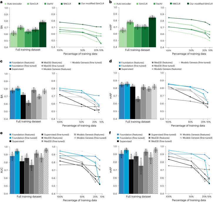
- Modified SimCLR pretraining outperformed auto-encoder, SimCLR, SwAV, and NNCLR on lesion anatomical site classification, achieving balanced accuracy 0.779 (95% CI 0.750–0.810) and mAP 0.847 (95% CI 0.750–0.810); SimCLR was second best with balanced accuracy 0.696 (95% CI 0.663–0.728) and mAP 0.779 (95% CI 0.749–0.811).
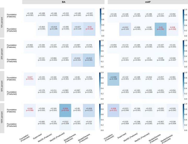
- For lesion anatomical site classification (use case 1), Foundation (fine-tuned) reached mAP 0.857 (95% CI 0.828–0.886) and balanced accuracy 0.804 (95% CI 0.775–0.835), significantly outperforming baselines on mAP and most baselines on balanced accuracy; Foundation (features) was especially robust under reduced training data.
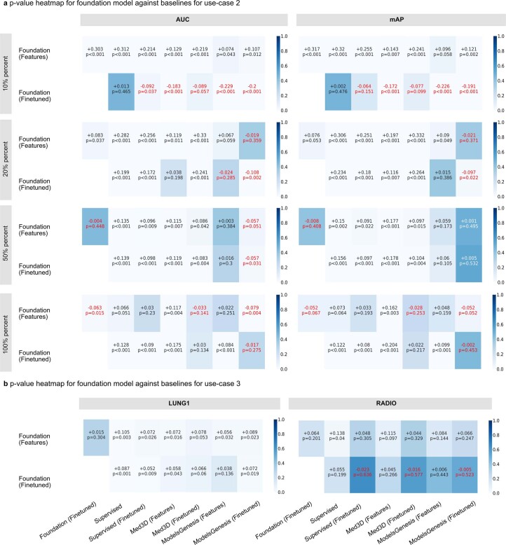
- For lung nodule malignancy (use case 2), Foundation (fine-tuned) achieved AUC 0.944 (95% CI 0.907–0.972) and mAP 0.953 (95% CI 0.915–0.979); Foundation (features) dominated baselines at 50%, 20%, and 10% training data (n = 254, 101, and 51).
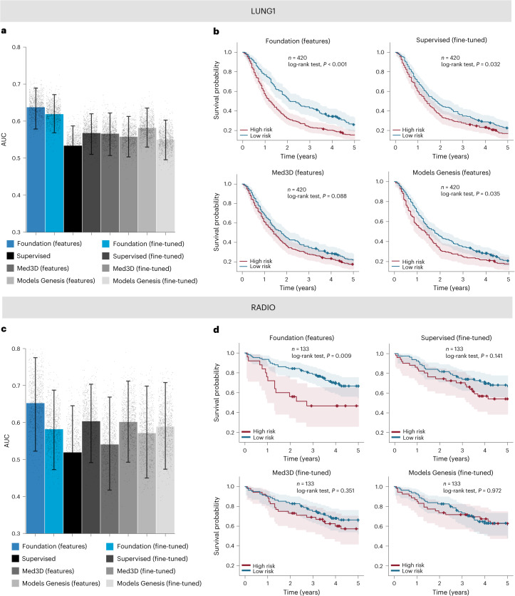
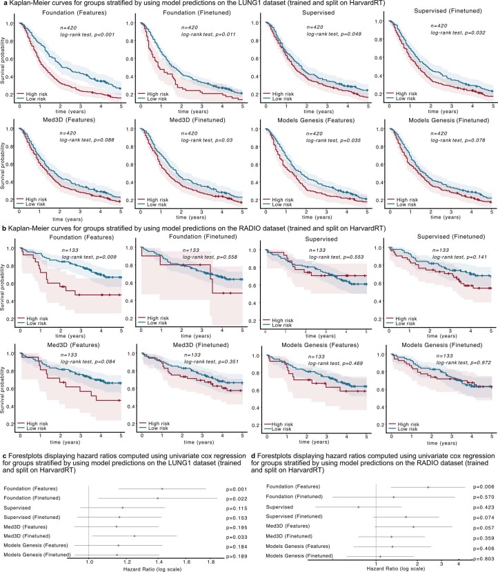
- For NSCLC prognostication (use case 3), Foundation (features) achieved AUC 0.638 (95% CI 0.584–0.692) on LUNG1 and AUC 0.653 (95% CI 0.532–0.771) on RADIO; it was the only model with significant Kaplan–Meier stratification on RADIO (P = 0.009), had test–retest ICC 0.984 versus 0.966 for Supervised (fine-tuned), and showed significantly higher absolute gene-expression correlations (P = 0.008) with immune-associated pathway enrichment.
Methods
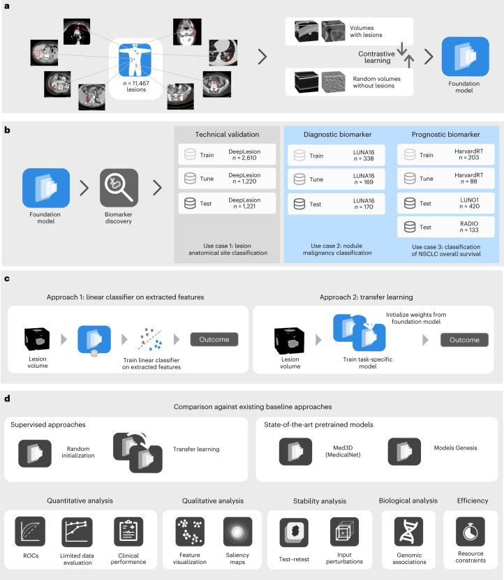
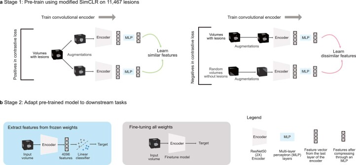
- Design: self-supervised contrastive pretraining of a 3D ResNet50 encoder using a modified SimCLR framework on 11,467 unlabelled CT lesions from 5,513 scans across 2,312 patients; downstream tasks used either frozen features with a logistic regression classifier or full fine-tuning.
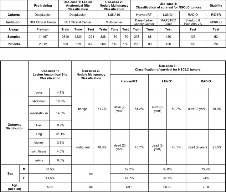
- Data: five datasets—DeepLesion, LUNA16, HarvardRT, LUNG1, and RADIO—covering three use cases: anatomical site classification (3,830 train/tune lesions; 1,221 test), nodule malignancy (338 train, 169 tune, 170 test nodules), and NSCLC 2-year overall survival (HarvardRT n = 291 train/tune; LUNG1 n = 420 and RADIO n = 133 test).
- Analysis: balanced accuracy and mAP for anatomical site, AUC-ROC for malignancy and prognosis, permutation tests with 1,000 permutations, bootstrap 95% CIs with 1,000 resamples, Kaplan–Meier/log-rank tests, stability analyses (ICC, MSE, perturbation trials), saliency maps, and gene-expression correlation plus gene-set enrichment analysis.
Limitations
- Stated by the paper: the study is retrospective; reliability across diverse demographic groups, distribution shifts, and biomarker tasks remains to be evaluated; larger pretraining data and integration of clinical covariates may be needed; deep-learning models remain “black box,” and saliency attribution methods have technical limitations; the biological association analysis is preliminary.
- Evident from the text: Models Genesis was excluded from use case 2 because it was pretrained on the same LUNA16 split, making comparison unfair due to overfitting; RADIO had a small sample size, so most model differences were not significant; reduced-training-data experiments were not performed for use case 3 because that use case already has inherently small sample sizes.
Model-written summary. Check it against the paper before relying on it.
Integrity signals
Registry
Cites
Statistics
Figures
Verification
Claims the paper makes
50 Own result · 16 Not checked yet · 16 Own interpretation · 6 No basis given · 4 Own method
A result the paper reports from its own data — it cites no other source for it, so there is none to check. The quote shows it is in the paper.
In the paper
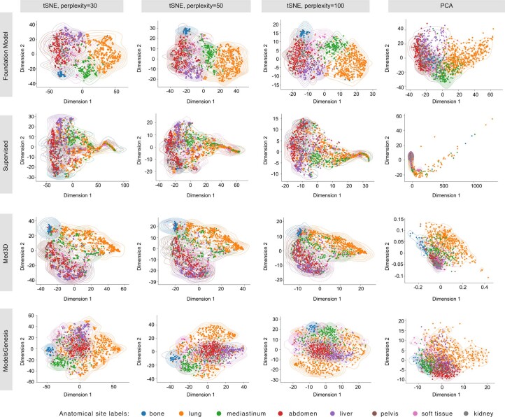
“We observed that features from the pretrained foundation model provided consistently interpretable and well-separated clusters across different settings.”
§ Foundation model for cancer imaging biomarkers > Results > Lesion anatomical site classification (use case 1)
plain- Asserted: passes — The authors put it forward as true.
- Checkable: passes — It can be checked against evidence.
- Load-bearing: passes — The paper’s conclusions depend on it.
- Based: passes — The paper gives a reason why it holds.
- Rests on an assumption: Access to large amounts of annotated data for specialized applications is often challenging and demands expertise, time and labour.
- Rests on an assumption: The quantity and quality of annotated data are strongly linked to the robustness of deep-learning models.
A result the paper reports from its own data — it cites no other source for it, so there is none to check. The quote shows it is in the paper.
In the paper
“Notably, none of the deep-learning fine-tuned baselines significantly improve over linear classification.”
§ Foundation model for cancer imaging biomarkers > Results > Nodule malignancy prediction (use case 2)
plain- Asserted: passes — The authors put it forward as true.
- Checkable: passes — It can be checked against evidence.
- Load-bearing: unclear — It is unclear whether the paper’s conclusions depend on it.
- Based: passes — The paper gives a reason why it holds.
- Rests on an assumption: Access to large amounts of annotated data for specialized applications is often challenging and demands expertise, time and labour.
- Rests on an assumption: The quantity and quality of annotated data are strongly linked to the robustness of deep-learning models.
A result the paper reports from its own data — it cites no other source for it, so there is none to check. The quote shows it is in the paper.
In the paper
“Moreover, when considering only mAP, the simple linear classification significantly (P < 0.05) outperformed all other implementations.”
§ Foundation model for cancer imaging biomarkers > Results > Lesion anatomical site classification (use case 1)
plaindirect- Asserted: passes — The authors put it forward as true.
- Checkable: passes — It can be checked against evidence.
- Load-bearing: passes — The paper’s conclusions depend on it.
- Based: passes — The paper gives a reason why it holds.
- Rests on an assumption: Access to large amounts of annotated data for specialized applications is often challenging and demands expertise, time and labour.
- Rests on an assumption: The quantity and quality of annotated data are strongly linked to the robustness of deep-learning models.
A result the paper reports from its own data — it cites no other source for it, so there is none to check. The quote shows it is in the paper.
In the paper
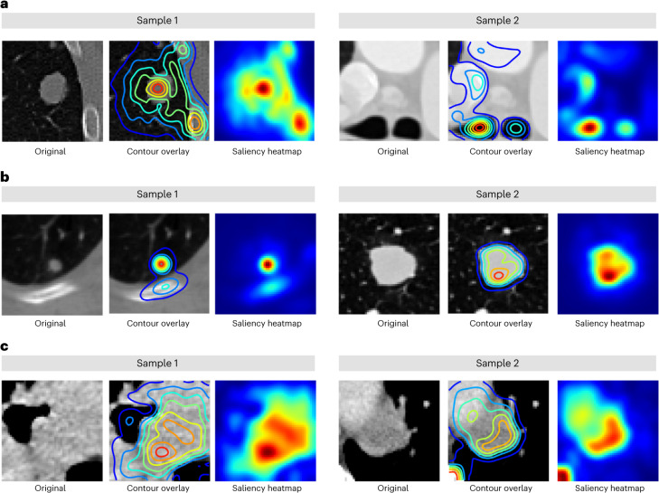
“Specifically, in use case 1 (Fig. 5a), the focus was mainly on areas surrounding the lesions, such as the parenchyma and bone regions in the lung and the trachea in mediastinal lesions.”
§ Foundation model for cancer imaging biomarkers > Results > Saliency maps for fine-tuned foundation models
plain- Asserted: passes — The authors put it forward as true.
- Checkable: passes — It can be checked against evidence.
- Load-bearing: unclear — It is unclear whether the paper’s conclusions depend on it.
- Based: passes — The paper gives a reason why it holds.
A result the paper reports from its own data — it cites no other source for it, so there is none to check. The quote shows it is in the paper.
In the paper
“Similar to the LUNG1 cohort, deep-learning implementations did not demonstrate superior performance (Fig. 3c).”
§ Foundation model for cancer imaging biomarkers > Results > NSCLC prognostication (use case 3)
plain- Asserted: passes — The authors put it forward as true.
- Checkable: passes — It can be checked against evidence.
- Load-bearing: passes — The paper’s conclusions depend on it.
- Based: passes — The paper gives a reason why it holds.
- Rests on an assumption: Access to large amounts of annotated data for specialized applications is often challenging and demands expertise, time and labour.
- Rests on an assumption: The quantity and quality of annotated data are strongly linked to the robustness of deep-learning models.
- Rests on an assumption: Image-biomarker studies for predicting endpoints such as overall survival on small cohorts largely rely on statistical feature extraction (engineered radiomics) and classical machine learning-based modelling.
- Rests on an assumption: These statistical feature extraction approaches require precise 3D segmentations, increasing the annotation burden of these studies.
A result the paper reports from its own data — it cites no other source for it, so there is none to check. The quote shows it is in the paper.
In the paper
“The auto-encoder approach, previously popular for pretraining, performed the worst compared to state-of-the-art contrastive SSL approaches.”
§ Foundation model for cancer imaging biomarkers > Results > Pretraining strategy selection
plain- Asserted: passes — The authors put it forward as true.
- Checkable: passes — It can be checked against evidence.
- Load-bearing: passes — The paper’s conclusions depend on it.
- Based: passes — The paper gives a reason why it holds.
- Rests on an assumption: Following pretraining, foundation models can be applied to task-specific problems, improving generalization, especially in tasks with small datasets.
A result the paper reports from its own data — it cites no other source for it, so there is none to check. The quote shows it is in the paper.
In the paper
“Regarding interpretability, we observed that models focused on varying regions of the tumour and surrounding tissue relevant to the associated use case.”
§ Foundation model for cancer imaging biomarkers > Discussion
plain- Asserted: passes — The authors put it forward as true.
- Checkable: passes — It can be checked against evidence.
- Load-bearing: unclear — It is unclear whether the paper’s conclusions depend on it.
- Based: passes — The paper gives a reason why it holds.
A result the paper reports from its own data — it cites no other source for it, so there is none to check. The quote shows it is in the paper.
In the paper
“Our analysis revealed that for each use case, the focus was primarily around tissues within or in proximity to the tumour”
§ Foundation model for cancer imaging biomarkers > Results > Saliency maps for fine-tuned foundation models
plain- Asserted: passes — The authors put it forward as true.
- Checkable: passes — It can be checked against evidence.
- Load-bearing: passes — The paper’s conclusions depend on it.
- Based: passes — The paper gives a reason why it holds.
None of the sources cited for this claim have been checked yet.
In the paper
“Features extracted from the foundation model, Foundation (features), when linearly classified, showed significantly improved performance in balanced accuracy and mAP over features extracted from Med3D (ref. 17) and Models Genesis18 baseline methods.”
§ Foundation model for cancer imaging biomarkers > Results > Lesion anatomical site classification (use case 1)
What the sources say
- Not checked yetcites [17] Chen, S., Ma, K. & Zheng, Y. Med3D: transfer learning for 3D medical image analysis. Preprint at https://arxiv.org/abs/1904.00625 (2019).
- Not checked yetcites [18] Zhou, Z. et al. Models Genesis: generic autodidactic models for 3D medical image analysis. Med. Image Comput. Comput. Assist. Interv. 11767, 384–393 (2019).
plaindirect- Asserted: passes — The authors put it forward as true.
- Checkable: passes — It can be checked against evidence.
- Load-bearing: passes — The paper’s conclusions depend on it.
- Based: passes — The paper gives a reason why it holds.
- Rests on an assumption: Access to large amounts of annotated data for specialized applications is often challenging and demands expertise, time and labour.
- Rests on an assumption: The quantity and quality of annotated data are strongly linked to the robustness of deep-learning models.
A result the paper reports from its own data — it cites no other source for it, so there is none to check. The quote shows it is in the paper.
In the paper
“Additionally, features and predictions from the foundation model features were found to be highly stable against inter-reader and test–retest variations.”
§ Foundation model for cancer imaging biomarkers > Discussion
plain- Asserted: passes — The authors put it forward as true.
- Checkable: passes — It can be checked against evidence.
- Load-bearing: passes — The paper’s conclusions depend on it.
- Based: passes — The paper gives a reason why it holds.
- It may state two separate claims as one. 25
- Probably not a substantial claim. 18
- The quote only partly states this claim. 2
A result the paper reports from its own data — it cites no other source for it, so there is none to check. The quote shows it is in the paper.
In the paper
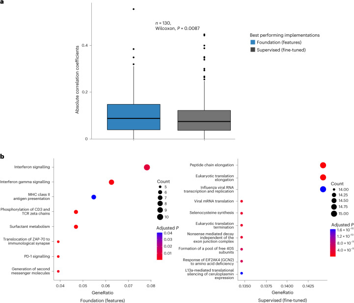
“Conversely, while the supervised model did show enrichment of individual pathways, no identifiable pattern was observed (Fig. 6b).”
§ Foundation model for cancer imaging biomarkers > Results > Underlying biological basis of the foundation model
plain- Asserted: passes — The authors put it forward as true.
- Checkable: passes — It can be checked against evidence.
- Load-bearing: unclear — It is unclear whether the paper’s conclusions depend on it.
- Based: passes — The paper gives a reason why it holds.
It may state two separate claims as one.
A result the paper reports from its own data — it cites no other source for it, so there is none to check. The quote shows it is in the paper.
In the paper
“Use case 3 (Fig. 5c) primarily attributed areas surrounding the centre of mass of the tumour, with some contribution from high-density bone regions.”
§ Foundation model for cancer imaging biomarkers > Results > Saliency maps for fine-tuned foundation models
plaincorrelational- Asserted: passes — The authors put it forward as true.
- Checkable: passes — It can be checked against evidence.
- Load-bearing: unclear — It is unclear whether the paper’s conclusions depend on it.
- Based: passes — The paper gives a reason why it holds.
It may state two separate claims as one.
A result the paper reports from its own data — it cites no other source for it, so there is none to check. The quote shows it is in the paper.
In the paper
“The implementation Med3D (fine-tuned), with AUC = 0.917 (95% CI 0.871–0.957) and mAP = 0.9307 (95% CI 0.888–0.964), performs slightly worse than our model, but this is not significant (P = 0.134).”
§ Foundation model for cancer imaging biomarkers > Results > Nodule malignancy prediction (use case 2)
plain- Asserted: passes — The authors put it forward as true.
- Checkable: passes — It can be checked against evidence.
- Load-bearing: passes — The paper’s conclusions depend on it.
- Based: passes — The paper gives a reason why it holds.
It may state two separate claims as one.
- Rests on an assumption: Access to large amounts of annotated data for specialized applications is often challenging and demands expertise, time and labour.
- Rests on an assumption: The quantity and quality of annotated data are strongly linked to the robustness of deep-learning models.
A result the paper reports from its own data — it cites no other source for it, so there is none to check. The quote shows it is in the paper.
In the paper
“Using these features provided the best performance on small cohorts in predicting prognosis and also demonstrated significant stratification of patients by their associated risk for each of the LUNG1 and RADIO cohorts (P < 0.01).”
§ Foundation model for cancer imaging biomarkers > Discussion
boostedcorrelational- Asserted: passes — The authors put it forward as true.
- Checkable: passes — It can be checked against evidence.
- Load-bearing: passes — The paper’s conclusions depend on it.
- Based: passes — The paper gives a reason why it holds.
It may state two separate claims as one.
- Rests on an assumption: Access to large amounts of annotated data for specialized applications is often challenging and demands expertise, time and labour.
- Rests on an assumption: The quantity and quality of annotated data are strongly linked to the robustness of deep-learning models.
- Rests on an assumption: Image-biomarker studies for predicting endpoints such as overall survival on small cohorts largely rely on statistical feature extraction (engineered radiomics) and classical machine learning-based modelling.
- Rests on an assumption: These statistical feature extraction approaches require precise 3D segmentations, increasing the annotation burden of these studies.
A result the paper reports from its own data — it cites no other source for it, so there is none to check. The quote shows it is in the paper.
In the paper
“With a balanced accuracy of 0.804 (95% CI 0.775–0.835), a significant (P < 0.01) improvement in balanced accuracy was also observed in comparison to all baselines except Med3D (fine-tuned), where the improvement was borderline (P = 0.059).”
§ Foundation model for cancer imaging biomarkers > Results > Lesion anatomical site classification (use case 1)
plaindirect- Asserted: passes — The authors put it forward as true.
- Checkable: passes — It can be checked against evidence.
- Load-bearing: passes — The paper’s conclusions depend on it.
- Based: passes — The paper gives a reason why it holds.
It may state two separate claims as one.
- Rests on an assumption: Access to large amounts of annotated data for specialized applications is often challenging and demands expertise, time and labour.
- Rests on an assumption: The quantity and quality of annotated data are strongly linked to the robustness of deep-learning models.
A result the paper reports from its own data — it cites no other source for it, so there is none to check. The quote shows it is in the paper.
In the paper
“achieving a balanced accuracy of 0.779 (95% confidence interval (CI) 0.750–0.810) and mAP = 0.847 (95% CI 0.750–0.810).”
§ Foundation model for cancer imaging biomarkers > Results > Pretraining strategy selection
plain- Asserted: passes — The authors put it forward as true.
- Checkable: passes — It can be checked against evidence.
- Load-bearing: passes — The paper’s conclusions depend on it.
- Based: passes — The paper gives a reason why it holds.
It may state two separate claims as one.
- Rests on an assumption: Following pretraining, foundation models can be applied to task-specific problems, improving generalization, especially in tasks with small datasets.
A result the paper reports from its own data — it cites no other source for it, so there is none to check. The quote shows it is in the paper.
In the paper
“All comparisons were significant (P < 0.05) except for Med3D (fine-tuned), where borderline significance was observed (P = 0.053).”
§ Foundation model for cancer imaging biomarkers > Results > NSCLC prognostication (use case 3)
plainn = 420 ·- Asserted: passes — The authors put it forward as true.
- Checkable: passes — It can be checked against evidence.
- Load-bearing: passes — The paper’s conclusions depend on it.
- Based: passes — The paper gives a reason why it holds.
It may state two separate claims as one.
- Rests on an assumption: Access to large amounts of annotated data for specialized applications is often challenging and demands expertise, time and labour.
- Rests on an assumption: The quantity and quality of annotated data are strongly linked to the robustness of deep-learning models.
- Rests on an assumption: Image-biomarker studies for predicting endpoints such as overall survival on small cohorts largely rely on statistical feature extraction (engineered radiomics) and classical machine learning-based modelling.
- Rests on an assumption: These statistical feature extraction approaches require precise 3D segmentations, increasing the annotation burden of these studies.
A result the paper reports from its own data — it cites no other source for it, so there is none to check. The quote shows it is in the paper.
In the paper
“Due to the small sample size, none of the models showed significant differences from the rest (P > 0.05) except for the Foundation (features) improving over the Supervised model, which had near-random performance (AUC = 0.520).”
§ Foundation model for cancer imaging biomarkers > Results > NSCLC prognostication (use case 3)
plain- Asserted: passes — The authors put it forward as true.
- Checkable: passes — It can be checked against evidence.
- Load-bearing: passes — The paper’s conclusions depend on it.
- Based: passes — The paper gives a reason why it holds.
It may state two separate claims as one.
- Rests on an assumption: Access to large amounts of annotated data for specialized applications is often challenging and demands expertise, time and labour.
- Rests on an assumption: The quantity and quality of annotated data are strongly linked to the robustness of deep-learning models.
- Rests on an assumption: Image-biomarker studies for predicting endpoints such as overall survival on small cohorts largely rely on statistical feature extraction (engineered radiomics) and classical machine learning-based modelling.
- Rests on an assumption: These statistical feature extraction approaches require precise 3D segmentations, increasing the annotation burden of these studies.
A result the paper reports from its own data — it cites no other source for it, so there is none to check. The quote shows it is in the paper.
In the paper
“The approach of fine-tuning the foundation model, Foundation (fine-tuned), with an area under the curve (AUC) = 0.944 (95% CI 0.907–0.972) and mAP = 0.953 (95% CI 0.915–0.979) resulted in significant (P < 0.01) superiority over most of the baseline implementations (Fig. 2e,f).”
§ Foundation model for cancer imaging biomarkers > Results > Nodule malignancy prediction (use case 2)
plaindirect- Asserted: passes — The authors put it forward as true.
- Checkable: passes — It can be checked against evidence.
- Load-bearing: passes — The paper’s conclusions depend on it.
- Based: passes — The paper gives a reason why it holds.
It may state two separate claims as one.
- Rests on an assumption: Access to large amounts of annotated data for specialized applications is often challenging and demands expertise, time and labour.
- Rests on an assumption: The quantity and quality of annotated data are strongly linked to the robustness of deep-learning models.
A result the paper reports from its own data — it cites no other source for it, so there is none to check. The quote shows it is in the paper.
In the paper
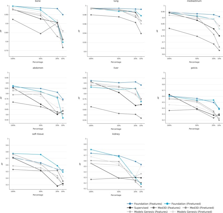
“More importantly, it remained robust as evidenced by the smallest decline in balanced accuracy and mAP of 9 and 12%, respectively, when reducing training data from 100 to 10%.”
§ Foundation model for cancer imaging biomarkers > Results > Pretraining strategy selection
boosteddirect- Asserted: passes — The authors put it forward as true.
- Checkable: passes — It can be checked against evidence.
- Load-bearing: passes — The paper’s conclusions depend on it.
- Based: passes — The paper gives a reason why it holds.
It may state two separate claims as one.
- Rests on an assumption: Following pretraining, foundation models can be applied to task-specific problems, improving generalization, especially in tasks with small datasets.
A result the paper reports from its own data — it cites no other source for it, so there is none to check. The quote shows it is in the paper.
In the paper
“Foundation (fine-tuned) showed a larger drop in performance and failed to improve significantly over baseline implementations as training data were decreased (losing significance from 20% onward).”
§ Foundation model for cancer imaging biomarkers > Results > Lesion anatomical site classification (use case 1)
plaindirectn = 1,010- Asserted: passes — The authors put it forward as true.
- Checkable: passes — It can be checked against evidence.
- Load-bearing: passes — The paper’s conclusions depend on it.
- Based: passes — The paper gives a reason why it holds.
It may state two separate claims as one.
- Rests on an assumption: Access to large amounts of annotated data for specialized applications is often challenging and demands expertise, time and labour.
- Rests on an assumption: The quantity and quality of annotated data are strongly linked to the robustness of deep-learning models.
The paper states this without a source or data behind it here, so there is nothing to check it against. The quote shows it is in the paper.
In the paper
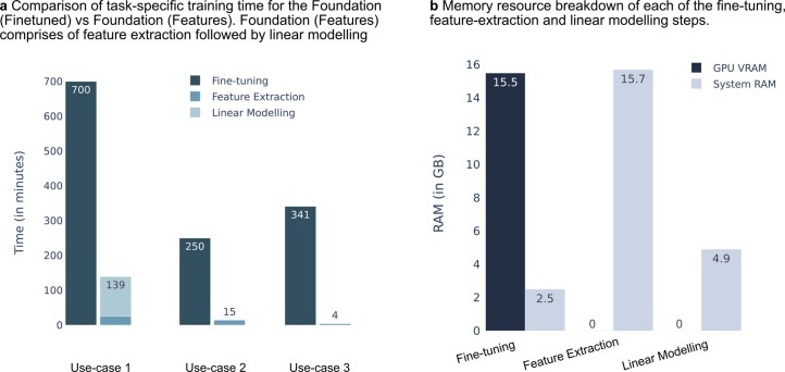
“Feature-based implementations were also computationally efficient when considering both time and memory.”
§ Foundation model for cancer imaging biomarkers > Discussion
plain- Asserted: passes — The authors put it forward as true.
- Checkable: passes — It can be checked against evidence.
- Load-bearing: fails — The paper’s conclusions may not depend on it.
- Based: passes — The paper gives a reason why it holds.
Probably not a substantial claim. The paper’s conclusions may not depend on it.
- Rests on an assumption: Access to large amounts of annotated data for specialized applications is often challenging and demands expertise, time and labour.
- Rests on an assumption: The quantity and quality of annotated data are strongly linked to the robustness of deep-learning models.
A result the paper reports from its own data — it cites no other source for it, so there is none to check. The quote shows it is in the paper.
In the paper
“For the RADIO cohort, Foundation (features) shows the best performance with an AUC of 0.653 (95% CI 0.532–0.771).”
§ Foundation model for cancer imaging biomarkers > Results > NSCLC prognostication (use case 3)
plaincorrelational- Asserted: passes — The authors put it forward as true.
- Checkable: passes — It can be checked against evidence.
- Load-bearing: passes — The paper’s conclusions depend on it.
- Based: passes — The paper gives a reason why it holds.
The quote only partly states this claim.
- Rests on an assumption: Access to large amounts of annotated data for specialized applications is often challenging and demands expertise, time and labour.
- Rests on an assumption: The quantity and quality of annotated data are strongly linked to the robustness of deep-learning models.
- Rests on an assumption: Image-biomarker studies for predicting endpoints such as overall survival on small cohorts largely rely on statistical feature extraction (engineered radiomics) and classical machine learning-based modelling.
- Rests on an assumption: These statistical feature extraction approaches require precise 3D segmentations, increasing the annotation burden of these studies.
A result the paper reports from its own data — it cites no other source for it, so there is none to check. The quote shows it is in the paper.
In the paper
“We found that absolute correlation coefficients between gene expression profiles and model predictions were significantly higher (P = 0.008) for the foundation model, indicating a stronger association with underlying tumour biology (Fig. 6a).”
§ Foundation model for cancer imaging biomarkers > Results > Underlying biological basis of the foundation model
hedgedcorrelationaln = 130 ·- Asserted: passes — The authors put it forward as true.
- Checkable: passes — It can be checked against evidence.
- Load-bearing: passes — The paper’s conclusions depend on it.
- Based: passes — The paper gives a reason why it holds.
It may state two separate claims as one.
A result the paper reports from its own data — it cites no other source for it, so there is none to check. The quote shows it is in the paper.
In the paper
“Modelling using features extracted from the foundation model was the most robust across these use cases when subjected to drops in training data, offering stable performance even when data sizes were considerably reduced, for example, using only 51 samples in use case 2.”
§ Foundation model for cancer imaging biomarkers > Discussion
plaincorrelationaln = 51- Asserted: passes — The authors put it forward as true.
- Checkable: passes — It can be checked against evidence.
- Load-bearing: passes — The paper’s conclusions depend on it.
- Based: passes — The paper gives a reason why it holds.
It may state two separate claims as one.
- Rests on an assumption: Access to large amounts of annotated data for specialized applications is often challenging and demands expertise, time and labour.
- Rests on an assumption: The quantity and quality of annotated data are strongly linked to the robustness of deep-learning models.
A result the paper reports from its own data — it cites no other source for it, so there is none to check. The quote shows it is in the paper.
In the paper
“When limited data (50, 20 and 10%) was used for downstream task training, our method demonstrated consistently improved performance.”
§ Foundation model for cancer imaging biomarkers > Results > Pretraining strategy selection
boosteddirect- Asserted: passes — The authors put it forward as true.
- Checkable: passes — It can be checked against evidence.
- Load-bearing: passes — The paper’s conclusions depend on it.
- Based: passes — The paper gives a reason why it holds.
The quote only partly states this claim.
- Rests on an assumption: Following pretraining, foundation models can be applied to task-specific problems, improving generalization, especially in tasks with small datasets.
A result the paper reports from its own data — it cites no other source for it, so there is none to check. The quote shows it is in the paper.
In the paper
“As expected, the second best-performing approach was SimCLR (balanced accuracy 0.696 (95% CI 0.663–0.728); mAP = 0.779 (95% CI 0.749–0.811)).”
§ Foundation model for cancer imaging biomarkers > Results > Pretraining strategy selection
plain- Asserted: passes — The authors put it forward as true.
- Checkable: passes — It can be checked against evidence.
- Load-bearing: unclear — It is unclear whether the paper’s conclusions depend on it.
- Based: passes — The paper gives a reason why it holds.
It may state two separate claims as one.
- Rests on an assumption: Following pretraining, foundation models can be applied to task-specific problems, improving generalization, especially in tasks with small datasets.
A result the paper reports from its own data — it cites no other source for it, so there is none to check. The quote shows it is in the paper.
In the paper
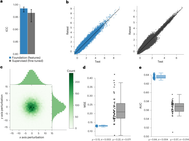
“We found that predictions from the overall best-performing models on LUNG1 and RADIO: Foundation (features) and Supervised (fine-tuned) had high stability with intraclass correlation coefficient (ICC) values of 0.984 and 0.966, respectively.”
§ Foundation model for cancer imaging biomarkers > Results > Stability of the foundation model
plainn = 26- Asserted: passes — The authors put it forward as true.
- Checkable: passes — It can be checked against evidence.
- Load-bearing: passes — The paper’s conclusions depend on it.
- Based: passes — The paper gives a reason why it holds.
It may state two separate claims as one.
A result the paper reports from its own data — it cites no other source for it, so there is none to check. The quote shows it is in the paper.
In the paper
“Foundation (fine-tuned) shows superior performance over all baselines at 50% but shows large drops in performance from a 20% reduction onward.”
§ Foundation model for cancer imaging biomarkers > Results > Nodule malignancy prediction (use case 2)
plaincorrelationaln = 254- Asserted: passes — The authors put it forward as true.
- Checkable: passes — It can be checked against evidence.
- Load-bearing: passes — The paper’s conclusions depend on it.
- Based: passes — The paper gives a reason why it holds.
It may state two separate claims as one.
- Rests on an assumption: Access to large amounts of annotated data for specialized applications is often challenging and demands expertise, time and labour.
- Rests on an assumption: The quantity and quality of annotated data are strongly linked to the robustness of deep-learning models.
The paper states this without a source or data behind it here, so there is nothing to check it against. The quote shows it is in the paper.
In the paper
“Furthermore, it surpasses current deep-learning techniques in various tasks while fitting conveniently into existing radiomic research methods.”
§ Foundation model for cancer imaging biomarkers > Discussion
plaindirect- Asserted: passes — The authors put it forward as true.
- Checkable: passes — It can be checked against evidence.
- Load-bearing: passes — The paper’s conclusions depend on it.
- Based: fails — The paper may give no reason why it holds.
Probably not a substantial claim. The paper may give no reason why it holds. It may state two separate claims as one.
The paper states this without a source or data behind it here, so there is nothing to check it against. The quote shows it is in the paper.
In the paper
“However, the original value of 0.1 proposed by the original paper was the most effective.”
§ Foundation model for cancer imaging biomarkers > Methods > Task-agnostic pretraining of the foundation model
plaindirect- Asserted: passes — The authors put it forward as true.
- Checkable: passes — It can be checked against evidence.
- Load-bearing: fails — The paper’s conclusions may not depend on it.
- Based: fails — The paper may give no reason why it holds.
Probably not a substantial claim. The paper’s conclusions may not depend on it. The paper may give no reason why it holds.
What the paper did — it cites no source for this, so there is none to check. The quote shows it is in the paper.
In the paper
“The baseline Models Genesis implementation was excluded in this analysis as this model was pretrained on the same dataset and, therefore, does not indicate a fair comparison.”
§ Foundation model for cancer imaging biomarkers > Results > Nodule malignancy prediction (use case 2)
plaindirect- Asserted: passes — The authors put it forward as true.
- Checkable: passes — It can be checked against evidence.
- Load-bearing: passes — The paper’s conclusions depend on it.
- Based: passes — The paper gives a reason why it holds.
Probably not a substantial claim. It may state two separate claims as one.
A conclusion the paper draws from its own results — it cites no other source for it, so there is none to check. The quote shows it is in the paper.
In the paper
“By contrast, our approach is generalizable to multiple use cases, and for nodule malignancy, we obtain high performance using significantly lesser training data, only 338 nodules (due to our more stringent exclusion criteria).”
§ Foundation model for cancer imaging biomarkers > Discussion
plaindirect- Asserted: passes — The authors put it forward as true.
- Checkable: passes — It can be checked against evidence.
- Load-bearing: passes — The paper’s conclusions depend on it.
- Based: passes — The paper gives a reason why it holds.
It may state two separate claims as one.
- Rests on an assumption: Access to large amounts of annotated data for specialized applications is often challenging and demands expertise, time and labour.
- Rests on an assumption: The quantity and quality of annotated data are strongly linked to the robustness of deep-learning models.
In the paper
“which is consistent with research demonstrating the tumour microenvironment’s influence on cancer development22 and prognosis.”
§ Foundation model for cancer imaging biomarkers > Results > Saliency maps for fine-tuned foundation models
What the sources say
- Not checked yetcites [22] Hinshaw, D. C. & Shevde, L. A. The tumor microenvironment innately modulates cancer progression. Cancer Res. 79, 4557–4566 (2019). source
boosteddirect- Asserted: passes — The authors put it forward as true.
- Checkable: passes — It can be checked against evidence.
- Load-bearing: fails — The paper’s conclusions may not depend on it.
- Based: passes — The paper gives a reason why it holds.
Probably not a substantial claim. The paper’s conclusions may not depend on it. It may state two separate claims as one.
A conclusion the paper draws from its own results — it cites no other source for it, so there is none to check. The quote shows it is in the paper.
In the paper
“as expressing saliency maps over 4,096-dimensional outputs remains challenging in practice.”
§ Foundation model for cancer imaging biomarkers > Methods > Feature visualization and saliency maps
plain- Asserted: passes — The authors put it forward as true.
- Checkable: passes — It can be checked against evidence.
- Load-bearing: passes — The paper’s conclusions depend on it.
- Based: fails — The paper may give no reason why it holds.
Probably not a substantial claim. The paper may give no reason why it holds.
A conclusion the paper draws from its own results — it cites no other source for it, so there is none to check. The quote shows it is in the paper.
In the paper
“This approach allows for significantly higher throughput than manual segmentation, which can be more tedious.”
§ Foundation model for cancer imaging biomarkers > Methods > Data preprocessing
plaindirect- Asserted: passes — The authors put it forward as true.
- Checkable: passes — It can be checked against evidence.
- Load-bearing: fails — The paper’s conclusions may not depend on it.
- Based: fails — The paper may give no reason why it holds.
Probably not a substantial claim. The paper’s conclusions may not depend on it. The paper may give no reason why it holds.
A conclusion the paper draws from its own results — it cites no other source for it, so there is none to check. The quote shows it is in the paper.
In the paper
“In conclusion, our foundation model offers a powerful and reliable framework for discovering cancer imaging biomarkers, especially in small datasets.”
§ Foundation model for cancer imaging biomarkers > Discussion
plaindirect- Asserted: passes — The authors put it forward as true.
- Checkable: passes — It can be checked against evidence.
- Load-bearing: passes — The paper’s conclusions depend on it.
- Based: fails — The paper may give no reason why it holds.
Probably not a substantial claim. The paper may give no reason why it holds.
In the paper
“Existing literature12 has suggested several strategies, such as image reconstruction, to pretrain networks to learn these representations.”
§ Foundation model for cancer imaging biomarkers > Main
What the sources say
- Not checked yetcites [12] Balestriero, R. et al. A cookbook of self-supervised learning. Preprint at https://arxiv.org/abs/2304.12210 (2023).
hedgeddirect- Asserted: passes — The authors put it forward as true.
- Checkable: passes — It can be checked against evidence.
- Load-bearing: fails — The paper’s conclusions may not depend on it.
- Based: passes — The paper gives a reason why it holds.
Probably not a substantial claim. The paper’s conclusions may not depend on it.
In the paper
“This point largely depends on the nature and sizes of training data in the downstream task.”
§ Foundation model for cancer imaging biomarkers > Discussion
What the sources say
- Not checked yetcites Cole et al.38 Cole, E., Yang, X., Wilber, K., Aodha, O. M. & Belongie, S. When does contrastive visual representation learning work? In Proc. 2022 IEEE/CVF Conference on Computer Vision and Pattern Recognition (CVPR) 14755–14764 (IEEE, 2022). source
plaincorrelational- Asserted: passes — The authors put it forward as true.
- Checkable: passes — It can be checked against evidence.
- Load-bearing: fails — The paper’s conclusions may not depend on it.
- Based: passes — The paper gives a reason why it holds.
Probably not a substantial claim. The paper’s conclusions may not depend on it.
In the paper
“Deep-learning methods, in comparison, are robust to differences in acquisition and segmentation variability and provide improved performance10.”
§ Foundation model for cancer imaging biomarkers > Discussion
What the sources say
- Not checked yetcites [10] Hosny, A. et al. Deep learning for lung cancer prognostication: a retrospective multi-cohort radiomics study. PLoS Med. 15, e1002711 (2018). source
plaindirect- Asserted: passes — The authors put it forward as true.
- Checkable: passes — It can be checked against evidence.
- Load-bearing: passes — The paper’s conclusions depend on it.
- Based: passes — The paper gives a reason why it holds.
It may state two separate claims as one.
A conclusion the paper draws from its own results — it cites no other source for it, so there is none to check. The quote shows it is in the paper.
In the paper
“as it is typical to have inherently small sample sizes in such use cases when compared to task complexity due to study-specific inclusion criteria.”
§ Foundation model for cancer imaging biomarkers > Methods > Task-specific training of the foundation model
plaindirect- Asserted: passes — The authors put it forward as true.
- Checkable: passes — It can be checked against evidence.
- Load-bearing: passes — The paper’s conclusions depend on it.
- Based: fails — The paper may give no reason why it holds.
Probably not a substantial claim. The paper may give no reason why it holds.
A conclusion the paper draws from its own results — it cites no other source for it, so there is none to check. The quote shows it is in the paper.
In the paper
“Third, a significant challenge with deep-learning models, including ours, is their ‘black box’ nature, which limits interpretability and explainability.”
§ Foundation model for cancer imaging biomarkers > Discussion
plaindirect- Asserted: passes — The authors put it forward as true.
- Checkable: passes — It can be checked against evidence.
- Load-bearing: fails — The paper’s conclusions may not depend on it.
- Based: passes — The paper gives a reason why it holds.
Probably not a substantial claim. The paper’s conclusions may not depend on it.
A conclusion the paper draws from its own results — it cites no other source for it, so there is none to check. The quote shows it is in the paper.
In the paper
“In our study, we pretrained on 11,467 lesion volumes and randomly sampled volumes, from 5,513 unique CT scans, leveraging not only one of the largest lesion-specific datasets but also one of the largest pretraining 3D CT datasets.”
§ Foundation model for cancer imaging biomarkers > Discussion
plain- Asserted: passes — The authors put it forward as true.
- Checkable: passes — It can be checked against evidence.
- Load-bearing: passes — The paper’s conclusions depend on it.
- Based: passes — The paper gives a reason why it holds.
It may state two separate claims as one.
None of the sources cited for this claim have been checked yet.
In the paper
“While many studies investigating imaging-based biomarkers incorporate supervised deep-learning algorithms into their models9–11, they are typically applied in scenarios where large datasets are available for training and testing.”
§ Foundation model for cancer imaging biomarkers > Main
What the sources say
- Not checked yetcites [9] Mahajan, A. et al. Deep learning-based predictive imaging biomarker model for EGFR mutation status in non-small cell lung cancer from CT imaging. J. Clin. Orthod. 38, 3106 (2020).
- Not checked yetcites [10] Hosny, A. et al. Deep learning for lung cancer prognostication: a retrospective multi-cohort radiomics study. PLoS Med. 15, e1002711 (2018). source
- Not checked yetcites [11] Braghetto, A., Marturano, F., Paiusco, M., Baiesi, M. & Bettinelli, A. Radiomics and deep learning methods for the prediction of 2-year overall survival in LUNG1 dataset. Sci. Rep. 12, 14132 (2022). source
plaincorrelational- Asserted: passes — The authors put it forward as true.
- Checkable: passes — It can be checked against evidence.
- Load-bearing: passes — The paper’s conclusions depend on it.
- Based: passes — The paper gives a reason why it holds.
It may state two separate claims as one.
In the paper
“They show a saturation point associated with pretraining dataset size and diminishing returns beyond this point.”
§ Foundation model for cancer imaging biomarkers > Discussion
What the sources say
- Not checked yetcites Cole et al.38 Cole, E., Yang, X., Wilber, K., Aodha, O. M. & Belongie, S. When does contrastive visual representation learning work? In Proc. 2022 IEEE/CVF Conference on Computer Vision and Pattern Recognition (CVPR) 14755–14764 (IEEE, 2022). source
plaincorrelational- Asserted: passes — The authors put it forward as true.
- Checkable: passes — It can be checked against evidence.
- Load-bearing: passes — The paper’s conclusions depend on it.
- Based: passes — The paper gives a reason why it holds.
Probably not a substantial claim.
What the paper did — it cites no source for this, so there is none to check. The quote shows it is in the paper.
In the paper
“In our comparative analysis, we maintained uniformity in batch sizes and dataset parameters across all methods, while optimizer and loss-specific settings were aligned with each method’s original configuration.”
§ Foundation model for cancer imaging biomarkers > Methods > Task-agnostic pretraining of the foundation model
plain- Asserted: passes — The authors put it forward as true.
- Checkable: passes — It can be checked against evidence.
- Load-bearing: fails — The paper’s conclusions may not depend on it.
- Based: passes — The paper gives a reason why it holds.
Probably not a substantial claim. The paper’s conclusions may not depend on it. It may state two separate claims as one.
A conclusion the paper draws from its own results — it cites no other source for it, so there is none to check. The quote shows it is in the paper.
In the paper
“Furthermore, our initial biological association analysis, aimed at explaining the model’s decisions, is preliminary and requires more rigorous investigation for a concrete understanding.”
§ Foundation model for cancer imaging biomarkers > Discussion
plain- Asserted: passes — The authors put it forward as true.
- Checkable: fails — It may be too vague to check against evidence.
- Load-bearing: fails — The paper’s conclusions may not depend on it.
- Based: passes — The paper gives a reason why it holds.
Probably not a substantial claim. It may be too vague to check against evidence. The paper’s conclusions may not depend on it.
A conclusion the paper draws from its own results — it cites no other source for it, so there is none to check. The quote shows it is in the paper.
In the paper
“due to the high proportion of negatives relative to positives, which results in consistently low false-positive rates and might overestimate the AUC.”
§ Foundation model for cancer imaging biomarkers > Methods > Performance analysis
hedgedconditional- Asserted: passes — The authors put it forward as true.
- Checkable: passes — It can be checked against evidence.
- Load-bearing: passes — The paper’s conclusions depend on it.
- Based: passes — The paper gives a reason why it holds.
It may state two separate claims as one.
None of the sources cited for this claim have been checked yet.
In the paper
“Several studies23–25 have demonstrated the efficacy of SSL in medicine where only limited data might be available for training deep-learning networks.”
§ Foundation model for cancer imaging biomarkers > Discussion
What the sources say
- Not checked yetcites [23] Azizi, S. et al. Big self-supervised models advance medical image classification. In 2021 IEEE/CVF International Conference on Computer Vision (ICCV) 3458–3468 (IEEE, 2021). source
- Not checked yetcites [24] Krishnan, R., Rajpurkar, P. & Topol, E. J. Self-supervised learning in medicine and healthcare. Nat. Biomed. Eng. 6, 1346–1352 (2022). source
- Not checked yetcites [25] Ghesu, F. C. et al. Contrastive self-supervised learning from 100 million medical images with optional supervision. J. Med. Imaging 9, 064503 (2022). source
boosteddirect- Asserted: passes — The authors put it forward as true.
- Checkable: passes — It can be checked against evidence.
- Load-bearing: fails — The paper’s conclusions may not depend on it.
- Based: passes — The paper gives a reason why it holds.
Probably not a substantial claim. The paper’s conclusions may not depend on it.
None of the sources cited for this claim have been checked yet.
In the paper
“We used scans from 26 patients from the RIDER dataset19, routinely used for test–retest robustness analysis in tumour imaging19–21.”
§ Foundation model for cancer imaging biomarkers > Results > Stability of the foundation model
What the sources say
- Not checked yetcites [19] Zhao, B. et al. Evaluating variability in tumor measurements from same-day repeat CT scans of patients with non-small cell lung cancer. Radiology 252, 263–272 (2009). source
- Not checked yetcites [20] Aerts, H. J. W. L. et al. Decoding tumour phenotype by noninvasive imaging using a quantitative radiomics approach. Nat. Commun. 5, 4006 (2014). source
- Not checked yetcites [21] Hosny, A. et al. Clinical validation of deep learning algorithms for radiotherapy targeting of non-small-cell lung cancer: an observational study. Lancet Digit. Health 4, e657–e666 (2022). source
plain- Asserted: passes — The authors put it forward as true.
- Checkable: passes — It can be checked against evidence.
- Load-bearing: fails — The paper’s conclusions may not depend on it.
- Based: passes — The paper gives a reason why it holds.
Probably not a substantial claim. The paper’s conclusions may not depend on it.
A conclusion the paper draws from its own results — it cites no other source for it, so there is none to check. The quote shows it is in the paper.
In the paper
“Therefore, experiments involving further data reduction in this case do not provide any valuable insights.”
§ Foundation model for cancer imaging biomarkers > Methods > Task-specific training of the foundation model
plain- Asserted: passes — The authors put it forward as true.
- Checkable: passes — It can be checked against evidence.
- Load-bearing: passes — The paper’s conclusions depend on it.
- Based: passes — The paper gives a reason why it holds.
Probably not a substantial claim.
The paper relies on this without support.
Access to large amounts of annotated data for specialized applications is often challenging and demands expertise, time and labour.
“However, access to large amounts of annotated data for specialized applications is often challenging and demands expertise, time and labour.”
The paper relies on this without support.
There is still limited evidence investigating whether SSL can help train foundation models that learn general, robust and transferrable representations that can act as imaging biomarkers, especially prognostic, for tasks of clinical relevance.
“There is still limited evidence investigating whether SSL can help train foundation models that learn general, robust and transferrable representations that can act as imaging biomarkers, especially prognostic, for tasks of clinical relevance.”
The paper relies on this without support.
The quantity and quality of annotated data are strongly linked to the robustness of deep-learning models.
“The quantity and quality of annotated data are strongly linked to the robustness of deep-learning models.”
The paper relies on this without support.
In scenarios with limited annotated data, many investigators fall back on traditional handcrafted or engineered approaches that limit the scope of discovery.
“In such scenarios, many investigators fall back on traditional handcrafted or engineered approaches based on defined mathematical and statistical algorithms that analyse attributes such as the shape and texture of objects in images, which limit the scope of discovery.”
The paper relies on this without support.
Following pretraining, foundation models can be applied to task-specific problems, improving generalization, especially in tasks with small datasets.
“Following pretraining, foundation models can be applied to task-specific problems, improving generalization, especially in tasks with small datasets.”
The paper relies on this without support.
Image-biomarker studies for predicting endpoints such as overall survival on small cohorts largely rely on statistical feature extraction (engineered radiomics) and classical machine learning-based modelling.
“Image-biomarker studies for predicting endpoints, such as overall survival on small cohorts, largely rely on statistical feature extraction (engineered radiomics) and classical machine learning-based modelling.”
The paper relies on this without support.
These statistical feature extraction approaches require precise 3D segmentations, increasing the annotation burden of these studies.
“These require precise 3D segmentations for feature extraction, increasing the annotation burden of these studies.”
Claims other papers make about this one
Each is checked against this paper’s own text: does it say what the citing paper says it does?
The cited source says what the claim attributes to it.
In the citing paper
“Built on rich knowledge representations, these models have shown impressive performance across medical fields such as ophthalmology20, radiology21 and pathology22–25.”
From A Multimodal Vision Foundation Model for Clinical Dermatology (2025) · cites this paper as [21]
In the citing paper
“In oncology, transformer-based methods support clinical text processing, whole-slide and radiologic image interpretation (vision transformers), and genomic sequence modeling [24,25,26,27].”
From Artificial Intelligence in Clinical Oncology: From Productivity Enhancement to Creative Discovery (2025) · cites this paper as [24]
In the citing paper
“Specialty-specific foundation models20,21,23 typically focus on single imaging modalities”
From A Multimodal Vision Foundation Model for Clinical Dermatology (2025) · cites this paper as [21]
Self-check
Percentages, sums and reported differences re-computed from the paper’s own numbers.
Each contradiction between two passages listed here was confirmed by an independent check.
Rated minor: it would not touch a result — a value no result rests on, a p-value that stays on the same side of the significance level, or a conflict that is clerical, resolved elsewhere in the paper, or outside its own findings.
In the paper
§ Foundation model for cancer imaging biomarkers > Results > Stability of the foundation model
“Each AUC and MSE measure in the box plots (d,e) have been computed on a dataset with n = 422 samples”
§ Foundation model for cancer imaging biomarkers > Methods > Study population
“For our study, we selected CT scans of 420 patients (right-censored for 2-year survival) with annotated primary gross tumour volumes and used these as an independent test set for prognostication networks.”
Rated minor: it would not touch a result — a value no result rests on, a p-value that stays on the same side of the significance level, or a conflict that is clerical, resolved elsewhere in the paper, or outside its own findings.
In the paper
§ Foundation model for cancer imaging biomarkers > Results > Pretraining strategy selection
“mAP = 0.847”
§ Foundation model for cancer imaging biomarkers > Results > Pretraining strategy selection
“95% CI 0.750–0.810”
Citations
Cites
Deep Residual Learning for Image Recognition
He, K., Zhang, X., Ren, S., & Sun, J. (2016) · 2016 IEEE Conference on Computer Vision and Pattern Recognition (CVPR) · doi:10.1109/cvpr.2016.90 · Cited by 235,474
Learning Transferable Visual Models From Natural Language Supervision
Radford, A., Kim, J. W., Hallacy, C., Ramesh, A., Goh, G., Agarwal, S., Sastry, G., Askell, A., Mishkin, P., Clark, J., et al. (2021) · arXiv · doi:10.48550/arxiv.2103.00020 · Cited by 15,293
Larian Studios Dev Team (2024) · doi:10.1145/3292500.3330701 · Cited by 8,618
Decoding tumour phenotype by noninvasive imaging using a quantitative radiomics approach
Aerts, H. J. W. L., Velazquez, E. R., Leijenaar, R. T. H., Parmar, C., Grossmann, P., Carvalho, S., Bussink, J., Monshouwer, R., Haibe-Kains, B., Rietveld, D., et al. (2014) · Nature Communications · doi:10.1038/ncomms5006 · Cited by 5,132
Self-citationTraining language models to follow instructions with human feedback
Ouyang, L., Wu, J., Jiang, X., Almeida, D., Wainwright, C. L., Mishkin, P., Zhang, C., Agarwal, S., Slama, K., Ray, A., et al. (2022) · arXiv · doi:10.48550/arxiv.2203.02155 · Cited by 4,208
The Tumor Microenvironment Innately Modulates Cancer Progression
Hinshaw, D. C., & Shevde, L. A. (2019) · Cancer Research · doi:10.1158/0008-5472.can-18-3962 · Cited by 3,270
On the Opportunities and Risks of Foundation Models
Bommasani, R., Hudson, D. A., Adeli, E., Altman, R., Arora, S., von Arx, S., Bernstein, M. S., Bohg, J., Bosselut, A., Brunskill, E., et al. (2021) · arXiv · doi:10.48550/arxiv.2108.07258 · Cited by 2,676
Unsupervised Learning of Visual Features by Contrasting Cluster Assignments
Caron, M., Misra, I., Mairal, J., Goyal, P., Bojanowski, P., & Joulin, A. (2020) · arXiv · doi:10.48550/arxiv.2006.09882 · Cited by 2,499
Foundation models for generalist medical artificial intelligence
Moor, M., Banerjee, O., Abad, Z. S. H., Krumholz, H. M., Leskovec, J., Topol, E. J., & Rajpurkar, P. (2023) · Nature · doi:10.1038/s41586-023-05881-4 · Cited by 2,028
SmoothGrad: removing noise by adding noise
Smilkov, D., Thorat, N., Kim, B., Viégas, F., & Wattenberg, M. (2017) · arXiv · doi:10.48550/arxiv.1706.03825 · Cited by 1,467
Research works printed but not linked: 8
- Devlin, J., Chang, M.-W., Lee, K. & Toutanova, K. BERT: Pre-training of deep bidirectional transformers for language understanding. In Proc. 2019 Conference of the North American Chapter of the Association for Computational Linguistics (eds Burstein, J. et al.) 4171–4186 (ACL, 2019).
- Chen, T., Kornblith, S., Norouzi, M. & Hinton, G. A simple framework for contrastive learning of visual representations. In Proc. 37th International Conference on Machine Learning (eds III, H.D. & Singh, A.) 1597–1607 (PMLR, 2020).
- Kumar, D. et al. in Image Analysis and Recognition (eds Karray, F. et al.) 54–62 (Springer, 2017).
- Taleb, A. et al. 3D self-supervised methods for medical imaging. Adv. Neural Inf. Process. Syst. 33, 18158–18172 (2020).
- Adebayo, J. et al. Sanity checks for saliency maps. In Advances in Neural Information Processing Systems 9505–9515 (Curran Associates, 2018).
- Sohn, K. Improved deep metric learning with multi-class N-pair loss objective. In Advances in Neural Information Processing Systems (eds Lee, D. et al.) 1857–1865 (Curran Associates, 2016).
- van der Maaten, L. & Hinton, G. Visualizing data using t-SNE. J. Mach. Learn. Res. 9, 2579–2605 (2008).
- Springenberg, J. T., Dosovitskiy, A., Brox, T. & Riedmiller, M. A. Striving for simplicity: the all convolutional net. In 3rd International Conference on Learning Representations Workshop (ICLR, 2015).
Cited by
Sorted by citations
Artificial intelligence in drug development
Zhang, K., Yang, X., Wang, Y., Yu, Y., Huang, N., Li, G. L., Li, X., Wu, J. C., & Yang, S. (2025) · Nature Medicine · doi:10.1038/s41591-024-03434-4 · Cited by 585
“Robust validation and responsible data curation will facilitate biomarker identification and application, supporting future drug development and disease treatment.”
Medical digital twins: enabling precision medicine and medical artificial intelligence
Sadée, C., Testa, S., Barba, T., Hartmann, K., Schuessler, M., Thieme, A. H., Church, G. M., Okoye, I. J., Hernandez‐Boussard, T., Hood, L., et al. (2025) · The Lancet Digital Health · doi:10.1016/j.landig.2025.02.004 · Cited by 167
“19,40 These abstract feature vectors are then concatenated in a data fusion approach and passed to the patient-in-silico.”
Acquired resistance in cancer: towards targeted therapeutic strategies
Soragni, A., Knudsen, E. S., O’Connor, T. N., Tognon, C. E., Tyner, J. W., Gini, B., Kim, D., Bivona, T. G., Zang, X., Witkiewicz, A. K., et al. (2025) · Nature reviews. Cancer · doi:10.1038/s41568-025-00824-9 · Cited by 151
“AI foundation models that link imaging and pathology provide new perspectives on spatial and temporal resistance trajectories to enhance cancer therapy and resistance monitoring 289 .”
Generative artificial intelligence in medicine
Teo, Z. L., Thirunavukarasu, A. J., Elangovan, K., Cheng, H., Moova, P., Soetikno, B., Nielsen, C., Pollreisz, A., Ting, D. S. J., Morris, R. J. T., et al. (2025) · Nature Medicine · doi:10.1038/s41591-025-03983-2 · Cited by 119
A Multimodal Vision Foundation Model for Clinical Dermatology
Yan, S., Yu, Z., Primiero, C., Vico-Alonso, C., Wang, Z., Yang, L., Tschandl, P., Hu, M., Ju, L., Tan, G., et al. (2025) · Nature Medicine · doi:10.1038/s41591-025-03747-y · Cited by 117
“Specialty-specific foundation models 20,21,23 typically focus on single imaging modalities, while general biomedical models, despite their broad scope, struggle with domain-specific data scarcity and integrating heterogeneous modalities for comprehensive clinical analysis.”
A semantic-enhanced multi-modal remote sensing foundation model for Earth observation
Wu, K., Zhang, Y., Ru, L., Dang, B., Lao, J., Yu, L., Luo, J., Zhu, Z., Sun, Y., Zhang, J., et al. (2025) · Nature Machine Intelligence · doi:10.1038/s42256-025-01078-8 · Cited by 130
Machine learning methods for histopathological image analysis: updates in 2024
Komura, D., Ochi, M., & Ishikawa, S. (2025) · Computational and Structural Biotechnology Journal · doi:10.1016/j.csbj.2024.12.033 · Cited by 76
Yuan, H., Yu, K., Xie, F., Liu, M., & Sun, S. (2024) · Medicine Advances · doi:10.1002/med4.75 · Cited by 61
Lambin, P., Woodruff, H. C., Mali, S. A., Zhong, X., Kuang, S., Lavrova, E., Khan, H., Lekadir, K., Zwanenburg, A., Deasy, J. O., et al. (2025) · Nature Reviews Clinical Oncology · doi:10.1038/s41571-025-01067-1 · Cited by 72
“For example, a foundation model trained on CT images was found to be more stable to input variations and outperformed conventional pre-training and supervised strategies 121 .”
Image-Based Generative Artificial Intelligence in Radiology: Comprehensive Updates
Jung, H. K., Kim, K., Park, J. E., & Kim, N. (2024) · Korean Journal of Radiology · doi:10.3348/kjr.2024.0392 · Cited by 45
“…basic theories of image-generative AI, the strengths and weaknesses of the evaluation metrics, and the diverse applications of image-generative AI in the field of radiology according to their foundation models, trained on a large and broad amount of data, across multiple downstream tasks [2,6,7].”
BackgroundLarge models in medical imaging: Advances and prospects
Fang, M., Wang, Z., Pan, S., Feng, X., Zhao, Y., Hou, D., Wu, L., Xie, X., Zhang, X.-Y., Tian, J., et al. (2025) · Chinese Medical Journal · doi:10.1097/cm9.0000000000003699 · Cited by 43
“Pai et al [79] proposed a framework for cancer imaging analysis, using self-supervised learning to effectively extract key features from images without requiring large-scale labeled data.”
Hou, J., Zhang, R., Xie, Y., Li, C., & Qin, W. (2025) · npj Digital Medicine · doi:10.1038/s41746-025-02257-y · Cited by 38
“With the evolution of foundation models, the features they extract exhibit strong generalization capabilities, and numerous studies have validated the effectiveness of foundation model embeddings in summarizing data across various tasks 23 – 25 .”
The Use of Artificial Intelligence for Cancer Therapeutic Decision-Making
Elemento, O., Khozin, S., & Sternberg, C. N. (2025) · NEJM AI · doi:10.1056/aira2401164 · Cited by 28
Multimodal generative AI for interpreting 3D medical images and videos
Lee, J.-O., Zhou, H.-Y., Berzin, T. M., Sodickson, D. K., & Rajpurkar, P. (2025) · npj Digital Medicine · doi:10.1038/s41746-025-01649-4 · Cited by 27
“Unlike existing 3D medical imaging foundation models 52 – 54 , which typically rely only on single-phase images during training, this approach incorporates multiple image sequences alongside reports, electronic health records, and relevant metadata.”
Ki, M., Kim, D. H., Abdelhamid, M. A. A., & Pack, S. P. (2025) · Biosensors · doi:10.3390/bios15110737 · Cited by 33
A generalizable foundation model for analysis of human brain MRI
Tak, D., Garomsa, B. A., Zapaishchykova, A., Chaunzwa, T. L., Pardo, J. C., Ye, Z., Zielke, J., Ravipati, Y., Pai, S., Vajapeyam, S., et al. (2026) · Nature Neuroscience · doi:10.1038/s41593-026-02202-6 · Cited by 66
“We implemented a self-supervised pretraining approach based on SimCLR 6 and MAE 5 , which has demonstrated great success in 3D radiological imaging analysis 12 , for the training of the foundation model.”
Self-citationMultimodal integration strategies for clinical application in oncology
Zhang, B., Wan, Z., Luo, Y., Zhao, X., Samayoa, J. A., Zhao, W., & Wu, S. (2025) · Frontiers in Pharmacology · doi:10.3389/fphar.2025.1609079 · Cited by 28
Krasniqi, E., Filomeno, L., Arcuri, T., Ferretti, G., Gasparro, S., Fulvi, A., Roselli, A., D’Onofrio, L., Pizzuti, L., Barba, M., et al. (2025) · Biology Direct · doi:10.1186/s13062-025-00661-8 · Cited by 21
From pretraining to privacy: federated ultrasound foundation model with self-supervised learning
Jiang, Y., Feng, C.-M., Ren, J., Wei, J., Zhang, Z., Hu, Y., Liu, Y., Sun, R., Tang, X., Du, J., et al. (2025) · npj Digital Medicine · doi:10.1038/s41746-025-02085-0 · Cited by 22
“Prior to UltraFedFM, several studiesexploredFMsinmedical imaging 43 – 47 ,covering variousmodalitiessuch as ophthalmic images, endoscopy, and CT scans.”
Muneer, A., Waqas, M. M., Saad, M. B., Showkatian, E., Bandyopadhyay, R., Xu, H. M., Li, W., Chang, J., Liao, Z., Haymaker, C., et al. (2026) · Artificial Intelligence Review · doi:10.1007/s10462-026-11522-9 · Cited by 24
End-to-end reproducible AI pipelines in radiology using the cloud
Bontempi, D., Nürnberg, L., Pai, S., Krishnaswamy, D., Thiriveedhi, V., Hosny, A., Mak, R. H., Farahani, K., Kikinis, R., Fedorov, A., et al. (2024) · Nature Communications · doi:10.1038/s41467-024-51202-2 · Cited by 23
Self-citationTan, M. J. T., Lichlyter, D. A., Maravilla, N. M. A. T., Schrock, W. J., Ting, F. I. L., Choa-Go, J. M., Francisco, K. K. Y., Byers, M. C., Karim, H. B. A., & AlDahoul, N. (2025) · Frontiers in Digital Health · doi:10.3389/fdgth.2025.1535018 · Cited by 19
“These models are not bound by the rigid assumptions of traditional biostatistics and are better equipped to handle the evolving nature of cancer treatment paradigms (105).”
Jiang, C., Jiang, Z., Zhang, X., et al. (2024) · European Journal of Nuclear Medicine and Molecular Imaging · doi:10.1007/s00259-024-07024-x · Cited by 14
Suo, X., Chen, M., Chen, L., Luo, C., Kemp, G. J., Lui, S., & Sun, H. (2025) · EBioMedicine · doi:10.1016/j.ebiom.2025.105773 · Cited by 15
“…imaging domains, such as fundus imaging, 25 detection of pathologies from unannotated chest X-ray images, 26 mammography classi fi cation, dermatology and pathology imaging, 38 cancer-related diagnostic/prognostic imaging biomarkers, 39 and brain disorder detection using functional MRI (fMRI).”
Toward general text-guided multimodal brain MRI synthesis for diagnosis and medical image analysis
Wang, Y., Xiong, H., Sun, K., Bai, S., Dai, L., Ding, Z., Liu, J., Wang, Q., Liu, Q., & Shen, D. G. (2025) · Cell Reports Medicine · doi:10.1016/j.xcrm.2025.102182 · Cited by 25
Radio DINO: A foundation model for advanced radiomics and AI-driven medical imaging analysis
Zedda, L., Loddo, A., & Ruberto, C. D. (2025) · Computers in Biology and Medicine · doi:10.1016/j.compbiomed.2025.110583 · Cited by 23
A lung CT vision foundation model facilitating disease diagnosis and medical imaging
Gao, Z., Zhang, G., Liang, H., Liu, J., Ma, L., Wang, T., Guo, Y., Chen, Y., Yan, Z., He, J., et al. (2025) · Nature Communications · doi:10.1038/s41467-025-66620-z · Cited by 23
“We conducted a comparative analysis of LCTfound, Foundation 25 , Med3D 42 , and MG 43 to investigate the performance disparities between models that were fully fi ne-tuned and those that only had their terminal linear classi fi er fi ne-tuned maintaining the feature extraction part frozen (Fig.”
InfluentialLeveraging large language models for word sense disambiguation
Yae, J. H., Skelly, N. C., Ranly, N. C., & LaCasse, P. M. (2024) · Neural Computing and Applications · doi:10.1007/s00521-024-10747-5 · Cited by 18
A foundation model for generalized brain MRI analysisPreprint
Tak, D., Garomsa, B. A., Chaunzwa, T. L., Zapaishchykova, A., Pardo, J. C. C., Ye, Z., Zielke, J., Ravipati, Y., Vajapeyam, S., Mahootiha, M., et al. (2024) · medRxiv · doi:10.1101/2024.12.02.24317992 · Cited by 13
Self-citationMoving the dial on diagnostics: an update from the Lancet Commission on diagnostics
Horton, S., Wilson, M. L., Cheung, A., DeStigter, K. K., Kohli, M., Sayed, S., Schroeder, L. F., Sullivan, R. J., Tan, B.-S., Alooh, M., et al. (2025) · The Lancet · doi:10.1016/s0140-6736(25)00804-9 · Cited by 13
Radiomics in clinical radiology: advances, challenges, and future directions
Linton‐Reid, K., Chen, M., Martell, M. B., Posma, J. M., & Aboagye, E. O. (2025) · Clinical Radiology · doi:10.1016/j.crad.2025.107165 · Cited by 16
Discovery of predictive biomarkers for cancer therapy through computational approaches
Wang, X., Nguyen, J., Nader, K. M., Miihkinen, M., Wall, P., Singhal, A., Bedard, P. L., Ideker, T., Aittokallio, T. A., & Haibe‐Kains, B. (2026) · Nature Reviews Clinical Oncology · doi:10.1038/s41571-025-01109-8 · Cited by 24
MicroRNAs in oncology: a translational perspective in the era of AI
Jurj, A. M., Dragomir, M. P., Li, Z., & Calin, G. A. (2026) · Nature Reviews Clinical Oncology · doi:10.1038/s41571-025-01114-x · Cited by 17
Bridging modalities with AI: a review of AI advances in multimodal biomedical imaging
Doan, L. M. T., Shahhosseini, K., Verma, S., Marefat, A., Locicero, G., Verma, S., Angione, C., & Occhipinti, A. (2026) · Communications Engineering · doi:10.1038/s44172-026-00602-x · Cited by 18
“Finally, we present the future directions, including multimodal large language models (MLLMs) and specialist AI agents,tofurtherenhancethesuccessfulapplicationsofAIinmultimodal biomedicalimaging.”
InfluentialAccurate phenotyping of luminal A breast cancer in magnetic resonance imaging: A new 3D CNN approach
Fontes, J. P. P., Raimundo, J. N. C., Magalhães, L., & Guevara, M. A. (2025) · Computers in Biology and Medicine · doi:10.1016/j.compbiomed.2025.109903 · Cited by 14
Foundation models in drug discovery: Phenomenal growth today, transformative potential tomorrow?
Delile, J., Mukherjee, S., Mueller, J., Khalil, I. G., Евгеньевич, Ж. Л., & Meier, C. (2025) · Drug Discovery Today · doi:10.1016/j.drudis.2025.104518 · Cited by 23
Scaling up self-supervised learning for improved surgical foundation models
Jaspers, T. J. M., de Jong, R. L. P. D., Li, Y., Kusters, C. H. J., Bakker, F. H. A., van Jaarsveld, R. C., Kuiper, G. M., van Hillegersberg, R., Ruurda, J. P. H., Brinkman, W. M., et al. (2025) · Medical Image Analysis · doi:10.1016/j.media.2025.103873 · Cited by 24
Farahani, S., Hejazi, M., Tabassum, M., Ieva, A. D., Mahdavifar, N., & Liu, S. (2025) · European Radiology · doi:10.1007/s00330-025-11898-2 · Cited by 20
“Pretrained on large, multi-institutional datasets, they tend to capture more stable and biologically meaningful features, making them more robust to variations in input data [123].”
AI can help to tailor drugs for Africa — but Africans should lead the way
Turón, G., Njoroge, M., Mulubwa, M., et al. (2024) · Nature · doi:10.1038/d41586-024-01001-y · Cited by 11
Tarhini, A., & Naqa, I. E. (2025) · International Journal of Molecular Sciences · doi:10.3390/ijms26168002 · Cited by 13
“For instance, another FM in this field was developed by Pai et al. This general predictive model employs SSL, utilizing a dataset of 11,467 radiographic lesions via CT scans from 2312 patients, and is developed for cancer imaging biomarker discovery [18].”
InfluentialArtificial intelligence in prostate cancer
Li, W., Hu, R., Zhang, Q., Yu, Z., Deng, L., Zhu, X., Xia, Y., Song, Z., Cimadamore, A., Chen, F., et al. (2025) · Chinese Medical Journal · doi:10.1097/cm9.0000000000003689 · Cited by 10
“[75] Such foundation model implementations showed superiority over compared base-line methods in lesion anatomical site classification (mean average precision [mAP] = 0.85, accuracy = 0.804).”
InfluentialA unified time-frequency foundation model for sleep decoding
Huang, W., Wang, Y., Cheng, H., Xu, W., Li, T., Wu, X., Xu, H., Liao, P., Cui, Z., Zou, Q., et al. (2026) · Nature Communications · doi:10.1038/s41467-025-67970-4 · Cited by 13
Foundation Models Meet Medical Image Interpretation
Jiao, L., Hao, J., Li, R., Li, L., Liu, X., Liu, F., Ma, W., Chen, P., Huang, Z., Yang, J., et al. (2026) · Research · doi:10.34133/research.1024 · Cited by 12
Marsilla, J., Weiss, J., Ye, X. Y., et al. (2024) · Radiotherapy and Oncology · doi:10.1016/j.radonc.2024.110463 · Cited by 9
Wang, Y., Gu, Y., Zhang, X., Wang, B., Wang, R., Li, X., Liu, Y., Qu, F., Ren, F., Yan, R., et al. (2025) · Chinese Medical Journal · doi:10.1097/cm9.0000000000003790 · Cited by 9
“[8,9] After training on vast datasets that include medical images, clinical notes, and biomedical literature, these foundation models can perform a range of functions, from diagnosing diseases to predicting patient outcomes and assisting in drug discovery.”
URFM: A general Ultrasound Representation Foundation Model for advancing ultrasound image diagnosis
Kang, Q., Lao, Q., Gao, J. Y., Bao, W., He, Z., Du, C., Lu, Q., & Li, K. (2025) · iScience · doi:10.1016/j.isci.2025.112917 · Cited by 18
CRCFound: A Colorectal Cancer CT Image Foundation Model Based on Self‐Supervised Learning
Yang, J., Cai, D., Liu, J., Zhuang, Z., Zhao, Y., Wang, F., Li, C., Hu, C., Gai, B., Chen, Y., et al. (2025) · Advanced Science · doi:10.1002/advs.202407339 · Cited by 12
Fu, J., Fang, M., Wu, L., Li, X., COBELLI, F. D., Palumbo, D., Xie, X., Feng, X., Zhang, X.-Y., Zhang, J., et al. (2025) · Chinese Medical Journal · doi:10.1097/cm9.0000000000003922 · Cited by 10
“[6,7] These models learn universal feature representations via self-supervised learning on large-scale medical imaging datasets, serving as a common initialization for multiple downstream tasks, demonstrating excellent generalization performance in scenarios characterized by data scarcity or…”
Zhan, Z., Zhou, S., Zhou, X., Xiao, Y., Wang, J., Deng, J., Zhu, H., Hou, Y., Song, Y., Lin, M., et al. (2026) · Journal of Biomedical Informatics · doi:10.1016/j.jbi.2026.105017 · Cited by 10
Gu, T., Feng, H., Li, M., Gu, W., & Wang, G. (2025) · Journal of Advanced Research · doi:10.1016/j.jare.2025.03.020 · Cited by 7
Artificial Intelligence in Pathology: Advancing Large Models for Scalable Applications
Xiao, Z., Feng, B., Yang, J., Sun, G., Shen, Y., Xu, S., Yang, L., Xu, H., Zhang, M., & Wang, S. (2025) · Annual Review of Biomedical Data Science · doi:10.1146/annurev-biodatasci-103123-095814 · Cited by 9
Chang, L., Li, H., Wu, W., Liu, X., Yan, J., Chen, Z., Wu, H., & Song, S. (2025) · Journal of Translational Medicine · doi:10.1186/s12967-025-07591-z · Cited by 10
Wu, J., Yan, D., Du, X., Wu, C., Lin, X., Xu, B., Xu, Y. C., Ye, J., & Shen, Y. (2025) · Acta Pharmaceutica Sinica B · doi:10.1016/j.apsb.2025.06.010 · Cited by 7
RetractedArtificial Intelligence in Clinical Oncology: From Productivity Enhancement to Creative Discovery
Kuno, M., Osumi, H., Udagawa, S., Yoshikawa, K., Ooki, A., Shinozaki, E., Ishikawa, T., Oba, J., Yamaguchi, K., & Sakurada, K. (2025) · Current Oncology · doi:10.3390/curroncol32110588 · Cited by 7
“In oncology, transformer-based methods support clinical text processing, whole-slide and radiologic image interpretation (vision transformers), and genomic sequence modeling [24–27].”
Yahaya, B. S., Osman, N. D., Karim, N. K. A., Appalanaido, G. K., & Isa, I. S. (2025) · Computers in Biology and Medicine · doi:10.1016/j.compbiomed.2025.110491 · Cited by 6
A vision–language pretrained transformer for versatile clinical respiratory disease applications
Ma, L., Liang, H., He, Y., Wang, W., Yan, Z., Li, W., Wang, R., Li, Y., Lizhu, Y., Liu, Y., et al. (2025) · Nature Biomedical Engineering · doi:10.1038/s41551-025-01544-z · Cited by 9
A Representation Fusion Framework for Decoupling Diagnostic Information in Multimodal Learning
Tonekaboni, S., Friedman, S. F., Zhang, X., Maddah, M., & Uhler, C. (2025) · npj Digital Medicine · doi:10.1038/s41746-025-02144-6 · Cited by 11
AI ‐Powered Laryngoscopy: Exploring the Future With Google Gemini
Setzen, S. A., Andreadis, K., Elemento, O., & Rameau, A. (2025) · The Laryngoscope · doi:10.1002/lary.32089 · Cited by 8
Wang, J., Wang, R., Chen, P., et al. (2025) · Journal of Materials Chemistry B · doi:10.1039/d5tb00647c · Cited by 6
Longitudinal risk prediction for pediatric glioma with temporal deep learningPreprint
Tak, D., Garomsa, B. A., Zapaishchykova, A., et al. (2024) · medRxiv · doi:10.1101/2024.06.04.24308434 · Cited by 6
Self-citationHuBERT-ECG as a self-supervised foundation model for broad and scalable cardiac applicationsPreprint
Coppola, E., Savardi, M., Massussi, M., Adamo, M., Metra, M., & Signoroni, A. (2024) · medRxiv · doi:10.1101/2024.11.14.24317328 · Cited by 18
Foundational artificial intelligence models and modern medical practice
Medetalibeyoğlu, A., Velichko, Y., Hart, E. M., & Bağcı, U. (2024) · BJR|Artificial Intelligence · doi:10.1093/bjrai/ubae018 · Cited by 6
Mei, H., Chen, H., Zheng, Q., Yang, R., Wang, N., Jiao, P., Wang, X., Chen, Z., & Liu, X. (2025) · Academic Radiology · doi:10.1016/j.acra.2025.03.002 · Cited by 5
Uncertainties in outcome modelling in radiation oncology
Dünger, L., Mäusel, E., Zwanenburg, A., & Löck, S. (2025) · Physics and Imaging in Radiation Oncology · doi:10.1016/j.phro.2025.100774 · Cited by 7
“(c) To use foundational models that capture the salient characteristics of the true data domain and use these for training in a transfer learning approach, e.g. to discover new imaging biomarkers [136].”
Modality-projection universal model for comprehensive full-body medical imaging segmentation
Chen, Y., Gao, L., Gao, Y., Wang, R., Lian, J., Meng, X., Duan, Y., Chai, L., Han, H., Cheng, Z., et al. (2025) · Nature Communications · doi:10.1038/s41467-025-64469-w · Cited by 8
“This standardization was essential to address variations in slice thickness and in-plane resolutions across different studies[11].”
A comprehensive foundation model for cryo-EM image processing
Yang, Y. X., Fan, S., Yuan, F., & Shen, H. (2025) · Nature Methods · doi:10.1038/s41592-025-02916-8 · Cited by 7
Yakdan, S. M., Warner, B., Ghogawala, Z., Ray, W. Z., Bydon, M., Steinmetz, M. P., Griffey, R. T., Foraker, R., Wilcox, A., Lu, C., et al. (2026) · npj Digital Medicine · doi:10.1038/s41746-026-02337-7 · Cited by 4
Tackling the complexity of cancer with generative models
Conard, A. M., Hughes, M., Hall, J., Tenenholtz, N., Zimmermann, E., Crawford, L. A., Amini, A. P., & Severson, K. (2026) · Cell · doi:10.1016/j.cell.2026.03.027 · Cited by 5
Multimodal learning for non-small cell lung cancer prognosis
Wu, Y., Wang, Y., Huang, X., Wang, H., Yang, F., Sun, W., Ling, S. H., & Su, S. W. (2025) · Biomedical Signal Processing and Control · doi:10.1016/j.bspc.2025.107663 · Cited by 6
Wang, Q., Li, Z., Zhang, J., et al. (2025) · BMC Cancer · doi:10.1186/s12885-025-14197-7 · Cited by 5
“The biomarkers based on medical imaging [12, 13] have demonstrated outstanding value in the clinical diagnosis of tumors.”
Upadhaya, T., Chetty, I. J., McKenzie, E. M., Bagher‐Ebadian, H., & Atkins, K. M. (2023) · BJR|Open · doi:10.1093/bjro/tzae038 · Cited by 6
Lam, N. F. D., Cai, J., & Ng, K. H. (2025) · Physical and Engineering Sciences in Medicine · doi:10.1007/s13246-025-01535-z · Cited by 5
Revealing the Impact of Pre-training Data on Medical Foundation ModelsPreprint
Zhou, Y., Wang, Z., Wu, Y.-L., et al. (2025) · Research Square · doi:10.21203/rs.3.rs-6080254/v1 · Cited by 5
A large-scale curated and filterable dataset for cryo-EM foundation model pre-training
Chen, Q., Xu, Z., Dai, H., Shen, Y., Zhang, J., Liu, Z., Pei, Y., & Jingyi Yu, A. (2025) · Scientific Data · doi:10.1038/s41597-025-05179-2 · Cited by 7
Wong, C., Yang, Q., Liang, Y., Wei, Z., Dai, Y., Xu, Z., Chen, X., Du, S., Han, C., Liang, C., et al. (2025) · Breast Cancer Research · doi:10.1186/s13058-025-02118-2 · Cited by 4
“Foundation models, pretrained on large-scale and diverse data through self-supervised learning (SSL), have demonstrated remarkable adaptability and generalizability across various medical imaging tasks [26, 32].”
Los, B., Rocha, B. A., da Silva, D. N., Ribeiro, V. M., Kohara, M. A., Rodrigues, R. A., Pintão, M. C. T., Pereira, O. J. E., Malvezzi, J. V. M., da Silva, F. H., et al. (2025) · International Journal of Colorectal Disease · doi:10.1007/s00384-025-05030-5 · Cited by 5
Chuter, B., Joshi, V., Hallaj, S., Walker, E., Bowd, C., Belghith, A., Goldbaum, M. H., Grzybowski, A., Fazio, M. A., Girkin, C. A., et al. (2025) · Ophthalmology Science · doi:10.1016/j.xops.2025.101012 · Cited by 5
“16,20,21 RETFound is an SSL-based foundation model that applies this approach to 2 ophthalmic imaging modalities: CFP and OCT. 22 RETFound was trained on roughly 1.7 million ophthalmic images and serves as a foundational model that can be adapted or fine-tuned for specific tasks without requiring…”
InfluentialMengistu, T. M., Faisal, K. M., Kim, T., Ullah, A., & Choi, W. (2025) · Computer Science Review · doi:10.1016/j.cosrev.2025.100870 · Cited by 4
McConnell, N., Vasudev, P., Yamada, D., Cheng, D., Azimbagirad, M., McCabe, J., Aslani, S., Shahin, A. H., Zhou, Y., Janes, S. M., et al. (2026) · Communications Medicine · doi:10.1038/s43856-025-01328-1 · Cited by 7
“WebenchmarkedTANGERINEagainstseveralwidelyusedpre-trainedmodels,includingCT-Foundation-Gemini,CT-CLIP,Med3D,and CT-Cancer-Foundation, as well as non-pretrained ViT and ResNet base-lines.”
InfluentialLi, J., Xing, Y., Gao, X., Ye, Z., Wang, M., & Song, F. (2026) · Radiology Imaging Cancer · doi:10.1148/rycan.250360 · Cited by 4
Understanding pre-training data effects in retinal foundation models using two large fundus cohorts
Zhou, Y., Wang, Z., Wu, Y., Ong, A. Y., Wagner, S. K., Ruffell, E., Chia, M. A., Guan, Z., Ju, L., Engelmann, J., et al. (2026) · Nature Communications · doi:10.1038/s41467-026-70077-z · Cited by 17
Foundation Models for Volumetric Medical Imaging: Opportunities, Challenges, and Future Directions
Ghosh, T., Sheikhi, F., Guo, J., Singh, Y., Younis, K., Kuanar, S. P., Faghani, S., Farina, E. M. J. D. M., Huo, Y., & Maleki, F. (2026) · Electronics · doi:10.3390/electronics15061245 · Cited by 8
Bioimaging and the future of whole-organismal developmental physiology
Tills, O., Ibbini, Z., & Spicer, J. I. (2024) · Comparative Biochemistry and Physiology Part A Molecular & Integrative Physiology · doi:10.1016/j.cbpa.2024.111783 · Cited by 4
Zhang, S., Richter, J., Veale, J., Phan, V. M. H., Candy, N. G., Poonnoose, S. I., Agzarian, M., & To, M. (2025) · Journal of Clinical Neuroscience · doi:10.1016/j.jocn.2025.111118 · Cited by 6
Lingshu: A Generalist Foundation Model for Unified Multimodal Medical Understanding and Reasoning
Xu, W., Chan, H. P., Li, L., Aljunied, M., Yuan, R., Wang, J., Xiao, C., Chen, G., Liu, C., Li, Z., et al. (2025) · arXiv · doi:10.48550/arxiv.2506.07044 · Cited by 4
From task-specific to foundation models: A paradigm shift in medical vision-language analysis
Ali, M. U., Zafar, A., Kim, S., Kim, K. S., & Lee, S. W. (2025) · Computer Science Review · doi:10.1016/j.cosrev.2025.100831 · Cited by 6
Foundation models: Insights and implications for gastrointestinal cancer
Shi, L., Huang, R., Zhao, L.-L., & Guo, A.-J. (2025) · World Journal of Gastroenterology · doi:10.3748/wjg.v31.i47.112921 · Cited by 3
Band folding unlocks high-density hidden modes for sub-terahertz cancer cell phenotyping
Xu, Z., Hua, Y., Kong, X., Wu, H., Chang, J., Wan, B., Shen, Y., & Cui, T. J. (2026) · PhotoniX · doi:10.1186/s43074-026-00229-3 · Cited by 5
Kashkooli, F. M., Zhan, W., Bhandari, A., Yusufaly, T., Kolios, M. C., Rahmim, A., & Soltani, M. (2026) · Frontiers in Radiology · doi:10.3389/fradi.2026.1737577 · Cited by 3
Dupont, J., Frau, F., Robic, V., Jimeno, M. M., Phan, L., Nougaret, S., Ammari, S., Cournède, P.-H., Italiano, A., Lassau, N., et al. (2026) · Diagnostic and Interventional Imaging · doi:10.1016/j.diii.2026.02.001 · Cited by 3
Li, J., Liu, R., Xing, Y., Gao, X., Yin, Q., & Su, Q. (2026) · JCO Precision Oncology · doi:10.1200/po-25-00930 · Cited by 9
A multi-modal foundation model for brain disease diagnosis and medical imaging
Zhang, G., Gao, Z., Duan, C., Liu, J., Lizhu, Y., Liu, Y., Chen, Q., Wang, L., Fei, K., Wang, T., et al. (2026) · Patterns · doi:10.1016/j.patter.2026.101538 · Cited by 4
Transforming Cancer Research through Informatics
Klemm, J. D., Singer, D. S., & Mesirov, J. P. (2024) · Cancer Discovery · doi:10.1158/2159-8290.cd-24-0604 · Cited by 3
“Foundation models, trained on enormous corpuses of biomedical data, are emerging as powerful resources for research that can be refined with more specific datasets through transfer learning to advance research in data-limited domains, such as rare tumors (14).”
BackgroundMeneghetti, A. R., Ligero, M., Kühn, J., Löck, S., Carrero, Z. I., Pérez-López, R., Bressem, K. K., Brinker, T. J., Pearson, A. T., Truhn, D., et al. (2025) · BMC Artificial Intelligence · doi:10.1186/s44398-025-00003-8 · Cited by 3
“Currently available FMs for feature extraction that utilize radiological images for training include BioMedClip [13], which uses 2D images from single views or the FM by Pai et al. [14] which employs 3D bounding boxes.”
Predicting ROS1 and ALK fusions in NSCLC from H&E slides with a two-step vision transformer approach
Amidi, E., Ramzanpour, M., Chen, M., Boucher, T., Varma, M., Samec, T., Lamon, B. D., Stransky, N., Miglarese, M. R., Oberley, M. J., et al. (2025) · npj Precision Oncology · doi:10.1038/s41698-025-01037-x · Cited by 4
A Lung CT Foundation Model Facilitating Disease Diagnosis and Medical ImagingPreprint
Gao, Z., Zhang, G., Liang, H., Liu, J., Ma, L., Wang, T., Guo, Y., Chen, Y., Yan, Z., Chen, X., et al. (2025) · medRxiv · doi:10.1101/2025.01.13.25320295 · Cited by 3
Cherezov, D., Dam, T., & Madabhushi, A. (2025) · SPIE Medical Imaging: Computer-Aided Diagnosis · doi:10.1117/12.3047396 · Cited by 2
Foundation Models in Medical Image Analysis: Overview and Prospects
Rashed, E. A., & Bekhit, M. (2024) · IEEE International Conference on Future Machine Learning and Data Science (FMLDS) · doi:10.1109/fmlds63805.2024.00011 · Cited by 4
“These models require intensive training with a variety of datasets to extend the training domain to a more general space [2], [3].”
3D-NASE: A Novel 3D CT Nasal Attention-Based Segmentation Ensemble
Pani, A., Zedda, L., Mura, D. A., et al. (2025) · Journal of Imaging · doi:10.3390/jimaging11050148 · Cited by 3
“These approaches are particularly promising for addressing the limitations posed by data scarcity in medical imaging [33,34].”
Liu, Y., Su, Y., Peng, J., et al. (2025) · Radiotherapy and Oncology · doi:10.1016/j.radonc.2025.111133 · Cited by 2
Metadata
- Authors
- Pai, S., Bontempi, D., Hadzic, I., Prudente, V., Sokač, M., Chaunzwa, T. L., Bernatz, S., Hosny, A., Mak, R. H., Birkbak, N. J., & Aerts, H. J. W. L.
- Affiliations
- Brigham and Women's Hospital (US)
- Aarhus University (DK)
- Harvard University (US)
- Corresponding author
- Aerts, Hugo J. W. L.
- ORCID
- Venue
- Nature Machine Intelligence
- Published
- 2024-03-15
- Volume
- 6
- Issue
- 3
- Pages
- 354-367
- ISSN
- 2522-5839 (ISSN-L)
- PMC
- PMC10957482
- OpenAlex
- W4392850630
- Semantic Scholar
- b0dc5a23b2f834660c53c436bd1c3dd9fa01d354
- Cited by
- 199
- References
- 54
- Citations by year
- 2026: 81 · 2025: 98 · 2024: 19
- FWCI
- 50.44
- Type
- journal article
- Language
- en
- Open access
- hybrid
- doi.org
- Locations
- Nature Machine Intelligence (publishedVersion, cc-by)
- PubMed Central (publishedVersion, cc-by)
- PubMed (publishedVersion)
- Research Publications (Maastricht University) (publishedVersion, cc-by)
- Funders
- European Commission
- Deutsche Forschungsgemeinschaft (502050303)
- National Institutes of Health (R35CA22052)
- National Cancer Institute (U01CA190234, U24CA194354, U54CA274516, U01CA209414)
- H2020 European Research Council (866504)
- Topics
- Health Sciences › Medicine › Radiology, Nuclear Medicine and Imaging › Radiomics and Machine Learning in Medical Imaging
- Physical Sciences › Computer Science › Artificial Intelligence › AI in cancer detection
- Physical Sciences › Computer Science › Artificial Intelligence › Domain Adaptation and Few-Shot Learning
- Keywords
- foundation models, cancer imaging biomarkers, self-supervised learning, convolutional encoder, radiographic lesions, limited labeled data, transfer learning, biomarker discovery, radiology
About these checks
Registry, retraction, Cites and statistics results are computed, not judged, with one exception: a printed reference that no index lists is read once by a model before it counts as a citation error. Claims are extracted by a model and kept only when their quote appears verbatim in the text; each cited source is then checked, with a model’s help, for whether it says what the claim relies on. Those verdicts can be wrong — read the claim’s quote and the cited source before relying on one.
A language model drew the claims from the paper on 2026-10-07, under version 3 of the claim definition (model deepseek-v4.1-flash, prompt extract-v3).
A second model labelled each claim on 2026-10-07 (model jev-1.13-20260917, definition revision gold-guideline-v0.2).
Statistics and arithmetic are recomputed, not judged.
Confidence bands are shown only for fields calibrated on our annotated test set. None is calibrated yet, so no band is shown.
What counts as a claim ›How we measure confidence ›How we check this ›
Are you an author, or do you see a problem with this page? Request a review
