

Distinct brain morphometry patterns revealed by deep learning improve prediction of post-stroke aphasia severity
Open accessThe DOI matches its Crossref record; 124 of the 124 references it cites were found in a registry or an index.
Abstract
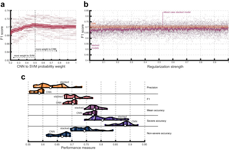
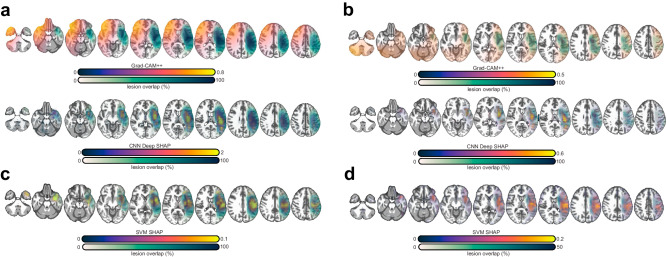
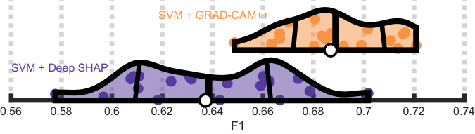
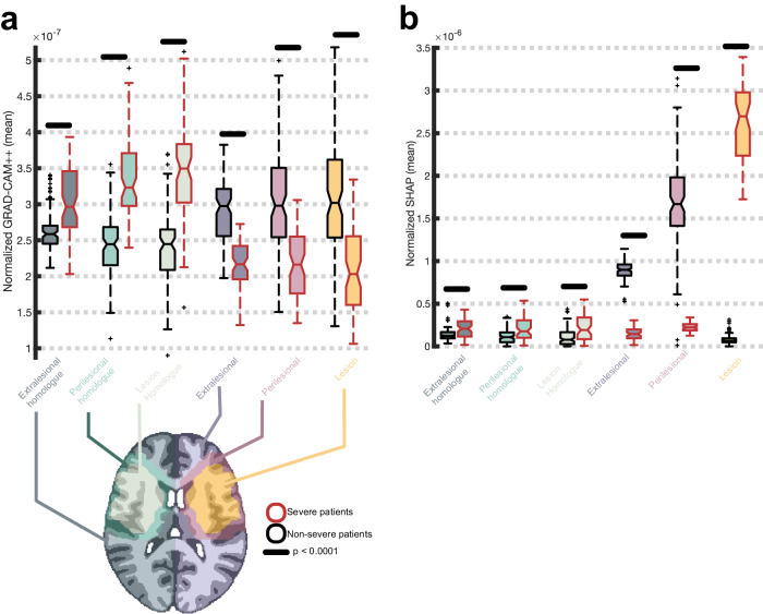
Background: Emerging evidence suggests that post-stroke aphasia severity depends on the integrity of the brain beyond the lesion. While measures of lesion anatomy and brain integrity combine synergistically to explain aphasic symptoms, substantial interindividual variability remains unaccounted. One explanatory factor may be the spatial distribution of morphometry beyond the lesion (e.g., atrophy), including not just specific brain areas, but distinct three-dimensional patterns. Methods: Here, we test whether deep learning with Convolutional Neural Networks (CNNs) on whole brain morphometry (i.e., segmented tissue volumes) and lesion anatomy better predicts chronic stroke individuals with severe aphasia (N = 231) than classical machine learning (Support Vector Machines; SVMs), evaluating whether encoding spatial dependencies identifies uniquely predictive patterns. Results: CNNs achieve higher balanced accuracy and F1 scores, even when SVMs are nonlinear or integrate linear or nonlinear dimensionality reduction. Parity only occurs when SVMs access features learned by CNNs. Saliency maps demonstrate that CNNs leverage distributed morphometry patterns, whereas SVMs focus on the area around the lesion. Ensemble clustering of CNN saliencies reveals distinct morphometry patterns unrelated to lesion size, consistent across individuals, and which implicate unique networks associated with different cognitive processes as measured by the wider neuroimaging literature. Individualized predictions depend on both ipsilateral and contralateral features outside the lesion. Conclusions: Three-dimensional network distributions of morphometry are directly associated with aphasia severity, underscoring the potential for CNNs to improve outcome prognostication from neuroimaging data, and highlighting the prospective benefits of interrogating spatial dependence at different scales in multivariate feature space.
Overview
The claims and statistics of this paper haven’t been checked yet.
Free: it reads the open-access text and checks the first 5 claims that cite a source we can check. Create a free account to check every claim.
Integrity signals
Registry
Cites
Statistics
Figures
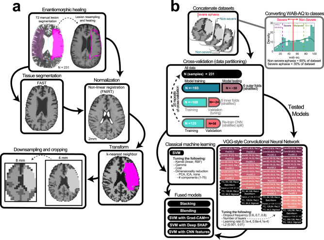
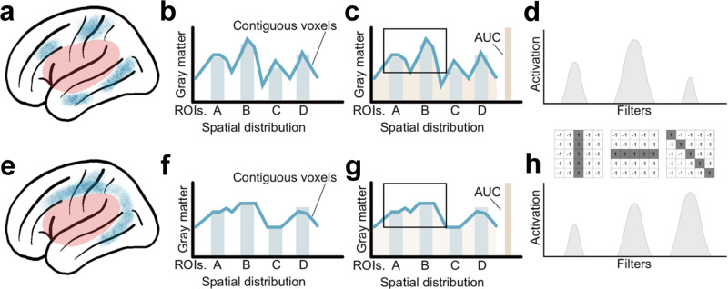
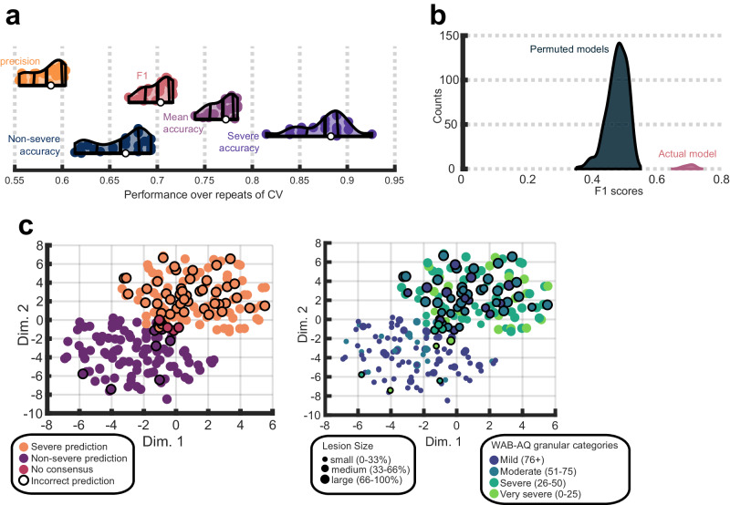
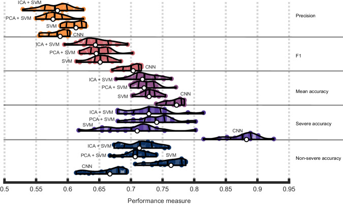
Verification
Claims the paper makes
The claims and statistics of this paper haven’t been checked yet.
Free: it reads the open-access text and checks the first 5 claims that cite a source we can check. Create a free account to check every claim.
Claims other papers make about this one
Each is checked against this paper’s own text: does it say what the citing paper says it does?
The source backs part of the claim, not all of it — or backs it for a different population, condition or comparison.
In the citing paper
“The analysis results showed that the machine learning algorithms used in post-stroke aphasia models were mainly supervised methods, including random forests, neural networks, and support vector machines.”
From Machine learning models in post-stroke aphasia: a scoping review (2026) · cites this paper as [24]
Experimental — accuracy not yet measuredThe source backs part of the claim, not all of it — or backs it for a different population, condition or comparison.
In the citing paper
“Five studies (21, 23, 24, 28, 36) constructed machine learning models for predicting the severity of aphasia by integrating multi-dimensional features such as neuroimaging and language samples.”
From Machine learning models in post-stroke aphasia: a scoping review (2026) · cites this paper as [24]
Experimental — accuracy not yet measuredThe source backs part of the claim, not all of it — or backs it for a different population, condition or comparison.
In the citing paper
“four studies (21, 23, 24, 36) focused on fine prediction in the chronic phase”
From Machine learning models in post-stroke aphasia: a scoping review (2026) · cites this paper as [24]
Experimental — accuracy not yet measuredThe source backs part of the claim, not all of it — or backs it for a different population, condition or comparison.
In the citing paper
“In terms of severity assessment and prediction, most models rely on single-center, small-sample data (21, 23, 24, 28, 36)”
From Machine learning models in post-stroke aphasia: a scoping review (2026) · cites this paper as [24]
Experimental — accuracy not yet measuredThe cited source says what the claim attributes to it.
In the citing paper
“Seven studies (19–21, 24, 27, 29, 31) used multiple algorithms to construct models and compare them.”
From Machine learning models in post-stroke aphasia: a scoping review (2026) · cites this paper as [24]
Experimental — accuracy not yet measured
8 more claims about this paper
Create a free account to see every claim other papers make about this one.
Self-check
Percentages, sums and reported differences re-computed from the paper’s own numbers.
The self-check needs the paper’s full text.
Citations
Cites
PyTorch: An Imperative Style, High-Performance Deep Learning Library
Paszke, A., Gross, S., Massa, F., Lerer, A., Bradbury, J., Chanan, G., Killeen, T., Lin, Z., Gimelshein, N., Antiga, L., et al. (2019) · arXiv · doi:10.48550/arxiv.1912.01703 · Cited by 31,473 · cited 1×
“Cross validation of the CNN was implemented in PyTorch 80 and cross validation of the SVM was performed with MATLAB ’ s machine learning toolbox 81 .”
Advances in neural information processing systems 7
Cios, K., & Shields, M. (1997) · Neurocomputing · doi:10.1016/s0925-2312(97)90018-7 · Cited by 22,059
Decoupled Weight Decay Regularization
Loshchilov, I., & Hutter, F. (2017) · arXiv · doi:10.48550/arxiv.1711.05101 · Cited by 12,522
Jenkinson, M., Beckmann, C. F., Behrens, T. E. J., Woolrich, M. W., & Smith, S. M. (2012) · NeuroImage · doi:10.1016/j.neuroimage.2011.09.015 · Cited by 11,314
Zhang, Y., Brady, M., & Smith, S. (2001) · IEEE Transactions on Medical Imaging · doi:10.1109/42.906424 · Cited by 6,982
Lipton, Z. C. (2018) · Queue · doi:10.1145/3236386.3241340 · Cited by 5,427
The Mythos of Model Interpretability
Lipton, Z. C. (2016) · arXiv · doi:10.48550/arxiv.1606.03490 · Cited by 5,422
A Survey of Convolutional Neural Networks: Analysis, Applications, and Prospects
Li, Z., Liu, F., Yang, W., Peng, S., & Zhou, J. (2022) · IEEE Transactions on Neural Networks and Learning Systems · doi:10.1109/tnnls.2021.3084827 · Cited by 5,273
What is a support vector machine?
Noble, W. S. (2006) · Nature Biotechnology · doi:10.1038/nbt1206-1565 · Cited by 4,301
Large-scale automated synthesis of human functional neuroimaging data.
Yarkoni, T., Poldrack, R. A., Nichols, T. E., Van Essen, D. C., & Wager, T. D. (2011) · Nature Methods · doi:10.1038/nmeth.1635 · Cited by 3,686
Grad-CAM++: Generalized Gradient-Based Visual Explanations for Deep Convolutional Networks
Chattopadhay, A., Sarkar, A., Howlader, P., & Balasubramanian, V. N. (2018) · 2018 IEEE Winter Conference on Applications of Computer Vision (WACV) · doi:10.1109/wacv.2018.00097 · Cited by 3,639
Advances in Neural Information Processing Systems 32 (NeurIPS 2019)
Wallach, H., Larochelle, H., Beygelzimer, A., d’Alché–Buc, F., Fox, E. B., & Garnett, R. (2020) · HAL · Cited by 2,451
Monti, S., Tamayo, P., Mesirov, J., & Golub, T. (2003) · Machine Learning · doi:10.1023/a:1023949509487 · Cited by 2,116
Hartigan, J. A., & Hartigan, P. M. (1985) · The Annals of Statistics · doi:10.1214/aos/1176346577 · Cited by 1,973 · cited 1×
“Brie fl y, Hartigan ’ s dip test of unimodality 89 was used to exclude solutions that showed signi fi cantly unimodal consensus distributions (i.e., no consensus because samples were not consistently being placed in either the same or differentclusters).”
Neuroinflammation: friend and foe for ischemic stroke
Jayaraj, R. L., Azimullah, S., Beiram, R., Jalal, F. Y., & Rosenberg, G. A. (2019) · Journal of Neuroinflammation · doi:10.1186/s12974-019-1516-2 · Cited by 1,428
Western Aphasia Battery--Revised
Kertesz, A. (2012) · PsycTESTS Dataset · doi:10.1037/t15168-000 · Cited by 1,367
Combining multiple clusterings using evidence accumulation
Fred, A. L. N., & Jain, A. K. (2005) · IEEE Transactions on Pattern Analysis and Machine Intelligence · doi:10.1109/tpami.2005.113 · Cited by 1,195
Cross-validation pitfalls when selecting and assessing regression and classification models
Krstajic, D., Buturovic, L. J., Leahy, D. E., & Thomas, S. (2014) · Journal of Cheminformatics · doi:10.1186/1758-2946-6-10 · Cited by 1,090
Aas, K., Jullum, M., & Løland, A. (2021) · Artificial Intelligence · doi:10.1016/j.artint.2021.103502 · Cited by 1,023
Patterns of brain atrophy in frontotemporal dementia and semantic dementia
Rosen, H. J., Gorno–Tempini, M. L., Goldman, W. P., Perry, R. J., Schuff, N., Weiner, M., Feiwell, R., Kramer, J. H., & Miller, B. L. (2002) · Neurology · doi:10.1212/wnl.58.2.198 · Cited by 841 · cited 1×
“Indeed,brainatrophyisknowntooccurwithinspeci fi c patterns across several neurological conditions, for example, Alzheimer ’ s disease, fronto-temporaldementia,epilepsy, amongothers 25 – 27 .”
Assessing and tuning brain decoders: Cross-validation, caveats, and guidelines
Varoquaux, G., Raamana, P. R., Engemann, D. A., Hoyos-Idrobo, A., Schwartz, Y., & Thirion, B. (2017) · NeuroImage · doi:10.1016/j.neuroimage.2016.10.038 · Cited by 792 · cited 1×
“Partitions used to tune models were nested to estimate model generalizability more reliably and mitigate the risk of over fi tting 65 – 67 .”
CATANI, M., & MESULAM, M. (2008) · Cortex · doi:10.1016/j.cortex.2008.04.002 · Cited by 760 · cited 2×
“Atthesametime,strokeinjurycantriggerprocessesthat have a widespread impacton the brain (e.g., neuroin fl ammation, Wallerian degeneration 104 – 106 ), inducing changes to morphology and functional capacity of regions distal to the injury 28 – 31 .”
Establishment of Best Practices for Evidence for Prediction: A Review
Poldrack, R. A., Huckins, G., & Varoquaux, G. (2020) · JAMA Psychiatry · doi:10.1001/jamapsychiatry.2019.3671 · Cited by 738
Aphasia after Stroke: Type, Severity and Prognosis
Pedersen, P. M., Vinter, K., & Olsen, T. S. (2004) · Cerebrovascular Diseases · doi:10.1159/000073896 · Cited by 622
Defining functional areas in individual human brains using resting functional connectivity MRI
Cohen, A. L., Fair, D. A., Dosenbach, N. U. F., Miezin, F. M., Dierker, D., Van Essen, D. C., Schlaggar, B. L., & Petersen, S. E. (2008) · NeuroImage · doi:10.1016/j.neuroimage.2008.01.066 · Cited by 582
The (Un)reliability of Saliency Methods
Kindermans, P.-J., Hooker, S., Adebayo, J., Alber, M., Schütt, K. T., Dähne, S., Erhan, D., & Kim, B. (2019) · Lecture Notes in Computer Science · doi:10.1007/978-3-030-28954-6_14 · Cited by 580
Computational anatomy with the SPM software
Ashburner, J. (2009) · Magnetic Resonance Imaging · doi:10.1016/j.mri.2009.01.006 · Cited by 564 · cited 1×
“1) 58 and SPM8 59 , then re fi ned for any necessary corrections in the case that any additional information about lesion extent was revealed by the T1-weighted image.”
A Review of Feature Reduction Techniques in Neuroimaging
Mwangi, B., Tian, T. S., & Soares, J. C. (2014) · Neuroinformatics · doi:10.1007/s12021-013-9204-3 · Cited by 559 · cited 1×
“Reducing the number of featuresinthedatapriortotrainingamodelforclassi fi cationorregressionis a commonly employed strategy (e.g., principal component regression) and PCA/ICAareoftenrecommendedtobeusedpreciselyinthiswaywithSVM inneuroimaging 72 .”
Aphasia in acute stroke and relation to outcome
Laska, A. C., Hellblom, A., Murray, V., Kahan, T., & Von Arbin, M. (2001) · Journal of Internal Medicine · doi:10.1046/j.1365-2796.2001.00812.x · Cited by 528
The Shapley Value: Essays in Honor of Lloyd S. Shapley
(1988) · Cambridge University Press eBooks · doi:10.1017/cbo9780511528446 · Cited by 527 · cited 1×
“Kernel SHAP leverages Shapley values, which are a game theoretic approach for quantifying the average marginal contribution of a player in a cooperative game 75 .”
RECOVERY PATTERNS AND PROGNOSIS IN APHASIA
KERTESZ, A., & MCCABE, P. (1977) · Brain · doi:10.1093/brain/100.1.1 · Cited by 514
Language-Selective and Domain-General Regions Lie Side by Side within Broca’s Area
Fedorenko, E., Duncan, J., & Kanwisher, N. (2012) · Current Biology · doi:10.1016/j.cub.2012.09.011 · Cited by 469 · cited 2×
“Impairments to different higher-level language functions are observed following damage to IFG 112,113 , damage to FO can result in aphasia 114 , and resection of MFG can cause apraxia of speech 115 .”
Interpretability of deep learning models: A survey of results
Chakraborty, S., Tomsett, R., Raghavendra, R., Harborne, D., Alzantot, M., Cerutti, F., Srivastava, M., Preece, A., Julier, S., Rao, R. M., et al. (2017) · 2017 IEEE SmartWorld, Ubiquitous Intelligence & Computing, Advanced & Trusted Computed, Scalable Computing & Communications, Cloud & Big Data Computing, Internet of People and Smart City Innovation (SmartWorld/SCALCOM/UIC/ATC/CBDCom/IOP/SCI) · doi:10.1109/uic-atc.2017.8397411 · Cited by 422
Critical limitations of consensus clustering in class discovery
Șenbabaoğlu, Y., Michailidis, G., & Li, J. Z. (2014) · Scientific Reports · doi:10.1038/srep06207 · Cited by 379
Declarative memory impairments in Alzheimer's disease and semantic dementia
Nestor, P. J., Fryer, T. D., & Hodges, J. R. (2006) · NeuroImage · doi:10.1016/j.neuroimage.2005.10.008 · Cited by 370 · cited 1×
“With respect to regions implicated in temporal cortex, atrophy in TP correlates with semantic dementia 116 , stimulation of pITG can disrupt naming 117 , and temporooccipital cortex supports reading 118 .”
A proposed regional hierarchy in recovery of post-stroke aphasia
Heiss, W.-D., & Thiel, A. (2006) · Brain and Language · doi:10.1016/j.bandl.2006.02.002 · Cited by 358 · cited 4×
“When language speci fi c areas aren ’ t spared, contralateral homotopic regions, but also right hemisphere regions more broadly, provide the substrate for recovery, typically with reduced likelihoodofsubstantialrecovery 13,15,18,19, .”
InfluentialThe impact of stroke: are people with aphasia different to those without?
Hilari, K. (2011) · Disability and Rehabilitation · doi:10.3109/09638288.2010.508829 · Cited by 321
Schulz, M.-A., Yeo, B. T. T., Vogelstein, J. T., Mourao-Miranada, J., Kather, J. N., Kording, K., Richards, B., & Bzdok, D. (2020) · Nature Communications · doi:10.1038/s41467-020-18037-z · Cited by 315 · cited 1×
“Access to larger sample sizes, and especially more patients with severe aphasia, may be helpful for improving performance 119 .”
Grad-CAM: Why did you say that?
Selvaraju, R. R., Das, A., Vedantam, R., Cogswell, M., Parikh, D., & Batra, D. (2016) · arXiv (Cornell University) · doi:10.48550/arxiv.1611.07450 · Cited by 309 · cited 1×
“The Grad-CAM ++ algorithm 77 improvesontheoriginalGrad-CAMmethodof explaining CNN predictions, which involves computing the weighted sum of the gradients of the predicted class score with respect to the feature maps of the last convolutional layer in the network 78 .”
Non-metric affinity propagation for unsupervised image categorization
Dueck, D., & Frey, B. J. (2007) · 2007 IEEE 11th International Conference on Computer Vision · doi:10.1109/iccv.2007.4408853 · Cited by 297 · cited 2×
“Clusters for the selected solution were extracted by applying af fi nity propagation 90 to the corresponding consensus matrix.”
Improvement in Aphasia Scores After Stroke Is Well Predicted by Initial Severity
Lazar, R. M., Minzer, B., Antoniello, D., Festa, J. R., Krakauer, J. W., & Marshall, R. S. (2010) · Stroke · doi:10.1161/strokeaha.109.577338 · Cited by 291
Enantiomorphic normalization of focally lesioned brains
Nachev, P., Coulthard, E., Jäger, H. R., Kennard, C., & Husain, M. (2008) · NeuroImage · doi:10.1016/j.neuroimage.2007.10.002 · Cited by 276 · cited 1×
“Anatomical deformation during normalization in the presence of large lesions was avoided using enantio-morphic healing 60 .”
Lam, J. M. C., & Wodchis, W. P. (2010) · Medical Care · doi:10.1097/mlr.0b013e3181ca2647 · Cited by 272
Hartwigsen, G., & Saur, D. (2019) · NeuroImage · doi:10.1016/j.neuroimage.2017.11.056 · Cited by 272
Task-induced brain activity in aphasic stroke patients: what is driving recovery?
Geranmayeh, F., Brownsett, S. L. E., & Wise, R. J. S. (2014) · Brain · doi:10.1093/brain/awu163 · Cited by 238 · cited 2×
“Further, some of the morphometry patterns predicting aphasia severity may be associated with domain-general regions that support language and language recovery after stroke 19 – 21 .”
Machine learning in neuroimaging: Progress and challenges
Davatzikos, C. (2019) · NeuroImage · doi:10.1016/j.neuroimage.2018.10.003 · Cited by 225 · cited 2×
“Support Vector Machines (SVMs) were one of the fi rst machine learning methods introduced to neuroimaging 65 and remain the most popular machine learning method in the fi eld (Supplementary Fig.”
Broca's area aphasias: Aphasia after lesions including the frontal operculum
Alexander, M. P., Naeser, M. A., & Palumbo, C. (1990) · Neurology · doi:10.1212/wnl.40.2.353 · Cited by 208
Butler, R. A., Lambon Ralph, M. A., & Woollams, A. M. (2014) · Brain · doi:10.1093/brain/awu286 · Cited by 199 · cited 1×
“The heterogeneity of integrity patterns associated with aphasia severity fi cits 109,110 emerging from varied damage along large-scale networks 111 .”
Activation and Role of Astrocytes in Ischemic Stroke
Shen, X.-Y., Gao, Z.-K., Han, Y., Yuan, M., Guo, Y.-S., & Bi, X. (2021) · Frontiers in Cellular Neuroscience · doi:10.3389/fncel.2021.755955 · Cited by 186
Qi, K., Yang, H., Li, C., Liu, Z., Wang, M., Liu, Q., & Wang, S. (2019) · Lecture notes in computer science · doi:10.1007/978-3-030-32248-9_28 · Cited by 183 · cited 1×
“Inthestrokeliterature,CNNshavebeenmainlyusedfor lesionsegmentation 37 – 41 .”
Dynamics of language reorganization after left temporo-parietal and frontal stroke
Stockert, A., Wawrzyniak, M., Klingbeil, J., Wrede, K., Kümmerer, D., Hartwigsen, G., Kaller, C. P., Weiller, C., & Saur, D. (2020) · Brain · doi:10.1093/brain/awaa023 · Cited by 183 · cited 3×
“Further, some of the morphometry patterns predicting aphasia severity may be associated with domain-general regions that support language and language recovery after stroke 19 – 21 .”
Alzheimer's Disease Detection Through Whole-Brain 3D-CNN MRI
Folego, G., Weiler, M., Casseb, R. F., Pires, R., & Rocha, A. D. R. (2020) · Frontiers in Bioengineering and Biotechnology · doi:10.3389/fbioe.2020.534592 · Cited by 181 · cited 1×
“This kind of architecture has been shown to perform as well or better than alternatives in similar tasks to ours 69 .”
Yu, Y., Xie, Y., Thamm, T., Gong, E., Ouyang, J., Huang, C., Christensen, S., Marks, M. P., Lansberg, M. G., Albers, G. W., et al. (2020) · JAMA Network Open · doi:10.1001/jamanetworkopen.2020.0772 · Cited by 175
Yourganov, G., Fridriksson, J., Rorden, C., Gleichgerrcht, E., & Bonilha, L. (2016) · The Journal of Neuroscience · doi:10.1523/jneurosci.4396-15.2016 · Cited by 174 · cited 2×
“Atthesametime,strokeinjurycantriggerprocessesthat have a widespread impacton the brain (e.g., neuroin fl ammation, Wallerian degeneration 104 – 106 ), inducing changes to morphology and functional capacity of regions distal to the injury 28 – 31 .”
Self-citationGray Matter Atrophy in Patients With Ischemic Stroke With Cognitive Impairment
Stebbins, G. T., Nyenhuis, D. L., Wang, C., Cox, J. L., Freels, S., Bangen, K., deToledo-Morrell, L., Sripathirathan, K., Moseley, M., Turner, D. A., et al. (2008) · Stroke · doi:10.1161/strokeaha.107.507392 · Cited by 168 · cited 1×
“Stebbins et al. 23 demonstrated that cognitive impair-mentsafterstrokewereassociatedwithgraymatteratrophyinthethalamus, cingulategyrusanddistributedregionsacrossfrontal,temporal,parietal, andoccipitallobesamongstrokesurvivorswithorwithoutmildaphasia.”
Consensus clustering and functional interpretation of gene-expression data
Swift, S., Tucker, A., Vinciotti, V., Martin, N., Orengo, C. A., Liu, X., & Kellam, P. (2004) · Genome biology · doi:10.1186/gb-2004-5-11-r94 · Cited by 167 · cited 1×
“A similarity-based clustering algorithm can be used to retrieve the consensus clustering solution embedded in this matrix 82 – 85 .”
Heterogeneous patterns of brain atrophy in Alzheimer's disease
Poulakis, K., Pereira, J. B., Mecocci, P., Vellas, B., Tsolaki, M., Kłoszewska, I., Soininen, H., Lovestone, S., Simmons, A., Wahlund, L.-O., et al. (2018) · Neurobiology of Aging · doi:10.1016/j.neurobiolaging.2018.01.009 · Cited by 166
The neural and neurocomputational bases of recovery from post-stroke aphasia
Stefaniak, J. D., Halai, A. D., & Lambon Ralph, M. A. (2020) · Nature Reviews Neurology · doi:10.1038/s41582-019-0282-1 · Cited by 162
A lexical semantic hub for heteromodal naming in middle fusiform gyrus
Forseth, K. J., Kadipasaoglu, C. M., Conner, C. R., Hickok, G., Knight, R. T., & Tandon, N. (2018) · Brain · doi:10.1093/brain/awy120 · Cited by 161
Structural Analysis and Optimization of Convolutional Neural Networks with a Small Sample Size
D’souza, R. N., Huang, P.-Y., & Yeh, F.-C. F. (2020) · Scientific Reports · doi:10.1038/s41598-020-57866-2 · Cited by 161 · cited 1×
“WhileCNNscanoutperformclassicalmachinelearningbydetecting patternsthatothermethodsareinsensitiveto,thisisnotguaranteed — such patterns might be absent and deep learning can be more likely to over fi t in small samples 96 .”
Discovering Relations Between Mind, Brain, and Mental Disorders Using Topic Mapping
Poldrack, R. A., Mumford, J. A., Schonberg, T., Kalar, D., Barman, B., & Yarkoni, T. (2012) · PLoS Computational Biology · doi:10.1371/journal.pcbi.1002707 · Cited by 151 · cited 1×
“Meta-analysesweregeneratedfor200topicsofanauthor-topicmodelofthe neuroimagingliterature 92 .”
Multiple Routes from Occipital to Temporal Cortices during Reading
Richardson, F. M., Seghier, M. L., Leff, A. P., Thomas, M. S. C., & Price, C. J. (2011) · The Journal of Neuroscience · doi:10.1523/jneurosci.6519-10.2011 · Cited by 149 · cited 1×
“With respect to regions implicated in temporal cortex, atrophy in TP correlates with semantic dementia 116 , stimulation of pITG can disrupt naming 117 , and temporooccipital cortex supports reading 118 .”
Geranmayeh, F., Chau, T. W., Wise, R. J. S., Leech, R., & Hampshire, A. (2017) · Brain · doi:10.1093/brain/awx134 · Cited by 146
Anglade, C., Thiel, A., & Ansaldo, A. I. (2014) · Brain Injury · doi:10.3109/02699052.2013.859734 · Cited by 145
The Prognosis for Aphasia in Stroke
Maas, M. B., Lev, M. H., Ay, H., Singhal, A. B., Greer, D. M., Smith, W. S., Harris, G. J., Halpern, E. F., Koroshetz, W. J., & Furie, K. L. (2012) · Journal of Stroke and Cerebrovascular Diseases · doi:10.1016/j.jstrokecerebrovasdis.2010.09.009 · Cited by 144 · cited 1×
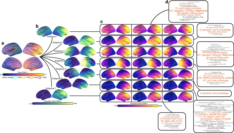
“In individuals with nonsevere aphasia, one subgroup showed right-lateralized saliency (subgroup 6) and multiplesubgroupsshowed modest-to-moderatesaliency in the non-lateralized hemisphere (subgroups 6, 4, and 2).”
Iglesias, J. E., Schleicher, R. L., Laguna, S., Billot, B., Schaefer, P. W., McKaig, B. N., Goldstein, J. N., Sheth, K. N., Rosen, M. S., & Kimberly, W. T. (2022) · Radiology · doi:10.1148/radiol.220522 · Cited by 133
Deep learning guided stroke management: a review of clinical applications
Feng, R., Badgeley, M., Mocco, J., & Oermann, E. K. (2018) · Journal of NeuroInterventional Surgery · doi:10.1136/neurintsurg-2017-013355 · Cited by 131 · cited 1×
“Two studies relied on CNNs to accelerate sequence acquisition or increase sequence resolution 40,41 , and another two studies leveraged CNNs for outcome prediction, but in the acute setting 54,55 .”
A hitchhiker's guide to lesion-behaviour mapping
de Haan, B., & Karnath, H.-O. (2018) · Neuropsychologia · doi:10.1016/j.neuropsychologia.2017.10.021 · Cited by 129
The PLORAS Database: A data repository for Predicting Language Outcome and Recovery After Stroke
Seghier, M. L., Patel, E., Prejawa, S., Ramsden, S., Selmer, A., Lim, L., Browne, R., Rae, J., Haigh, Z., Ezekiel, D., et al. (2016) · NeuroImage · doi:10.1016/j.neuroimage.2015.03.083 · Cited by 127
Group analyses of connectivity-based cortical parcellation using repeated k-means clustering
Nanetti, L., Cerliani, L., Gazzola, V., Renken, R. J., & Keysers, C. (2009) · NeuroImage · doi:10.1016/j.neuroimage.2009.06.014 · Cited by 121 · cited 1×
“Here, we subsampled 60% of the voxels in feature saliency maps 1000 times, each time performing k-means clustering using the k-means ++ algorithm with 250 replicates to improve solution stability 87 .”
Enlarged perivascular spaces and cognitive impairment after stroke and transient ischemic attack
Arba, F., Quinn, T. J., Hankey, G. J., Lees, K. R., Wardlaw, J. M., Ali, M., & Inzitari, D. (2016) · International Journal of Stroke · doi:10.1177/1747493016666091 · Cited by 121
Hilbert, A., Madai, V. I., Akay, E. M. Z., Aydin, O. U., Behland, J., Sobesky, J., Galinović, I., Khalil, A. A., Taha, A. A., Wuerfel, J. T., et al. (2020) · Frontiers in Artificial Intelligence · doi:10.3389/frai.2020.552258 · Cited by 119
Accurate nonlinear mapping between MNI volumetric and FreeSurfer surface coordinate systems
Wu, J., Ngo, G. H., Greve, D., Li, J., He, T., Fischl, B., Eickhoff, S. B., & Yeo, B. T. T. (2018) · Human Brain Mapping · doi:10.1002/hbm.24213 · Cited by 110
Sheth, S. A., Lopez-Rivera, V., Barman, A., Grotta, J. C., Yoo, A. J., Lee, S., Inam, M. E., Savitz, S. I., & Giancardo, L. (2019) · Stroke · doi:10.1161/strokeaha.119.026189 · Cited by 105
Incidence of Aphasia in Ischemic Stroke
Grönberg, A., Henriksson, I., Stenman, M., & Lindgren, A. G. (2022) · Neuroepidemiology · doi:10.1159/000524206 · Cited by 103
Thye, M., & Mirman, D. (2018) · NeuroImage: Clinical · doi:10.1016/j.nicl.2018.10.017 · Cited by 99
Convex Clustering with Exemplar-Based Models.
Lashkari, D., & Golland, P. (2007) · PubMed · Cited by 94
The multidimensional nature of aphasia recovery post-stroke
Stefaniak, J. D., Geranmayeh, F., & Ralph, M. A. L. (2022) · Brain · doi:10.1093/brain/awab377 · Cited by 94
Goubran, M., Ntiri, E. E., Akhavein, H., Holmes, M. F., Nestor, S. M., Ramirez, J., Adamo, S. M., Ozzoude, M., Scott, C. J. M., Gao, F., et al. (2019) · Human Brain Mapping · doi:10.1002/hbm.24811 · Cited by 92
Alyahya, R. S. W., Halai, A. D., Conroy, P., & Lambon Ralph, M. A. (2020) · Brain · doi:10.1093/brain/awaa074 · Cited by 91
Xue, Y., Farhat, F. G., Boukrina, O. V., Barrett, A. M., Binder, J. R., Roshan, U., & Graves, W. W. (2019) · NeuroImage Clinical · doi:10.1016/j.nicl.2019.102118 · Cited by 90
Nishi, H., Oishi, N., Ishii, A., Ono, I., Ogura, T., Sunohara, T., Chihara, H., Fukumitsu, R., Okawa, M., Yamana, N., et al. (2020) · Stroke · doi:10.1161/strokeaha.119.028101 · Cited by 90 · cited 1×
“TheseresultscorroborateandextendrecentworkdemonstratingthatCNNscanbesuccessfullydeployedoutsidetheircurrentnicheinthestrokelit-eratureforlesionsegmentationandcaneffectivelypredictstrokeoutcomes betterthanclassicalmethods 36,56 .”
Goldenberg, G., & Spatt, J. (1994) · Brain and Language · doi:10.1006/brln.1994.1063 · Cited by 87
The back and forth of axonal injury and repair after stroke
Hinman, J. D. (2014) · Current Opinion in Neurology · doi:10.1097/wco.0000000000000149 · Cited by 83 · cited 1×
“Atthesametime,strokeinjurycantriggerprocessesthat have a widespread impacton the brain (e.g., neuroin fl ammation, Wallerian degeneration 104 – 106 ), inducing changes to morphology and functional capacity of regions distal to the injury 28 – 31 .”
Automated Cerebral Hemorrhage Detection Using RAPID
Heit, J. J., Coelho, H., Lima, F. O., Granja, M., Aghaebrahim, A., Hanel, R., Kwok, K., Haerian, H., Cereda, C. W., Venkatasubramanian, C., et al. (2020) · American Journal of Neuroradiology · doi:10.3174/ajnr.a6926 · Cited by 72
Neuroplasticity in Aphasia: A Proposed Framework of Language Recovery
Kiran, S., Meier, E. L., & Johnson, J. P. (2019) · Journal of Speech, Language, and Hearing Research · doi:10.1044/2019_jslhr-l-rsnp-19-0054 · Cited by 67
Machine learning‐based multimodal prediction of language outcomes in chronic aphasia
Kristinsson, S., Zhang, W., Rorden, C., Newman‐Norlund, R., Basilakos, A., Bonilha, L., Yourganov, G., Xiao, F., Hillis, A., & Fridriksson, J. (2021) · Human Brain Mapping · doi:10.1002/hbm.25321 · Cited by 64
Self-citationNiMARE: Neuroimaging Meta-Analysis Research Environment
Salo, T., Yarkoni, T., Nichols, T. E., Poline, J., Bilgel, M., Bottenhorn, K. L., Eickhoff, S. B., Jarecka, D., Kent, J. D., Kimbler, A., et al. (2023) · Aperture Neuro · doi:10.52294/001c.87681 · Cited by 63
Machine learning and acute stroke imaging
Sheth, S. A., Giancardo, L., Colasurdo, M., Srinivasan, V. M., Niktabe, A., & Kan, P. (2023) · Journal of NeuroInterventional Surgery · doi:10.1136/neurintsurg-2021-018142 · Cited by 62
Thompson, C. K. (2000) · Journal of Communication Disorders · doi:10.1016/s0021-9924(00)00031-9 · Cited by 60 · cited 1×
“However,moderntheoreticalmodelsof aphasia neurobiology suggest that language recovery is contingent on the degree of preservation of hierarchically organized neuroanatomical systems beyond the lesion 13 – 17 .”
Cerebral White Matter Hyperintensities Predict Functional Stroke Outcome
Liou, L.-M., Chen, C.-F., Guo, Y.-C., Cheng, H.-L., Lee, H.-L., Hsu, J.-S., Lin, R.-T., & Lin, H.-F. (2010) · Cerebrovascular Diseases · doi:10.1159/000255970 · Cited by 60
Pure Apraxia of Speech After Resection Based in the Posterior Middle Frontal Gyrus
Chang, E. F., Kurteff, G., Andrews, J. P., Briggs, R. G., Conner, A. K., Battiste, J. D., & Sughrue, M. E. (2020) · Neurosurgery · doi:10.1093/neuros/nyaa002 · Cited by 58
Predictors beyond the lesion: Health and demographic factors associated with aphasia severity
Johnson, L., Nemati, S., Bonilha, L., Rorden, C., Busby, N., Basilakos, A., Newman-Norlund, R., Hillis, A. E., Hickok, G., & Fridriksson, J. (2022) · Cortex · doi:10.1016/j.cortex.2022.06.013 · Cited by 55 · cited 1×
“Beyond lesion size, the spatial location of stroke injury is predictive of bothaphasiaandstrokeseverity 10,12 .”
Self-citationThe role of the right hemisphere in the recovery of stroke-related aphasia: A systematic review
Cocquyt, E., Ley, L. D., Santens, P., Borsel, J. V., & Letter, M. D. (2017) · Journal of Neurolinguistics · doi:10.1016/j.jneuroling.2017.03.004 · Cited by 52
Right Hemisphere Grey Matter Volume and Language Functions in Stroke Aphasia
Lukic, S., Barbieri, E., Wang, X., Caplan, D., Kiran, S., Rapp, B. C., Parrish, T. B., & Thompson, C. K. (2017) · Neural Plasticity · doi:10.1155/2017/5601509 · Cited by 50 · cited 1×
“24 also demonstrated that preservation of right hemisphere volumes in the temporal gyrus and supplementary motor areas were associated with better language comprehension and production scores among individualswithaphasia.”
Mojiri Forooshani, P., Biparva, M., Ntiri, E. E., Ramirez, J., Boone, L., Holmes, M. F., Adamo, S., Gao, F., Ozzoude, M., Scott, C. J. M., et al. (2022) · Human Brain Mapping · doi:10.1002/hbm.25784 · Cited by 50
Chang, A. J., Roth, R., Bougioukli, E., Rüber, T., Keller, S. S., Drane, D. L., Gross, R. E., Welsh, J., Abrol, A., Calhoun, V. D., et al. (2023) · Communications Medicine · doi:10.1038/s43856-023-00262-4 · Cited by 47 · cited 1×
“Indeed,brainatrophyisknowntooccurwithinspeci fi c patterns across several neurological conditions, for example, Alzheimer ’ s disease, fronto-temporaldementia,epilepsy, amongothers 25 – 27 .”
Self-citationLeukoaraiosis is independently associated with naming outcome in poststroke aphasia
Wright, A. E., Tippett, D. C., Saxena, S., Sebastian, R., Breining, B. L., Faria, A. V., & Hillis, A. E. (2018) · Neurology · doi:10.1212/wnl.0000000000005945 · Cited by 45 · cited 1×
“Recent studies have shown that the accumulation of microvascular injuries, re fl ected in lower tissue volumes, is linked to both aphasia and response to treatment 32,107 .”
Sperber, C., Gallucci, L., Mirman, D., Arnold, M., & Umarova, R. M. (2023) · NeuroImage Clinical · doi:10.1016/j.nicl.2023.103511 · Cited by 45
Network dynamics of Broca’s area during word selection
Conner, C. R., Kadipasaoglu, C. M., Shouval, H. Z., Hickok, G., & Tandon, N. (2019) · PLOS ONE · doi:10.1371/journal.pone.0225756 · Cited by 43 · cited 1×
“Impairments to different higher-level language functions are observed following damage to IFG 112,113 , damage to FO can result in aphasia 114 , and resection of MFG can cause apraxia of speech 115 .”
White Matter Hyperintensities Predict Response to Language Treatment in Poststroke Aphasia
Varkanitsa, M., Peñaloza, C., Charidimou, A., Caplan, D., & Kiran, S. (2020) · Neurorehabilitation and neural repair · doi:10.1177/1545968320952809 · Cited by 41 · cited 2×
“…methods more sensitive to the effects of the stroke on brain regions outside of the immediate lesion (e.g., diaschisis) 28 – 31 , as well as other measures of overallbrainhealthassociatedwithsmallvesseldisease,suchaswhitematter hyperintensities, lacunes and enlarged perivascular spaces 32 – 35 .”
Brain age predicts long-term recovery in post-stroke aphasia
Kristinsson, S., Busby, N., Rorden, C., Newman‐Norlund, R., den Ouden, D., Magnúsdóttir, S., Hjaltason, H., Thors, H., Hillis, A. E., Kjartansson, Ó., et al. (2022) · Brain Communications · doi:10.1093/braincomms/fcac252 · Cited by 41 · cited 1×
“Further, acceleration of age-relatedpatterns of atrophy independently contributes to aphasia severity 33,108 .”
Self-citationMalla, C. U. P., Hernández, M. V., Rachmadi, M. F., & Komura, T. (2019) · Frontiers in Neuroinformatics · doi:10.3389/fninf.2019.00033 · Cited by 36
Ntiri, E. E., Holmes, M. F., Forooshani, P. M., Ramirez, J., Gao, F., Ozzoude, M., Adamo, S. M., Scott, C. J. M., Dowlatshahi, D. D., Lawrence‐Dewar, J. M., et al. (2021) · Neuroinformatics · doi:10.1007/s12021-021-09510-1 · Cited by 36
Advanced Brain Age and Chronic Poststroke Aphasia Severity
Busby, N., Wilmskoetter, J., Gleichgerrcht, E. L., Rorden, C., Roth, R., Newman‐Norlund, R., Hillis, A. E., Keller, S. S., de Bézenac, C. E., Kristinsson, S., et al. (2022) · Neurology · doi:10.1212/wnl.0000000000201693 · Cited by 35
Self-citationDeep learning prediction of motor performance in stroke individuals using neuroimaging data
Karakış, R., Gürkahraman, K., Mitsis, G. D., & Boudrias, M. (2023) · Journal of Biomedical Informatics · doi:10.1016/j.jbi.2023.104357 · Cited by 30 · cited 4×
“TheseresultscorroborateandextendrecentworkdemonstratingthatCNNscanbesuccessfullydeployedoutsidetheircurrentnicheinthestrokelit-eratureforlesionsegmentationandcaneffectivelypredictstrokeoutcomes betterthanclassicalmethods 36,56 .”
InfluentialDeep Learning for Neuroimaging Segmentation with a Novel Data Augmentation Strategy
Wu, W., Lu, Y., Mane, R., & Guan, C. (2020) · Annual International Conference of the IEEE Engineering in Medicine and Biology Society (EMBC) · doi:10.1109/embc44109.2020.9176537 · Cited by 28
Testing a convolutional neural network‐based hippocampal segmentation method in a stroke population
Zavaliangos‐Petropulu, A., Tubi, M. A., Haddad, E., Zhu, A. H., Braskie, M. N., Jahanshad, N., Thompson, P. M., & Liew, S. (2020) · Human Brain Mapping · doi:10.1002/hbm.25210 · Cited by 27 · cited 1×
“…a study treating Alzheimer ’ spatientsand astudy thatdid not use neuroimaging data for modeling, the majority of retrieved studies used CNNs for detecting stroke, segmenting lesions, white matter hyperintensities, or other regions of the brain that may be affected by atrophy ( N = 12) 42 – 53 .”
Detecting cerebral microbleeds via deep learning with features enhancement by reusing ground truth
Li, T., Zou, Y. L., Bai, P., Li, S., Wang, H., Chen, X., Meng, Z., Kang, Z., & Zhou, G. (2021) · Computer Methods and Programs in Biomedicine · doi:10.1016/j.cmpb.2021.106051 · Cited by 25
Structural Connectivity Remote From Lesions Correlates With Somatosensory Outcome Poststroke
Koh, C., Yeh, C., Liang, X., Vidyasagar, R., Seitz, R. J., Nilsson, M., Connelly, A., & Carey, L. M. (2021) · Stroke · doi:10.1161/strokeaha.120.031520 · Cited by 24
Syed, S., Anderssen, K. E., Stormo, S. K., & Kranz, M. (2023) · Scientific Reports · doi:10.1038/s41598-023-29665-y · Cited by 20 · cited 1×
“Some work has indicated that this can improve the reliability and localization of feature importance, including in MRI data, and particularly in the context of complex or nonlinear decision boundaries 77,79 .”
Recovering two languages with the right hemisphere
Marini, A., Galetto, V., Tatu, K., Duca, S., Geminiani, G. C., Sacco, K., & Zettin, M. (2016) · Brain and Language · doi:10.1016/j.bandl.2016.05.014 · Cited by 15
Grey and white matter atrophy 1 year after stroke aphasia
Egorova, N., Khlif, M. S., Werden, E., Bird, L., & Brodtmann, A. G. (2022) · Brain Communications · doi:10.1093/braincomms/fcac061 · Cited by 15 · cited 2×
“For example, Egorova-Brumley et al. 22 reported thatindividualswithpost-strokeaphasia have moreatrophy of the inferior frontal gyrus.”
Duan, C., Xiong, Y., Cheng, K., Xiao, S., Lyu, J., Wang, C., Bian, X., Zhang, J., Zhang, D., Chen, L., et al. (2022) · European Radiology · doi:10.1007/s00330-022-08638-1 · Cited by 14 · cited 1×
“Two studies relied on CNNs to accelerate sequence acquisition or increase sequence resolution 40,41 , and another two studies leveraged CNNs for outcome prediction, but in the acute setting 54,55 .”
rogiedodgie/nii_preprocess: Zenodo DOI release
Rorden, C., EmilieMcKinnon, Hanayik, T., Grigori77, & Reddydpusc (2020) · Zenodo (CERN European Organization for Nuclear Research) · doi:10.5281/zenodo.4027710 · Cited by 11
Self-citationAssessing the Capability of Deep-Learning Models in Parkinson’s Disease Diagnosis
West, C., Soltaninejad, S., & Cheng, I. (2020) · Lecture notes in computer science · doi:10.1007/978-3-030-54407-2_20 · Cited by 10 · cited 1×
“In the context of volumetric data, 2D CNNs are less capable of exploiting spatial dependencies compared to the 3D architecture that we deployherebecausetheyignoreanentirespatialdimension(i.e.,focusingon individualslices) 98 .”
Predicting Language Recovery after Stroke with Convolutional Networks on Stitched MRIPreprint
Roohani, Y., Sajid, N., Madhyastha, P., Price, C. J., & Hope, T. M. H. (2018) · arXiv (Cornell University) · doi:10.48550/arxiv.1811.10520 · Cited by 9 · cited 1×
“1 and Supplementary Table 1) has also reported successful application of 2D CNNs but to functional MRI data forpredicting language ability in chronic aphasia 97 .”
Aphasia Recovery Cohort (ARC) Dataset
Gibson, M., Newman‐Norlund, R., Bonilha, L., Fridriksson, J., Hickok, G. S., Hillis, A. E., den Ouden, D.-B., & Rorden, C. (2023) · OpenNeuro · doi:10.18112/openneuro.ds004884.v1.0.1 · Cited by 6
Self-citationTeghipco, A. (2022) · Zenodo (CERN European Organization for Nuclear Research) · doi:10.5281/zenodo.7271543 · Cited by 3
Self-citationVass, L., Moore, M. J., Hanayik, T., Mair, G., Pendlebury, S. T., Demeyere, N., & Jenkinson, M. (2021) · Neuroinformatics · doi:10.1007/s12021-021-09534-7 · Cited by 2
alexteghipco/StabilitySelection: v0.1-alpha
Teghipco, A. (2023) · Zenodo (CERN European Organization for Nuclear Research) · doi:10.5281/zenodo.7586724 · Cited by 1
Self-citationalexteghipco/volDNN: Release for zenodo
Teghipco, A. (2024) · Zenodo (CERN European Organization for Nuclear Research) · doi:10.5281/zenodo.10849304 · Cited by 1
Self-citationalexteghipco/consensusClustering: v0.1-alpha
Teghipco, A. (2023) · Zenodo (CERN European Organization for Nuclear Research) · doi:10.5281/zenodo.7586741 · Cited by 1
Self-citationTeghipco, A. (2024) · Figshare · doi:10.6084/m9.figshare.23579943 · Cited by 1
Self-citation
Research works printed but not linked: 3
- Kwon, Y., Won, J. H., Kim, B. J. & Paik, M. C. Uncertainty quantification using bayesian neural networks in classification: Application to ischemic stroke lesion segmentation. Medical Imaging with Deep Learning. 4, 2 (2018).
- Shrikumar, A., Greenside, P. & Kundaje, A. Learning important features through propagating activation differences. In International conference on machine learning, 3145–3153 (PMLR, 2017).
- Strehl A Ghosh J Cluster ensembles–a knowledge reuse framework for combining multiple partitions J. Mach. Learn. Res. 2002 3 583 617
Cited by
A Review on the Detection of Plant Disease Using Machine Learning and Deep Learning Approaches
Nyawose, T., Maswanganyi, R. C., & Khumalo, P. (2025) · Journal of Imaging · doi:10.3390/jimaging11100326 · Cited by 91
“While accuracy in Equation (1) is an intuitive measure of model performance, as it illustrates the correctly classified samples to the total number of samples in the dataset, it becomes less reliable in the presence of class imbalance [8].”
InfluentialAphasia severity prediction using a multi-modal machine learning approach
Hu, X., Varkanitsa, M., Kropp, E., Betke, M., Ishwar, P., & Kiran, S. (2025) · NeuroImage · doi:10.1016/j.neuroimage.2025.121300 · Cited by 8
Precision‐Optimised Post‐Stroke Prognoses
Hope, T. M. H., Bowman, H., Bruce, R. M., Leff, A. P., & Price, C. J. (2025) · Annals of Clinical and Translational Neurology · doi:10.1002/acn3.70077 · Cited by 2
“This could be done by varying the cut-off thresholds that we impose, as others have done in related work, such as: (a) distinguishing ‘severe im-pairment’ from ‘mild or moderate’ impairment [30]; or (b) distinguishing ‘excellent recovery’ from other types of recovery [31].”
Insights on Using Graph Neural Networks for Sulcal Graphs Predictive Models
Imbert, A., Gaüzère, B., Takerkart, S., Auzias, G., & Honeiné, P. (2025) · Lecture notes in computer science · doi:10.1007/978-3-031-94139-9_8 · Cited by 1
Korotkova, E. V., Kulikova, S., Meshkov, A. V., Lebedev, M., Tarasov, D., Kleeva, D. F., Kunnikova, K. I., & Lvova, O. A. (2026) · Communications in computer and information science · doi:10.1007/978-3-032-14674-8_46 · Cited by 1
Metadata
- Authors
- Teghipco, A., Newman-Norlund, R., Fridriksson, J., Rorden, C., & Bonilha, L.
- Affiliations
- University of South Carolina (US)
- Corresponding author
- Teghipco, Alex
- Venue
- Communications Medicine
- Published
- 2024-06-12
- Volume
- 4
- Issue
- 1
- Pages
- 115-115
- ISSN
- 2730-664X (ISSN-L)
- PMC
- PMC11169346
- OpenAlex
- W4399560892
- Cited by
- 11
- References
- 121
- Citations by year
- 2026: 6 · 2025: 4 · 2024: 1
- FWCI
- 2.67
- Type
- journal article
- Language
- en
- Open access
- gold
- doi.org
- Licence
- cc-by
- creativecommons.org/licenses/by/4.0 (tdm)
- creativecommons.org/licenses/by/4.0 (vor)
- Locations
- Communications Medicine (publishedVersion, cc-by)
- PubMed Central (publishedVersion, cc-by)
- PubMed (publishedVersion)
- DOAJ (DOAJ: Directory of Open Access Journals) (submittedVersion, cc-by-sa)
- Funders
- U.S. Department of Health & Human Services | NIH | National Institute on Deafness and Other Communication Disorders (P50 DC014664, R01 DC008355)
- Topics
- Life Sciences › Neuroscience › Cognitive Neuroscience › Neurobiology of Language and Bilingualism
- Health Sciences › Medicine › Psychiatry and Mental health › Dementia and Cognitive Impairment Research
- Health Sciences › Medicine › Clinical Neurology › Acute Ischemic Stroke Management
- Keywords
- post-stroke aphasia, brain morphometry, convolutional neural networks, brain atrophy, saliency maps, consensus clustering, interindividual variability
About these checks
Registry, retraction, Cites and statistics results are computed, not judged, with one exception: a printed reference that no index lists is read once by a model before it counts as a citation error. Claims are extracted by a model and kept only when their quote appears verbatim in the text; each cited source is then checked, with a model’s help, for whether it says what the claim relies on. Those verdicts can be wrong — read the claim’s quote and the cited source before relying on one.
Statistics and arithmetic are recomputed, not judged.
Confidence bands are shown only for fields calibrated on our annotated test set. None is calibrated yet, so no band is shown.
What counts as a claim ›How we measure confidence ›How we check this ›
Are you an author, or do you see a problem with this page? Request a review
Data sources
Bibliographic data: Semantic Scholar, OpenAlex, Crossref.
Contains information from the Semantic Scholar Academic Graph Datasets, made available under the ODC Attribution License (ODC-BY 1.0).
Semantic Scholar’s data is described in: Kinney, R., et al. (2023). The Semantic Scholar Open Data Platform. arXiv:2301.10140.
