

The DOI matches its Crossref record; 5 of the 5 references it cites were found in a registry or an index.
MicroRNA mir-34 provides robustness to environmental stress response via the DAF-16 network in C. elegans
Abstract
Diverse stresses and aging alter expression levels of microRNAs, suggesting a role for these posttranscriptional regulators of gene expression in stress modulation and longevity. Earlier studies demonstrated a central role for the miR-34 family in promoting cell cycle arrest and cell death following stress in human cells. However, the biological significance of this response was unclear. Here we show that in C. elegans mir-34 upregulation is necessary for developmental arrest, correct morphogenesis, and adaptation to a lower metabolic state to protect animals against stress-related damage. Either deletion or overexpression of mir-34 lead to an impaired stress response, which can largely be explained by perturbations in DAF-16/FOXO target gene expression. We demonstrate that mir-34 expression is regulated by the insulin signaling pathway via a negative feedback loop between miR-34 and DAF-16/FOXO. We propose that mir-34 provides robustness to stress response programs by controlling noise in the DAF-16/FOXO-regulated gene network.
Overview
The claims and statistics of this paper haven’t been checked yet.
Run the check — free, no account needed. It reads the open-access text and checks the sources it cites.
Integrity signals
Registry
Cites
Statistics
Figures
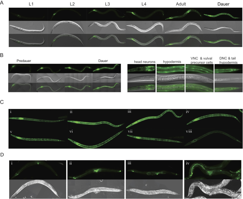
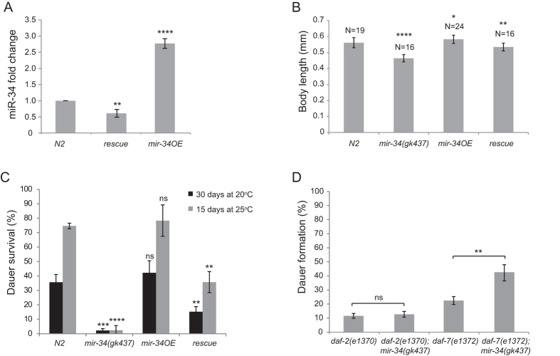
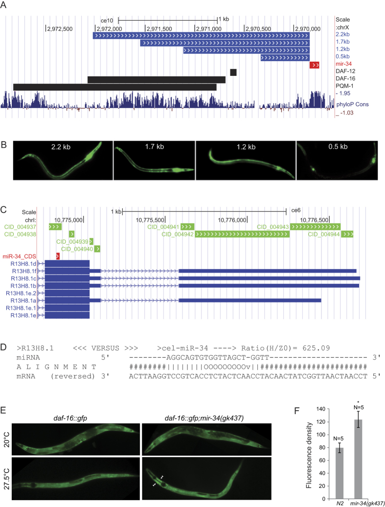
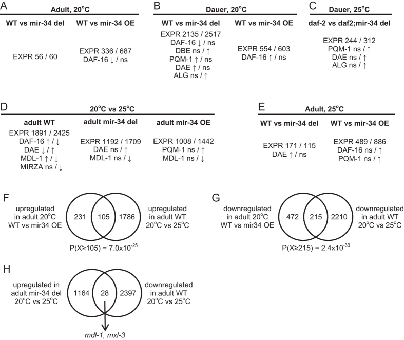
Verification
Claims the paper makes
The claims and statistics of this paper haven’t been checked yet.
Run the check — free, no account needed. It reads the open-access text and checks the sources it cites.
Claims other papers make about this one
Each is checked against this paper’s own text: does it say what the citing paper says it does?
In the citing paper
“One of the primary functions of miRNAs is to maintain cellular homeostasis in the presence of cellular stressors, perturbagens or disease conditions [11, 107–109].”
From microRNA-132 attenuates inflammation in induced pluripotent stem cell-derived microglia from Alzheimer’s disease patients (2026) · cites this paper as [107]
Self-check
Percentages, sums and reported differences re-computed from the paper’s own numbers.
The self-check needs the paper’s full text.
Citations
Cites
Transactivation of miR-34a by p53 broadly influences gene expression and promotes apoptosis
Chang, T.-C., Wentzel, E. A., Kent, O. A., Ramachandran, K., Mullendore, M., Lee, K. H., Feldmann, G., Yamakuchi, M., Ferlito, M., Lowenstein, C. J., et al. (2007) · Molecular Cell · doi:10.1016/j.molcel.2007.05.010 · Cited by 2,010
Ribosome Profiling Shows That miR-430 Reduces Translation Before Causing mRNA Decay in Zebrafish
Bazzini, A. A., Lee, M. T., & Giraldez, A. J. (2012) · Science · doi:10.1126/science.1215704 · Cited by 709
MicroRNAs in the p53 network: micromanagement of tumour suppression
Hermeking, H. (2012) · Nature Reviews Cancer · doi:10.1038/nrc3318 · Cited by 518
Most Caenorhabditis elegans microRNAs Are Individually Not Essential for Development or Viability
Miska, E. A., Alvarez-Saavedra, E., Abbott, A. L., Lau, N. C., Hellman, A. B., McGonagle, S. M., Bartel, D. P., Ambros, V. R., & Horvitz, H. R. (2007) · PLoS Genetics · doi:10.1371/journal.pgen.0030215 · Cited by 484
Intact p53-Dependent Responses in miR-34–Deficient Mice
Concepcion, C. P., Han, Y.-C., Mu, P., Bonetti, C., Yao, E., D'Andrea, A., Vidigal, J. A., Maughan, W. P., Ogrodowski, P., & Ventura, A. (2012) · PLoS Genetics · doi:10.1371/journal.pgen.1002797 · Cited by 197
Research works printed but not linked: 73
- Leung, A. K. L. & Sharp, P. A. MicroRNA functions in stress responses. Mol. Cell 40, 205–215 (2010). · DOI · Paper page
- Bushati, N. & Cohen, S. M. MicroRNA Functions. Annual Review of Cell and Developmental Biology 23, 175–205 (2007). · DOI · Paper page
- Leaman, D. et al. Antisense-mediated depletion reveals essential and specific functions of microRNAs in Drosophila development. Cell 121, 1097–1108 (2005). · DOI
- Li, X., Cassidy, J. J., Reinke, C. A., Fischboeck, S. & Carthew, R. W. A microRNA imparts robustness against environmental fluctuation during development. Cell 137, 273–282 (2009). · DOI · Paper page
- van Rooij, E. et al. Control of stress-dependent cardiac growth and gene expression by a microRNA. Science 316, 575–579 (2007). · DOI
- Flynt, A. S. et al. miR-8 microRNAs regulate the response to osmotic stress in zebrafish embryos. J Cell Biol 185, 115–127 (2009). · DOI
- Garcia-Segura, L. et al. High-Throughput Profiling of Caenorhabditis elegans Starvation-Responsive microRNAs. Plos One 10, e0142262 (2015). · DOI
- Ibáñez-Ventoso, C. et al. Modulated microRNA expression during adult lifespan in Caenorhabditis elegans. Aging Cell 5, 235–246 (2006). · DOI
- Karp, X., Hammell, M., Ow, M. C. & Ambros, V. Effect of life history on microRNA expression during C. elegans development. RNA 17, 639–651 (2011). · DOI
- de Lencastre, A. et al. MicroRNAs both promote and antagonize longevity in C. elegans. Curr. Biol. 20, 2159–2168 (2010). · DOI
- Khanna, A., Muthusamy, S., Liang, R., Sarojini, H. & Wang, E. Gain of survival signaling by down-regulation of three key miRNAs in brain of calorie-restricted mice. Aging (Albany NY) 3, 223–236 (2011). · DOI
- Li, X., Khanna, A., Li, N. & Wang, E. Circulatory miR34a as an RNAbased, noninvasive biomarker for brain aging. Aging (Albany NY) 3, 985–1002 (2011). · DOI
- Liu, N. et al. The microRNA miR-34 modulates ageing and neurodegeneration in Drosophila. Nature 482, 519–523 (2012). · DOI · Paper page
- Achari, C., Winslow, S., Ceder, Y. & Larsson, C. Expression of miR-34c induces G2/M cell cycle arrest in breast cancer cells. BMC Cancer 14, (2014). · DOI
- Chakraborty, C., George Priya Doss, C. & Bandyopadhyay, S. miRNAs in insulin resistance and diabetes-associated pancreatic cancer: the ‘minute and miracle’ molecule moving as a monitor in the ‘genomic galaxy’. Curr Drug Targets 14, 1110–1117 (2013). · DOI
- Chen, F. & Hu, S.-J. Effect of microRNA-34a in cell cycle, differentiation, and apoptosis: a review. J. Biochem. Mol. Toxicol. 26, 79–86 (2012). · DOI
- Cole, K. A. et al. A Functional Screen Identifies miR-34a as a Candidate Neuroblastoma Tumor Suppressor Gene. Mol Cancer Res 6, 735–742 (2008). · DOI
- Esguerra, J. L. S., Mollet, I. G., Salunkhe, V. A., Wendt, A. & Eliasson, L. Regulation of Pancreatic Beta Cell Stimulus-Secretion Coupling by microRNAs. Genes (Basel) 5, 1018–1031 (2014). · DOI
- Misso, G. et al. Mir-34: A New Weapon Against Cancer? Mol Ther Nucleic Acids 3, e194 (2014). · DOI
- Rokavec, M., Li, H., Jiang, L. & Hermeking, H. The p53/miR-34 axis in development and disease. J Mol Cell Biol 6, 214–230 (2014). · DOI
- Rottiers, V. & Näär, A. M. MicroRNAs in metabolism and metabolic disorders. Nat Rev Mol Cell Biol 13, 239–250 (2012). · DOI · Paper page
- Xu, Y. et al. A metabolic stress-inducible miR-34a-HNF4α pathway regulates lipid and lipoprotein metabolism. Nat Commun 6, 7466 (2015). · DOI
- Soni, K. et al. miR-34 is maternally inherited in Drosophila melanogaster and Danio rerio. Nucleic Acids Res. 41, 4470–4480 (2013). · DOI
- Kato, M. et al. The mir-34 microRNA is required for the DNA damage response in vivo in C. elegans and in vitro in human breast cancer cells. Oncogene 28, 2419–2424 (2009). · DOI
- Murakami, S. & Johnson, T. E. A Genetic Pathway Conferring Life Extension and Resistance to UV Stress in Caenorhabditis elegans. Genetics 143, 1207–1218 (1996). · DOI
- Riddle, D. L. & Albert, P. S. In C. elegans II (eds. Riddle, D. L., Blumenthal, T., Meyer, B. J. & Priess, J. R. ) (Cold Spring Harbor Laboratory Press, 1997).
- Karp, X., Hammell, M., Ow, M. C. & Ambros, V. Effect of life history on microRNA expression during C. elegans development. RNA 17, 639–651 (2011). · DOI
- Smith-Vikos, T. & Slack, F. J. MicroRNAs and their roles in aging. J Cell Sci 125, 7–17 (2012). · DOI · Paper page
- Ogg, S. et al. The Fork head transcription factor DAF-16 transduces insulin-like metabolic and longevity signals in C. elegans. Nature 389, 994–999 (1997). · DOI
- Lin, K. daf-16: An HNF-3/forkhead Family Member That Can Function to Double the Life-Span of Caenorhabditis elegans. Science 278, 1319–1322 (1997). · DOI
- Tepper, R. G. et al. PQM-1 Complements DAF-16 as a Key Transcriptional Regulator of DAF-2-Mediated Development and Longevity. Cell 154, 676–690 (2013). · DOI
- Contrino, S. et al. modMine: flexible access to modENCODE data. Nucleic Acids Res 40, D1082–D1088 (2012). · DOI
- Henderson, S. T. & Johnson, T. E. daf-16 integrates developmental and environmental inputs to mediate aging in the nematode Caenorhabditis elegans. Current Biology 11, 1975–1980 (2001). · DOI
- Wolf, M., Nunes, F., Henkel, A., Heinick, A. & Paul, R. J. The MAP kinase JNK-1 of Caenorhabditis elegans: Location, activation, and influences over temperature-dependent insulin-like signaling, stress responses, and fitness. J. Cell. Physiol. 214, 721–729 (2008). · DOI
- Leiser, S. F., Begun, A. & Kaeberlein, M. HIF-1 modulates longevity and healthspan in a temperature-dependent manner. Aging Cell 10, 318–326 (2011). · DOI
- Grosswendt, S. et al. Unambiguous identification of miRNA:target site interactions by different types of ligation reactions. Mol. Cell 54, 1042–1054 (2014). · DOI · Paper page
- Khorshid, M., Hausser, J., Zavolan, M. & van Nimwegen, E. A biophysical miRNA-mRNA interaction model infers canonical and noncanonical targets. Nature Methods 10, 253–255 (2013). · DOI
- Murphy, C. T. et al. Genes that act downstream of DAF-16 to influence the lifespan of Caenorhabditis elegans. Nature 424, 277–283 (2003). · DOI · Paper page
- Jones, S. J. et al. Changes in gene expression associated with developmental arrest and longevity in Caenorhabditis elegans. Genome Res. 11, 1346–1352 (2001). · DOI
- Wang, J. & Kim, S. K. Global analysis of dauer gene expression in Caenorhabditis elegans. Development 130, 1621–1634 (2003). · DOI
- Malmevik, J. et al. Distinct cognitive effects and underlying transcriptome changes upon inhibition of individual miRNAs in hippocampal neurons. Scientific Reports 6, 19879 (2016). · DOI
- Zhou, K. I., Pincus, Z. & Slack, F. J. Longevity and stress in Caenorhabditis elegans. Aging (Albany NY) 3, 733–753 (2011). · DOI
- Riesen, M. et al. MDL-1, a growth- and tumor-suppressor, slows aging and prevents germline hyperplasia and hypertrophy in C. elegans. Aging (Albany NY) 6, 98–117 (2014). · DOI
- Yuan, J., Tirabassi, R. S., Bush, A. B. & Cole, M. D. The C. elegans MDL-1 and MXL-1 proteins can functionally substitute for vertebrate MAD and MAX. Oncogene 17, 1109–1118 (1998). · DOI
- Johnson, D. W. et al. The Caenorhabditis elegans Myc-Mondo/Mad complexes integrate diverse longevity signals. PLoS Genet. 10, e1004278 (2014). · DOI
- Hausser, J. & Zavolan, M. Identification and consequences of miRNA-target interactions — beyond repression of gene expression. Nat Rev Genet 15, 599–612 (2014). · DOI
- Selbach, M. et al. Widespread changes in protein synthesis induced by microRNAs. Nature 455, 58–63 (2008). · DOI · Paper page
- Baek, D. et al. The impact of microRNAs on protein output. Nature 455, 64–71 (2008). · DOI · Paper page
- Djuranovic, S., Nahvi, A. & Green, R. miRNA-Mediated Gene Silencing by Translational Repression Followed by mRNA Deadenylation and Decay. Science 336, 237–240 (2012). · DOI · Paper page
- He, L. et al. A microRNA component of the p53 tumour suppressor network. Nature 447, 1130–1134 (2007). · DOI · Paper page
- Raver-Shapira, N. et al. Transcriptional Activation of miR-34a Contributes to p53-Mediated Apoptosis. Molecular Cell 26, 731–743 (2007). · DOI
- Burke, S. L., Hammell, M. & Ambros, V. Robust Distal Tip Cell Pathfinding in the Face of Temperature Stress Is Ensured by Two Conserved microRNAS in Caenorhabditis elegans. Genetics 200, 1201–1218 (2015). · DOI
- Yang, J. et al. MiR-34 modulates Caenorhabditis elegans lifespan via repressing the autophagy gene atg9. AGE 35, 11–22 (2013). · DOI
- Lee, R. Y. N., Hench, J. & Ruvkun, G. Regulation of C. elegans DAF-16 and its human ortholog FKHRL1 by the daf-2 insulin-like signaling pathway. Current Biology 11, 1950–1957 (2001). · DOI
- de Lencastre, A. et al. MicroRNAs Both Promote and Antagonize Longevity in C. elegans. Current Biology 20, 2159–2168 (2010). · DOI
- Bansal, A. et al. Transcriptional regulation of Caenorhabditis elegans FOXO/DAF-16 modulates lifespan. Longevity & Healthspan 3, 5 (2014). · DOI
- Kress, T. R. et al. The MK5/PRAK kinase and Myc form a negative feedback loop that is disrupted during colorectal tumorigenesis. Mol. Cell 41, 445–457 (2011). · DOI
- Bader, A. G. miR-34 – a microRNA replacement therapy is headed to the clinic. Frontiers in Genetics 3 (2012). · DOI
- Henrich, K.-O., Schwab, M. & Westermann, F. 1p36 tumor suppression–a matter of dosage? Cancer Res. 72, 6079–6088 (2012). · DOI
- Brenner, S. The Genetics of Caenorhabditis Elegans. Genetics 77, 71–94 (1974). · DOI
- Isik, M. & Berezikov, E. Biolistic transformation of Caenorhabditis elegans. Methods Mol. Biol. 940, 77–86 (2013). · DOI
- Ailion, M. & Thomas, J. H. Dauer formation induced by high temperatures in Caenorhabditis elegans. Genetics 156, 1047–1067 (2000). · DOI
- Ritchie, M. E. et al. limma powers differential expression analyses for RNA-sequencing and microarray studies. Nucleic Acids Research 43, e47–e47 (2015). · DOI · Paper page
- Kim, D., Langmead, B. & Salzberg, S. L. HISAT: a fast spliced aligner with low memory requirements. Nature Methods 12, 357–360 (2015). · DOI · Paper page
- Hartley, S. W. & Mullikin, J. C. QoRTs: a comprehensive toolset for quality control and data processing of RNA-Seq experiments. BMC Bioinformatics 16 (2015). · DOI · Paper page
- Robinson, M. D., McCarthy, D. J. & Smyth, G. K. edgeR: a Bioconductor package for differential expression analysis of digital gene expression data. Bioinformatics 26, 139–140 (2010). · DOI · Paper page
- Forman, J. J., Legesse-Miller, A. & Coller, H. A. A search for conserved sequences in coding regions reveals that the let-7 microRNA targets Dicer within its coding sequence. Proceedings of the National Academy of Sciences 105, 14879–14884 (2008). · DOI · Paper page
- Qin, W. et al. miR-24 Regulates Apoptosis by Targeting the Open Reading Frame (ORF) Region of FAF1 in Cancer Cells. Plos One 5, e9429 (2010). · DOI
- Ott, C. E. et al. MicroRNAs differentially expressed in postnatal aortic development downregulate elastin via 3′ UTR and coding-sequence binding sites. PLoS ONE 6, e16250 (2011). · DOI
- Huang, F. W. D., Qin, J., Reidys, C. M. & Stadler, P. F. Target prediction and a statistical sampling algorithm for RNA-RNA interaction. Bioinformatics 26, 175–181 (2010). · DOI
- Hausser, J., Syed, A. P., Bilen, B. & Zavolan, M. Analysis of CDS-located miRNA target sites suggests that they can effectively inhibit translation. Genome Research 23, 604–615 (2013). · DOI · Paper page
- Huang, D. W., Sherman, B. T. & Lempicki, R. A. Bioinformatics enrichment tools: paths toward the comprehensive functional analysis of large gene lists. Nucleic Acids Research 37, 1–13 (2009). · DOI · Paper page
- Huang, D. W., Sherman, B. T. & Lempicki, R. A. Systematic and integrative analysis of large gene lists using DAVID bioinformatics resources. Nature Protocols 4, 44–57 (2008). · DOI · Paper page
Cited by
Sorted by citations
Penning, A., Snoeck, S., Ormaechea, O. R., Ayyildiz, D., Polzer, O., Buitrago-Arango, M., Capobianco, R., de Winter, F., Balusu, S., Verhaagen, J., et al. (2026) · Acta Neuropathologica Communications · doi:10.1186/s40478-026-02228-8 · Cited by 6
Metadata
- Authors
- Isik, M., Blackwell, T. K., & Berezikov, E.
- Venue
- Scientific Reports
- Published
- 2016-12-01
- Volume
- 6
- Issue
- 1
- ISSN
- 2045-2322
- PMC
- PMC5131338
- Cited by
- 50
- References
- 5
- Language
- en
About these checks
Registry, retraction, Cites and statistics results are computed, not judged, with one exception: a printed reference that no index lists is read once by a model before it counts as a citation error. Claims are extracted by a model and kept only when their quote appears verbatim in the text; each cited source is then checked, with a model’s help, for whether it says what the claim relies on. Those verdicts can be wrong — read the claim’s quote and the cited source before relying on one.
Statistics and arithmetic are recomputed, not judged.
Confidence bands are shown only for fields calibrated on our annotated test set. None is calibrated yet, so no band is shown.
What counts as a claim ›How we measure confidence ›How we check this ›
Are you an author, or do you see a problem with this page? Request a review
