Cited by 53

The DOI matches its Crossref record.

Recycling of the Epidermal Growth Factor Receptor Is Mediated by a Novel Form of the Clathrin Adaptor Protein Eps15

Chi, Susan, Cao, Hong, Wang, Yu, McNiven, Mark A.·2011·Journal of Biological Chemistry

Overview

The claims and statistics of this paper haven’t been checked yet.

Run the check — free, no account needed. It reads the open-access text and checks the sources it cites.

A few free checks a day · papers already verified open instantly · results are public

Integrity signals

Registry

Crossref · 10.1074/jbc.m111.247577

Cites

Statistics

Figures

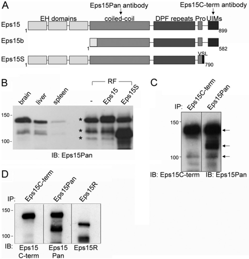
FIGURE 1 Identification of new Eps15 short isoform Eps15S. A , schematic representation of three isoforms of Eps15 as follows: conventional Eps15, Eps15b, and the newly identified Eps15S reported here. The indicated Eps15 protein domains include three Eps15 homology ( EH ) domains, a coiled-coil domain, a DPF repeat domain, a proline-rich region ( Pro ), and UIMs. The Eps15S isoform lacks the 111 amino acids at the C terminus of Eps15, including the UIMs, and has three unique amino acids (VSL) at the C-terminal ( C-term ) end. Two Eps15-specific polyclonal antibodies were generated to conserved and nonconserved domains, as indicated ( arrows ). B, protein extracts from rat brain, liver, and spleen or cell lysates from rat fibroblasts ( RF ) transfected with untagged Eps15 or Eps15S were subjected to SDS-PAGE and immunoblotting with the anti-Eps15Pan antibody. Asterisks label the three identified forms, including Eps15, Eps15S, and Eps15b, sequentially. C, protein extracts from rat brain were subjected to immunoprecipitation ( IP ) and immunoblotting ( IB ) using two anti-Eps15 antibodies ( Eps15 C-term or Eps15Pan ). Arrows indicate the predicted Eps15, Eps15S, and Eps15b forms. D, protein extracts from rat liver were subjected to immunoprecipitation and immunoblotting using three anti-Eps15 antibodies ( Eps15 C-term , Eps15Pan , or Eps15R ). Eps15 and Eps15S clearly differ from Eps15R.
FIGURE 1 Identification of new Eps15 short isoform Eps15S. A , schematic representation of three isoforms of Eps15 as follows: conventional Eps15, Eps15b, and the newly identified Eps15S reported here. The indicated Eps15 protein domains include three Eps15 homology ( EH ) domains, a coiled-coil domain, a DPF repeat domain, a proline-rich region ( Pro ), and UIMs. The Eps15S isoform lacks the 111 amino acids at the C terminus of Eps15, including the UIMs, and has three unique amino acids (VSL) at the C-terminal ( C-term ) end. Two Eps15-specific polyclonal antibodies were generated to conserved and nonconserved domains, as indicated ( arrows ). B, protein extracts from rat brain, liver, and spleen or cell lysates from rat fibroblasts ( RF ) transfected with untagged Eps15 or Eps15S were subjected to SDS-PAGE and immunoblotting with the anti-Eps15Pan antibody. Asterisks label the three identified forms, including Eps15, Eps15S, and Eps15b, sequentially. C, protein extracts from rat brain were subjected to immunoprecipitation ( IP ) and immunoblotting ( IB ) using two anti-Eps15 antibodies ( Eps15 C-term or Eps15Pan ). Arrows indicate the predicted Eps15, Eps15S, and Eps15b forms. D, protein extracts from rat liver were subjected to immunoprecipitation and immunoblotting using three anti-Eps15 antibodies ( Eps15 C-term , Eps15Pan , or Eps15R ). Eps15 and Eps15S clearly differ from Eps15R.CC-BY · From the PDF
Image 2
CC-BY · From the PDF
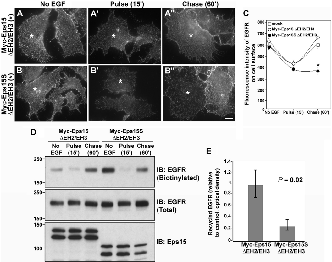
FIGURE 3 Overexpression of a mutant Eps15S specifically blocks recycling of EGFR to the cell surface. A-B ″ , HuH7-cells were transfected with either Myc-Eps15 𝚫 EH2/EH3 ( A-A ″ ) or Myc-Eps15S 𝚫 EH2/EH3 ( B-B ″ ). Cells were then serum-starved for 4 h ( A and B ) and pretreated with cycloheximide for 1 h. EGF (20 ng/ml) binding occurred for 30 min at 4 °C in the presence of cycloheximide. Subsequently, cells were shifted to 37 °C for 15 min (pulse 15 min, A ′ and B ′ ), washed twice with HBSS, and further incubated in serum-free medium for 60 min (chase 60 ′ , A ″ and B ″ ). After fixation, cells were immunostained for EGFR without permeabilization to visualize only surface EGFR (EGFR recycling). Subsequently, cells were permeabilized and immunostained for Myc to confirm expression of the Myc-Eps15 proteins (*). EGFR internalization was not reduced in cells expressing either Myc-Eps15 𝚫 EH2/EH3 or Myc-Eps15S 𝚫 EH2/EH3 (* in A ′ and B ′ ). However, recycling of EGFR to the cell surface was reduced significantly in Myc-Eps15S 𝚫 EH2/EH3-expressing cells (* in B ″ ) but not in Myc-Eps15 𝚫 EH2/EH3-expressingcells (* in A ″ ). C, quantitation of the average fluorescence intensity of EGFR at the cell surface at the indicated time points. Forty cells were measuredfor each condition. Overexpression of Myc-Eps15S 𝚫 EH2/EH3causedasignificant delay in EGFR recycling to the cell surface (*, p < 0.0001), whereas Myc-Eps15 𝚫 EH2/EH3 did not affect EGFR recycling. Error bars indicate the standard error. Scale bars, 10 𝛍 m( A-B ″ ). D, HuH-7 cells were subjected to a surface biotinylation assay, and immunoblotting ( IB ) was performed using an anti-EGFR antibody to detect surface EGFR (biotinylated EGFR). Less surface EGFR was detected after the 60-min chase in Eps15S 𝚫 EH2/EH3-expressing cells compared with Eps15 𝚫 EH2/EH3-expressing cells. E , ratio of recycled EGFR determined from densitometry of Western blot bands from three independent experiments. Biotinylated EGFR after the 60-min chase was normalized to total EGFR and then divided by biotinylated, normalized EGFR at the control time point. The level of recycled EGFR is significantly less in Myc-Eps15S 𝚫 EH2/EH3-expressing cells compared with Myc-Eps15 𝚫 EH2/EH3-expressing cells ( p = 0.02). Error bars indicate standard error.
FIGURE 3 Overexpression of a mutant Eps15S specifically blocks recycling of EGFR to the cell surface. A-B ″ , HuH7-cells were transfected with either Myc-Eps15 𝚫 EH2/EH3 ( A-A ″ ) or Myc-Eps15S 𝚫 EH2/EH3 ( B-B ″ ). Cells were then serum-starved for 4 h ( A and B ) and pretreated with cycloheximide for 1 h. EGF (20 ng/ml) binding occurred for 30 min at 4 °C in the presence of cycloheximide. Subsequently, cells were shifted to 37 °C for 15 min (pulse 15 min, A ′ and B ′ ), washed twice with HBSS, and further incubated in serum-free medium for 60 min (chase 60 ′ , A ″ and B ″ ). After fixation, cells were immunostained for EGFR without permeabilization to visualize only surface EGFR (EGFR recycling). Subsequently, cells were permeabilized and immunostained for Myc to confirm expression of the Myc-Eps15 proteins (*). EGFR internalization was not reduced in cells expressing either Myc-Eps15 𝚫 EH2/EH3 or Myc-Eps15S 𝚫 EH2/EH3 (* in A ′ and B ′ ). However, recycling of EGFR to the cell surface was reduced significantly in Myc-Eps15S 𝚫 EH2/EH3-expressing cells (* in B ″ ) but not in Myc-Eps15 𝚫 EH2/EH3-expressingcells (* in A ″ ). C, quantitation of the average fluorescence intensity of EGFR at the cell surface at the indicated time points. Forty cells were measuredfor each condition. Overexpression of Myc-Eps15S 𝚫 EH2/EH3causedasignificant delay in EGFR recycling to the cell surface (*, p < 0.0001), whereas Myc-Eps15 𝚫 EH2/EH3 did not affect EGFR recycling. Error bars indicate the standard error. Scale bars, 10 𝛍 m( A-B ″ ). D, HuH-7 cells were subjected to a surface biotinylation assay, and immunoblotting ( IB ) was performed using an anti-EGFR antibody to detect surface EGFR (biotinylated EGFR). Less surface EGFR was detected after the 60-min chase in Eps15S 𝚫 EH2/EH3-expressing cells compared with Eps15 𝚫 EH2/EH3-expressing cells. E , ratio of recycled EGFR determined from densitometry of Western blot bands from three independent experiments. Biotinylated EGFR after the 60-min chase was normalized to total EGFR and then divided by biotinylated, normalized EGFR at the control time point. The level of recycled EGFR is significantly less in Myc-Eps15S 𝚫 EH2/EH3-expressing cells compared with Myc-Eps15 𝚫 EH2/EH3-expressing cells ( p = 0.02). Error bars indicate standard error.CC-BY · From the PDF
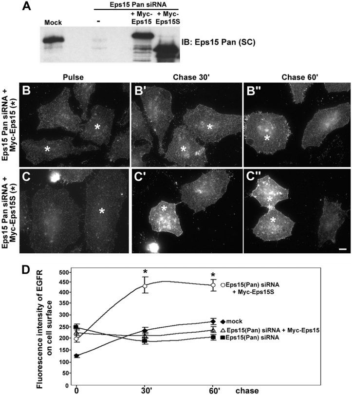
FIGURE 4 Eps15S, but not Eps15, promotes EGFR recycling. A, HeLa cells were treated with siRNA targeted to the coiled-coil domain of the three known Eps15 isoforms ( Eps15 Pan siRNA ). The cells were then transfected to re-express Myc-Eps15 or Myc-Eps15S. Cell lysates were subjected to SDS-PAGE and immunoblotting ( IB ) with the anti-Eps15Pan ( SC ) antibody. B-C ″ , HeLa cells treated with Eps15 Pan siRNA were transfected to re-express Myc-Eps15 ( B-B ″ ) or Myc-Eps15S ( C-C ″ ). EGFR recycling was assayed as described under 'Experimental Procedures.' Expression of either Myc-Eps15 or Myc-Eps15S was confirmed by immunostaining for Myc (data not shown). At 30 min ( B ′ and C ′ ) or 60 min ( B ″ and C ″ ) of chase following the 15-min pulse ( B and C ), EGFR recycling to the cell surface was minimal in cells expressing the Eps15 form (*) and was indistinguishable from untransfected adjacent cells ( B ′ and B ″ ). In comparison, substantial levels of EGFR were recycled back to the cell surface in Myc-Eps15S-re-expressing cells (* in C ′ and C ″ ). D, quantitation of the average fluorescence intensity of EGFR at the cell surface at the indicated time points. Twenty five cells were measured for each condition. Statistical analysis revealed that re-expression of Myc-Eps15S WT significantly promoted EGFR recycling ( p < 0.0001), whereas Myc-Eps15 WT had little effect on EGFR recycling. Error bars indicate the standard error. Scale bars, 10 𝛍 m( B-C ″ ).
FIGURE 4 Eps15S, but not Eps15, promotes EGFR recycling. A, HeLa cells were treated with siRNA targeted to the coiled-coil domain of the three known Eps15 isoforms ( Eps15 Pan siRNA ). The cells were then transfected to re-express Myc-Eps15 or Myc-Eps15S. Cell lysates were subjected to SDS-PAGE and immunoblotting ( IB ) with the anti-Eps15Pan ( SC ) antibody. B-C ″ , HeLa cells treated with Eps15 Pan siRNA were transfected to re-express Myc-Eps15 ( B-B ″ ) or Myc-Eps15S ( C-C ″ ). EGFR recycling was assayed as described under 'Experimental Procedures.' Expression of either Myc-Eps15 or Myc-Eps15S was confirmed by immunostaining for Myc (data not shown). At 30 min ( B ′ and C ′ ) or 60 min ( B ″ and C ″ ) of chase following the 15-min pulse ( B and C ), EGFR recycling to the cell surface was minimal in cells expressing the Eps15 form (*) and was indistinguishable from untransfected adjacent cells ( B ′ and B ″ ). In comparison, substantial levels of EGFR were recycled back to the cell surface in Myc-Eps15S-re-expressing cells (* in C ′ and C ″ ). D, quantitation of the average fluorescence intensity of EGFR at the cell surface at the indicated time points. Twenty five cells were measured for each condition. Statistical analysis revealed that re-expression of Myc-Eps15S WT significantly promoted EGFR recycling ( p < 0.0001), whereas Myc-Eps15 WT had little effect on EGFR recycling. Error bars indicate the standard error. Scale bars, 10 𝛍 m( B-C ″ ).CC-BY · From the PDF
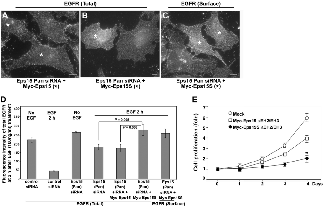
FIGURE 5 Eps15S-expressing cells target EGFR for recycling rather than degradation. A-C, HeLa cells were treated with siRNA targeted to the coiled-coil domain of three Eps15 isoforms ( Eps15 Pan siRNA ) and then transfected to re-express siRNA-resistant Myc-Eps15 ( A ) or Myc-Eps15S ( B and C ). Cells were serum-starved for 4 h, pretreated with cycloheximide for 1 h, and then incubated with 100 ng/ml EGF for 30 min at 4 °C in the presence of cycloheximide. Subsequently, cells were shifted to 37 °C for 15 min, washed twice with HBSS, and further incubated in serum-free medium for 2 h. After fixation, cells were permeabilized and immunostained for EGFR to examine total EGFR ( A and B ). To facilitate viewing of EGFR recycled back to the cell surface, cells were not permeabilized ( C ). The level of total EGFR remaining after 2 h of chase was much higher in Myc-Eps15S-re-expressing cells (* in B ) compared with Myc-Eps15re-expressing cells (* in A ). Expression of Myc-Eps15 and Myc-Eps15S was confirmed by immunostaining for Myc (data not shown). Scale bars, 10 𝛍 m. D, quantitation of the average fluorescence intensity of EGFR remaining after 2 h of chase for each condition. Knockdown of all forms of Eps15 ( Eps15PansiRNA ) showed an ∼ 3.5-fold higher total EGFR level after EGF treatment compared with control ( control siRNA ). Re-expression of Myc-Eps15S ( Eps15 Pan siRNA + Myc-Eps15S ), but not Myc-Eps15 ( Eps15 Pan siRNA + Myc-Eps15 ), led to an even higher ( ∼ 5.5-fold) level of EGFR ( p = 0.005). A high ratio of surface EGFR to total EGFR in Myc-Eps15S-re-expressing cells indicates that Eps15S targets EGFR for recycling rather than degradation. Twenty five cells were measured for each condition. Error bars indicate the standard error. E, HeLa cells were transfected with mock, Myc-Eps15 𝚫 EH2/EH3, or Myc-Eps15S 𝚫 EH2/EH3. After 6 h posttransfection, 2 × 10 5 of cells were plated in six wells in complete medium (10% FBS, day 0). After 24 h (day 1), cells were harvested and counted daily during 4 days. Values are expressed as a fold of control ( day 0 ). The data represent the average of three independent experiments. Myc-Eps15S 𝚫 EH2/EH3-expressing cells showed a significant delay in proliferation compared with mock-treated cells ( p = 0.002). Error bars indicate the standard deviation.
FIGURE 5 Eps15S-expressing cells target EGFR for recycling rather than degradation. A-C, HeLa cells were treated with siRNA targeted to the coiled-coil domain of three Eps15 isoforms ( Eps15 Pan siRNA ) and then transfected to re-express siRNA-resistant Myc-Eps15 ( A ) or Myc-Eps15S ( B and C ). Cells were serum-starved for 4 h, pretreated with cycloheximide for 1 h, and then incubated with 100 ng/ml EGF for 30 min at 4 °C in the presence of cycloheximide. Subsequently, cells were shifted to 37 °C for 15 min, washed twice with HBSS, and further incubated in serum-free medium for 2 h. After fixation, cells were permeabilized and immunostained for EGFR to examine total EGFR ( A and B ). To facilitate viewing of EGFR recycled back to the cell surface, cells were not permeabilized ( C ). The level of total EGFR remaining after 2 h of chase was much higher in Myc-Eps15S-re-expressing cells (* in B ) compared with Myc-Eps15re-expressing cells (* in A ). Expression of Myc-Eps15 and Myc-Eps15S was confirmed by immunostaining for Myc (data not shown). Scale bars, 10 𝛍 m. D, quantitation of the average fluorescence intensity of EGFR remaining after 2 h of chase for each condition. Knockdown of all forms of Eps15 ( Eps15PansiRNA ) showed an ∼ 3.5-fold higher total EGFR level after EGF treatment compared with control ( control siRNA ). Re-expression of Myc-Eps15S ( Eps15 Pan siRNA + Myc-Eps15S ), but not Myc-Eps15 ( Eps15 Pan siRNA + Myc-Eps15 ), led to an even higher ( ∼ 5.5-fold) level of EGFR ( p = 0.005). A high ratio of surface EGFR to total EGFR in Myc-Eps15S-re-expressing cells indicates that Eps15S targets EGFR for recycling rather than degradation. Twenty five cells were measured for each condition. Error bars indicate the standard error. E, HeLa cells were transfected with mock, Myc-Eps15 𝚫 EH2/EH3, or Myc-Eps15S 𝚫 EH2/EH3. After 6 h posttransfection, 2 × 10 5 of cells were plated in six wells in complete medium (10% FBS, day 0). After 24 h (day 1), cells were harvested and counted daily during 4 days. Values are expressed as a fold of control ( day 0 ). The data represent the average of three independent experiments. Myc-Eps15S 𝚫 EH2/EH3-expressing cells showed a significant delay in proliferation compared with mock-treated cells ( p = 0.002). Error bars indicate the standard deviation.CC-BY · From the PDF
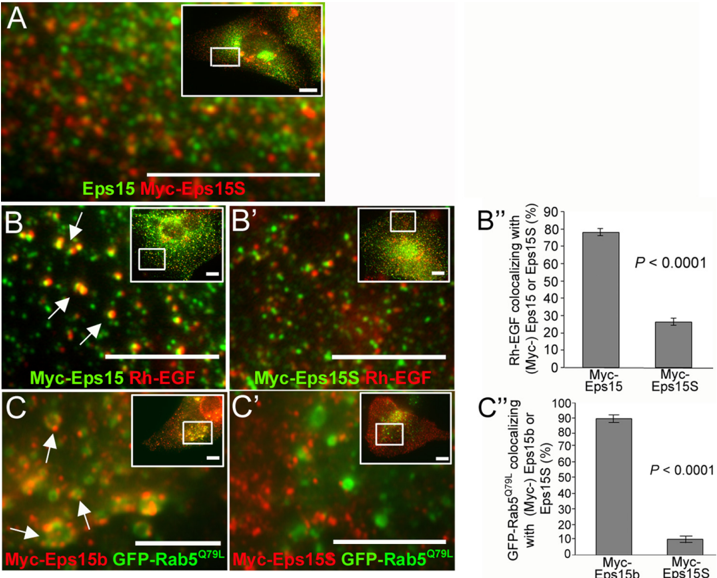
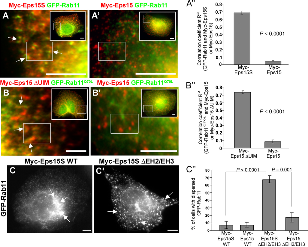
FIGURE 6 Eps15S localizes to the Rab11-positive ERC. A and A ′ , HuH-7 cells were transfected with GFP-Rab11 to label the ERC ( green ) and Myc-Eps15S ( A ) or Myc-Eps15( A ′ ). Cells were then immunostainedforMyc( red ). Higher magnificationimagesshowthatonlyMyc-Eps15S( A, arrows ), not Myc-Eps15( A ′ ), associates with the GFP-Rab11-positive ERC. Scale bars, 10 𝛍 m. A ″ , quantitation of GFP-Rab11 colocalizing with Myc-Eps15S or Myc-Eps15. Correlation coefficient R 2 between GFPRab11 and Myc-Eps15S or Myc-Eps15 was obtained from ≥ 30 random areas as shown in boxes drawn in each image from ≥ 10 cells. GFP-Rab11 colocalization with Myc-Eps15S is significantly higher than with Myc-Eps15 ( p < 0.0001). B and B ′ , HuH-7 cells were transfected with constitutively active Rab11 (GFP-Rab11 Q70L ; green ) andMyc-Eps15 𝚫 UIM( B ) or Myc-Eps15WT( B ′ ). Cells were then immunostainedforMyc( red ). Higher magnification imagesshowthatMyc-Eps15 𝚫 UIM( B, arrows ), but not Myc-Eps15 WT ( B ′ ), colocalizes with GFP-Rab11 Q70L . Scale bars, 5 𝛍 m. B ″ , quantitation of GFP-Rab11 Q70L colocalizing with Myc-Eps15 𝚫 UIM or Myc-Eps15. A correlation coefficient R 2 betweenGFP-Rab11 Q70L andMyc-Eps15 𝚫 UIMorMyc-Eps15wasobtainedfrom ≥ 30randomareasasshownin boxes drawnineachimage from ≥ 10 cells. GFP-Rab11 Q70L colocalizes with Myc-Eps15 𝚫 UIM significantly higher than Myc-Eps15 ( p < 0.0001). C and C ′ , HuH-7 cells were transfected with GFP-Rab11 and wild-type ( wt ) Myc-Eps15S ( C ) or the mutant truncated form, Myc-Eps15S 𝚫 EH2/EH3 ( C ′ ). GFP-Rab11 was distributed to a condensed perinuclear localizationinWTMyc-Eps15S-expressingcell( arrows in C ) butappearedmarkedlyfragmentedanddispersedinMyc-Eps15S 𝚫 EH2/EH3-expressingcells( arrows in C ′ ). Expression levels of WT Myc-Eps15S and Myc-Eps15S 𝚫 EH2/EH3 were confirmed by immunostaining for Myc (data not shown). Scale bars, 10 𝛍 m. C ″ , percentage of cells with dispersed localization of the GFP-Rab11 compartment in response to expression of the different Eps15 forms. From three independent experiments, ≥ 40 cells were counted for each experimental variable. Myc-Eps15S 𝚫 EH2/EH3 induced a significant dispersion of the GFP-Rab11 compared with Myc-Eps15S WT ( p < 0.0001) or to Myc-Eps15 𝚫 EH2/EH3 ( p = 0.001). Error bars indicate the standard error.
FIGURE 6 Eps15S localizes to the Rab11-positive ERC. A and A ′ , HuH-7 cells were transfected with GFP-Rab11 to label the ERC ( green ) and Myc-Eps15S ( A ) or Myc-Eps15( A ′ ). Cells were then immunostainedforMyc( red ). Higher magnificationimagesshowthatonlyMyc-Eps15S( A, arrows ), not Myc-Eps15( A ′ ), associates with the GFP-Rab11-positive ERC. Scale bars, 10 𝛍 m. A ″ , quantitation of GFP-Rab11 colocalizing with Myc-Eps15S or Myc-Eps15. Correlation coefficient R 2 between GFPRab11 and Myc-Eps15S or Myc-Eps15 was obtained from ≥ 30 random areas as shown in boxes drawn in each image from ≥ 10 cells. GFP-Rab11 colocalization with Myc-Eps15S is significantly higher than with Myc-Eps15 ( p < 0.0001). B and B ′ , HuH-7 cells were transfected with constitutively active Rab11 (GFP-Rab11 Q70L ; green ) andMyc-Eps15 𝚫 UIM( B ) or Myc-Eps15WT( B ′ ). Cells were then immunostainedforMyc( red ). Higher magnification imagesshowthatMyc-Eps15 𝚫 UIM( B, arrows ), but not Myc-Eps15 WT ( B ′ ), colocalizes with GFP-Rab11 Q70L . Scale bars, 5 𝛍 m. B ″ , quantitation of GFP-Rab11 Q70L colocalizing with Myc-Eps15 𝚫 UIM or Myc-Eps15. A correlation coefficient R 2 betweenGFP-Rab11 Q70L andMyc-Eps15 𝚫 UIMorMyc-Eps15wasobtainedfrom ≥ 30randomareasasshownin boxes drawnineachimage from ≥ 10 cells. GFP-Rab11 Q70L colocalizes with Myc-Eps15 𝚫 UIM significantly higher than Myc-Eps15 ( p < 0.0001). C and C ′ , HuH-7 cells were transfected with GFP-Rab11 and wild-type ( wt ) Myc-Eps15S ( C ) or the mutant truncated form, Myc-Eps15S 𝚫 EH2/EH3 ( C ′ ). GFP-Rab11 was distributed to a condensed perinuclear localizationinWTMyc-Eps15S-expressingcell( arrows in C ) butappearedmarkedlyfragmentedanddispersedinMyc-Eps15S 𝚫 EH2/EH3-expressingcells( arrows in C ′ ). Expression levels of WT Myc-Eps15S and Myc-Eps15S 𝚫 EH2/EH3 were confirmed by immunostaining for Myc (data not shown). Scale bars, 10 𝛍 m. C ″ , percentage of cells with dispersed localization of the GFP-Rab11 compartment in response to expression of the different Eps15 forms. From three independent experiments, ≥ 40 cells were counted for each experimental variable. Myc-Eps15S 𝚫 EH2/EH3 induced a significant dispersion of the GFP-Rab11 compared with Myc-Eps15S WT ( p < 0.0001) or to Myc-Eps15 𝚫 EH2/EH3 ( p = 0.001). Error bars indicate the standard error.CC-BY · From the PDF
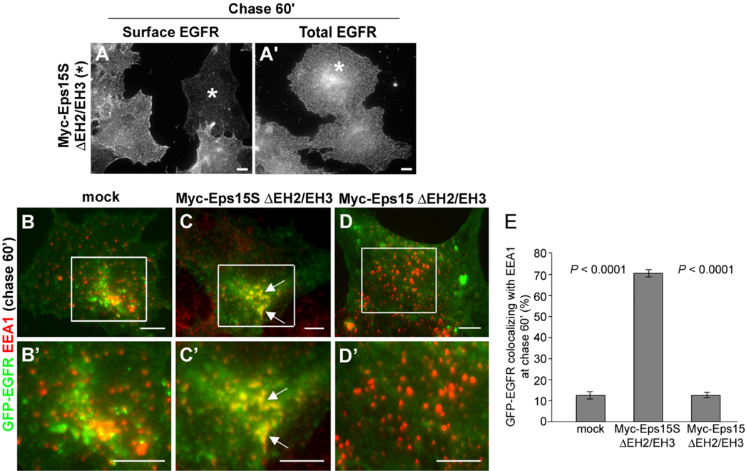
FIGURE 7 Expression of the Eps15S 𝚫 EH2/EH3 induces retention of EGFR in the early endosome. A and A ′ , surface only ( A ) or total cellular EGFR ( A ′ ) following a 60-min chase with 20 ng/ml EGF was visualized as described in Fig. 5. Cells expressing the Eps15 short form mutant protein ( A ) display very modest levels of EGFR at the plasma membrane. The retained receptor in these mutant cells accumulates within a perinuclear compartment ( A ′ ). Expression of Myc-Eps15S 𝚫 EH2/EH3 was confirmed by immunostaining for Myc (transfected cells indicated by *, data not shown). B-D ′ , to define the compartment to which EGFR is retained in the mutant-expressing cells, HuH-7 cells were cotransfected to express the GFP-EGFR under mock conditions ( B and B ′ ) or with Myc-Eps15S 𝚫 EH2/EH3 ( C and C ′ ) or Myc-Eps15 𝚫 EH2/EH3 ( D and D ′ ). Total EGFR (GFP-EGFR; green ) following a 60-min chase with 20 ng/ml EGF was examined as described in Fig. 5. Cells were then fixed and immunostained for EEA1 to label early endosomes ( red ). Higher magnification images of boxed regions show that GFP-EGFR colocalized markedly with EEA1 ( arrows in C and C ′ ) in Myc-Eps15S 𝚫 EH2/EH3-expressing cells, indicating aberrant retention in the early endosome ( yellow ). Little overlap was observed in the mock ( B and B ′ ) or Eps15 mutant-expressing cells ( D and D ′ ) because EGFR was either recycled back to the cell surface or degraded. Expression of Myc-Eps15S 𝚫 EH2/EH3 and Myc-Eps15 𝚫 EH2/EH3 was confirmed by immunostaining for Myc (data not shown). E, quantitation of GFP-EGFR colocalizing with EEA1. For each condition, ≥ 30 cells were measured. GFP-EGFR colocalized with EEA1 in Myc-Eps15S 𝚫 EH2/EH3-expressing cells significantly more than in mock-treated or Myc-Eps15 𝚫 EH2/EH3-expressing cells ( p < 0.0001). Error bars indicate standard error. Scale bars, 10 𝛍 m.
FIGURE 7 Expression of the Eps15S 𝚫 EH2/EH3 induces retention of EGFR in the early endosome. A and A ′ , surface only ( A ) or total cellular EGFR ( A ′ ) following a 60-min chase with 20 ng/ml EGF was visualized as described in Fig. 5. Cells expressing the Eps15 short form mutant protein ( A ) display very modest levels of EGFR at the plasma membrane. The retained receptor in these mutant cells accumulates within a perinuclear compartment ( A ′ ). Expression of Myc-Eps15S 𝚫 EH2/EH3 was confirmed by immunostaining for Myc (transfected cells indicated by *, data not shown). B-D ′ , to define the compartment to which EGFR is retained in the mutant-expressing cells, HuH-7 cells were cotransfected to express the GFP-EGFR under mock conditions ( B and B ′ ) or with Myc-Eps15S 𝚫 EH2/EH3 ( C and C ′ ) or Myc-Eps15 𝚫 EH2/EH3 ( D and D ′ ). Total EGFR (GFP-EGFR; green ) following a 60-min chase with 20 ng/ml EGF was examined as described in Fig. 5. Cells were then fixed and immunostained for EEA1 to label early endosomes ( red ). Higher magnification images of boxed regions show that GFP-EGFR colocalized markedly with EEA1 ( arrows in C and C ′ ) in Myc-Eps15S 𝚫 EH2/EH3-expressing cells, indicating aberrant retention in the early endosome ( yellow ). Little overlap was observed in the mock ( B and B ′ ) or Eps15 mutant-expressing cells ( D and D ′ ) because EGFR was either recycled back to the cell surface or degraded. Expression of Myc-Eps15S 𝚫 EH2/EH3 and Myc-Eps15 𝚫 EH2/EH3 was confirmed by immunostaining for Myc (data not shown). E, quantitation of GFP-EGFR colocalizing with EEA1. For each condition, ≥ 30 cells were measured. GFP-EGFR colocalized with EEA1 in Myc-Eps15S 𝚫 EH2/EH3-expressing cells significantly more than in mock-treated or Myc-Eps15 𝚫 EH2/EH3-expressing cells ( p < 0.0001). Error bars indicate standard error. Scale bars, 10 𝛍 m.CC-BY · From the PDF
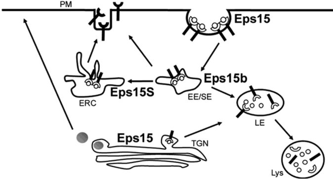
FIGURE 8 Modeldepicting roles of three Eps15 forms in EGFR trafficking. Eps15 functions in internalization of EGFR at the plasma membrane, and Eps15b localized on early endosomes mediates sorting of EGFR to the late endosome/lysosome for degradation. Eps15S directs the internalized EGFR to the recycling endosome for recycling back to the cell surface.
FIGURE 8 Modeldepicting roles of three Eps15 forms in EGFR trafficking. Eps15 functions in internalization of EGFR at the plasma membrane, and Eps15b localized on early endosomes mediates sorting of EGFR to the late endosome/lysosome for degradation. Eps15S directs the internalized EGFR to the recycling endosome for recycling back to the cell surface.CC-BY · From the PDF

Verification

Claims the paper makes

The claims and statistics of this paper haven’t been checked yet.

Run the check — free, no account needed. It reads the open-access text and checks the sources it cites.

A few free checks a day · papers already verified open instantly · results are public

Claims other papers make about this one

Each is checked against this paper’s own text: does it say what the citing paper says it does?

Self-check

Percentages, sums and reported differences re-computed from the paper’s own numbers.

The self-check needs the paper’s full text.

Citations

Cites

No references could be resolved yet.

Research works printed but not linked: 51

  1. Maxfield, 2004, Nat. Rev. Mol. Cell Biol., 5, 121, 10.1038/nrm1315 · DOI · Paper page
  2. Jones, 2006, Curr. Opin. Cell Biol., 18, 549, 10.1016/j.ceb.2006.08.003 · DOI
  3. Doherty, 2009, Annu. Rev. Biochem., 78, 857, 10.1146/annurev.biochem.78.081307.110540 · DOI · Paper page
  4. Gruenberg, 2004, Nat. Rev. Mol. Cell Biol., 5, 317, 10.1038/nrm1360 · DOI
  5. Slagsvold, 2006, Trends Cell Biol., 16, 317, 10.1016/j.tcb.2006.04.004 · DOI · Paper page
  6. Woodman, 2008, Curr. Opin. Cell Biol., 20, 408, 10.1016/j.ceb.2008.04.001 · DOI
  7. Sigismund, 2008, Dev. Cell, 15, 209, 10.1016/j.devcel.2008.06.012 · DOI
  8. Naslavsky, 2005, J. Cell Sci., 118, 4093, 10.1242/jcs.02595 · DOI · Paper page
  9. Grant, 2008, Traffic, 9, 2043, 10.1111/j.1600-0854.2008.00834.x · DOI · Paper page
  10. Grant, 2009, Nat. Rev. Mol. Cell Biol., 10, 597, 10.1038/nrm2755 · DOI · Paper page
  11. van der Sluijs, 1992, Cell, 70, 729, 10.1016/0092-8674(92)90307-X · DOI
  12. Sheff, 1999, J. Cell Biol., 145, 123, 10.1083/jcb.145.1.123 · DOI · Paper page
  13. Kouranti, 2006, Curr. Biol., 16, 1719, 10.1016/j.cub.2006.07.020 · DOI · Paper page
  14. Ren, 1998, Proc. Natl. Acad. Sci. U.S.A., 95, 6187, 10.1073/pnas.95.11.6187 · DOI · Paper page
  15. Naslavsky, 2006, Mol. Biol. Cell, 17, 163, 10.1091/mbc.e05-05-0466 · DOI · Paper page
  16. D'Souza-Schorey, 2006, Nat. Rev. Mol. Cell Biol., 7, 347, 10.1038/nrm1910 · DOI
  17. Sharma, 2009, Mol. Biol. Cell, 20, 5181, 10.1091/mbc.e09-06-0535 · DOI · Paper page
  18. Horgan, 2009, Biochem. Soc. Trans., 37, 1032, 10.1042/BST0371032 · DOI
  19. Carbone, 1997, Cancer Res., 57, 5498
  20. Benmerah, 1999, J. Cell Sci., 112, 1303, 10.1242/jcs.112.9.1303 · DOI
  21. Benmerah, 2000, J. Biol. Chem., 275, 3288, 10.1074/jbc.275.5.3288 · DOI
  22. van Bergen En Henegouwen, 2009, Cell Commun. Signal., 7, 24, 10.1186/1478-811X-7-24 · DOI
  23. Haffner, 1997, FEBS Lett., 419, 175, 10.1016/S0014-5793(97)01451-8 · DOI
  24. Chen, 1998, Nature, 394, 793, 10.1038/29555 · DOI · Paper page
  25. Tebar, 1996, J. Biol. Chem., 271, 28727, 10.1074/jbc.271.46.28727 · DOI
  26. Iannolo, 1997, Cancer Res., 57, 240
  27. Klapisz, 2002, J. Biol. Chem., 277, 30746, 10.1074/jbc.M203004200 · DOI
  28. Polo, 2002, Nature, 416, 451, 10.1038/416451a · DOI · Paper page
  29. de Melker, 2004, J. Biol. Chem., 279, 55465, 10.1074/jbc.M409765200 · DOI
  30. Sigismund, 2005, Proc. Natl. Acad. Sci. U.S.A., 102, 2760, 10.1073/pnas.0409817102 · DOI · Paper page
  31. Hoeller, 2006, Nat. Cell Biol., 8, 163, 10.1038/ncb1354 · DOI
  32. Roxrud, 2008, J. Cell Biol., 180, 1205, 10.1083/jcb.200708115 · DOI
  33. Cao, 2005, Nat. Cell Biol., 7, 483, 10.1038/ncb1246 · DOI · Paper page
  34. Chi, 2008, Mol. Biol. Cell, 19, 3564, 10.1091/mbc.e07-10-0997 · DOI
  35. Orth, 2006, Cancer Res., 66, 3603, 10.1158/0008-5472.CAN-05-2916 · DOI
  36. Cao, 2010, Mol. Cell. Biol., 30, 781, 10.1128/MCB.00330-09 · DOI
  37. Henley, 1998, J. Cell Biol., 141, 85, 10.1083/jcb.141.1.85 · DOI · Paper page
  38. Fazioli, 1993, Mol. Cell. Biol., 13, 5814
  39. Bache, 2003, J. Biol. Chem., 278, 12513, 10.1074/jbc.M210843200 · DOI · Paper page
  40. Coda, 1998, J. Biol. Chem., 273, 3003, 10.1074/jbc.273.5.3003 · DOI
  41. Poupon, 2002, J. Biol. Chem., 277, 8941, 10.1074/jbc.M108385200 · DOI
  42. Torrisi, 1999, Mol. Biol. Cell, 10, 417, 10.1091/mbc.10.2.417 · DOI
  43. Acconcia, 2009, Exp. Cell Res., 315, 1610, 10.1016/j.yexcr.2008.10.014 · DOI
  44. Ullrich, 1996, J. Cell Biol., 135, 913, 10.1083/jcb.135.4.913 · DOI · Paper page
  45. Schlierf, 2000, Exp. Cell Res., 259, 257, 10.1006/excr.2000.4947 · DOI
  46. Naslavsky, 2004, Mol. Biol. Cell, 15, 2410, 10.1091/mbc.e03-10-0733 · DOI
  47. Soldati, 2006, Nat. Rev. Mol. Cell Biol., 7, 897, 10.1038/nrm2060 · DOI
  48. Nedvetsky, 2007, Traffic, 8, 110, 10.1111/j.1600-0854.2006.00508.x · DOI
  49. Swiatecka-Urban, 2007, J. Biol. Chem., 282, 23725, 10.1074/jbc.M608531200 · DOI
  50. Wang, 2008, Cell, 135, 535, 10.1016/j.cell.2008.09.057 · DOI · Paper page
  51. Traer, 2007, Nat. Cell Biol., 9, 1370, 10.1038/ncb1656 · DOI · Paper page

Cited by

Sorted by citations

Metadata

Authors
Chi, S., Cao, H., Wang, Y., & McNiven, M. A.
Venue
Journal of Biological Chemistry
Publisher
Elsevier BV
Published
2011-10
Volume
286
Issue
40
Pages
35196-35208
ISSN
0021-9258
Cited by
53
References
0
Language
en

About these checks

Registry, retraction, Cites and statistics results are computed, not judged, with one exception: a printed reference that no index lists is read once by a model before it counts as a citation error. Claims are extracted by a model and kept only when their quote appears verbatim in the text; each cited source is then checked, with a model’s help, for whether it says what the claim relies on. Those verdicts can be wrong — read the claim’s quote and the cited source before relying on one.

Statistics and arithmetic are recomputed, not judged.

Confidence bands are shown only for fields calibrated on our annotated test set. None is calibrated yet, so no band is shown.

What counts as a claim ›How we measure confidence ›How we check this ›

Are you an author, or do you see a problem with this page? Request a review