

The DOI matches its Crossref record; 73 of the 74 references it cites were found in a registry or an index.
Regulation of Apoptosis in Myeloid Cells by Interferon Consensus Sequence–Binding Protein
Open accessAbstract
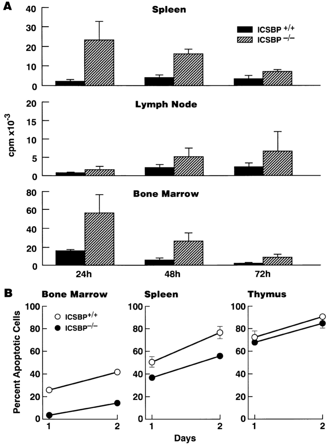
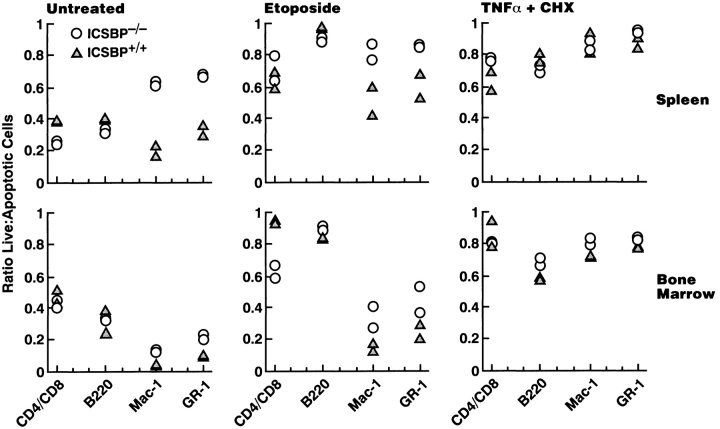
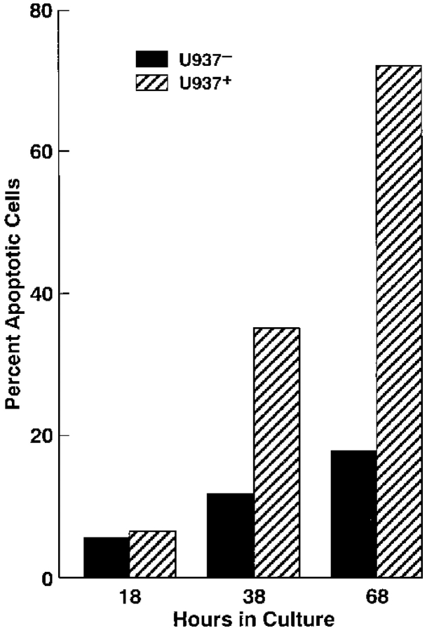
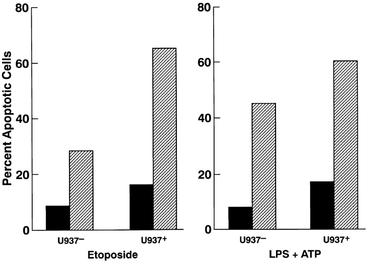
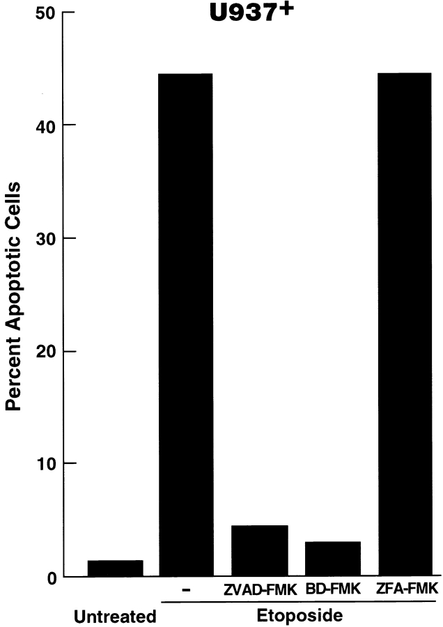
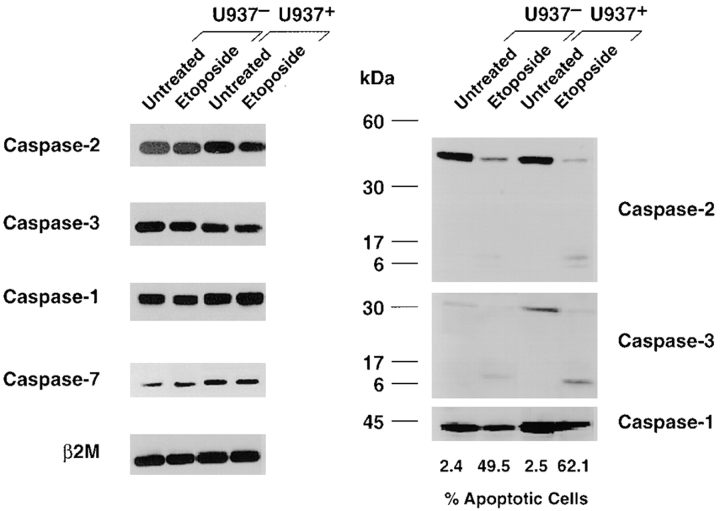
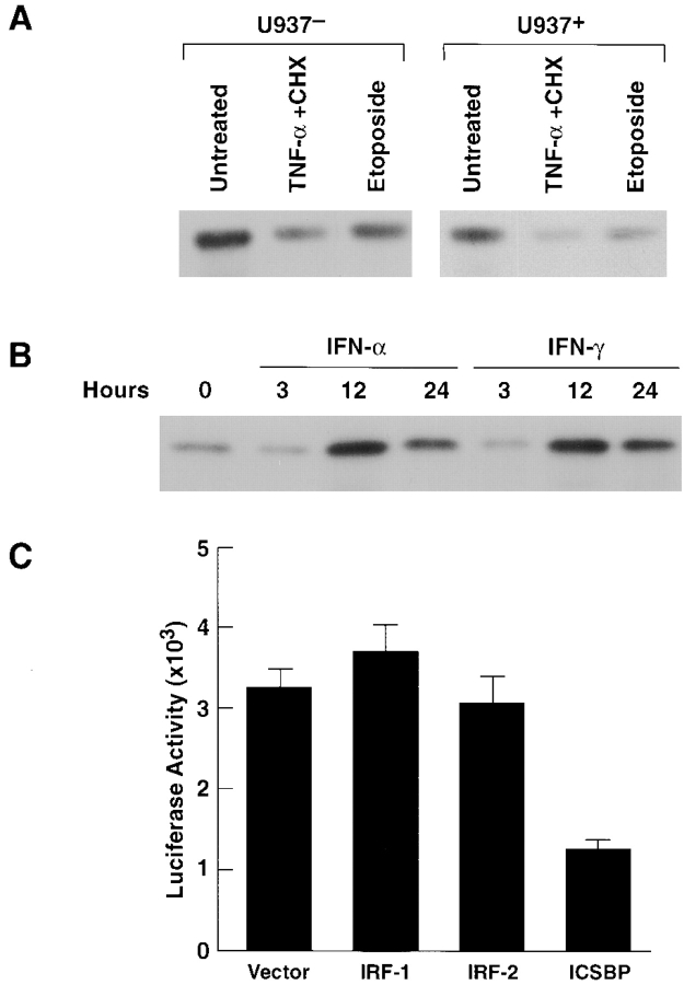
Mice with a null mutation of the gene encoding interferon consensus sequence–binding protein (ICSBP) develop a disease with marked expansion of granulocytes and macrophages that frequently progresses to a fatal blast crisis, thus resembling human chronic myelogenous leukemia (CML). One important feature of CML is decreased responsiveness of myeloid cells to apoptotic stimuli. Here we show that myeloid cells from mice deficient in ICSBP exhibit reduced spontaneous apoptosis and a significant decrease in sensitivity to apoptosis induced by DNA damage. In contrast, apoptosis in thymocytes from ICSBP-deficient mice is unaffected. We also show that overexpression of ICSBP in the human U937 monocytic cell line enhances the rate of spontaneous apoptosis and the sensitivity to apoptosis induced by etoposide, lipopolysaccharide plus ATP, or rapamycin. Programmed cell death induced by etoposide was specifically blocked by peptides inhibitory for the caspase-1 or caspase-3 subfamilies of caspases. Studies of proapoptotic genes showed that cells overexpressing ICSBP have enhanced expression of caspase-3 precursor protein. In addition, analyses of antiapoptotic genes showed that overexpression of ICSBP results in decreased expression of Bcl-XL. These data suggest that ICSBP modulates survival of myeloid cells by regulating expression of apoptosis-related genes.
Overview
The claims and statistics of this paper haven’t been checked yet.
Run the check — free, no account needed. It reads the open-access text and checks the sources it cites.
Integrity signals
Registry
Cites
Statistics
Figures
Verification
Claims the paper makes
The claims and statistics of this paper haven’t been checked yet.
Run the check — free, no account needed. It reads the open-access text and checks the sources it cites.
Claims other papers make about this one
Each is checked against this paper’s own text: does it say what the citing paper says it does?
In the citing paper
“Myeloid cells (Gr1 1 as well as Mac1 1 cells) from ICSBP 2 / 2 were shown to exhibit reduced spontaneous apoptosis.”
From Review: ICSBP/IRF-8: Its Regulatory Roles in the Development of Myeloid Cells (2002) · cites this paper as [56]
In the citing paper
“Programmed cell death induced by etoposide was specifically blocked by peptides inhibitory for the caspase-1 or caspase-3 subfamilies of caspases.”
From Review: ICSBP/IRF-8: Its Regulatory Roles in the Development of Myeloid Cells (2002) · cites this paper as [56]
In the citing paper
“Cells overexpressing ICSBP had enhanced expression of caspase-3 precursor protein and decreased expression of Bcl-X(L).”
From Review: ICSBP/IRF-8: Its Regulatory Roles in the Development of Myeloid Cells (2002) · cites this paper as [56]
In the citing paper
“Overexpression of ICSBP in the human U937 myelomonoblastic leukemia cell line enhanced the rate of spontaneous apoptosis and increased the sensitivity to apoptosis induced by etoposide, LPS plus ATP, or rapamycin.”
From Review: ICSBP/IRF-8: Its Regulatory Roles in the Development of Myeloid Cells (2002) · cites this paper as [56]
In the citing paper
“Overexpression of ICSBP in the human U937 myelomonoblastic leukemia cell line enhanced the rate of spontaneous apoptosis and increased the sensitivity to apoptosis induced by etoposide, LPS plus ATP, or rapamycin.”
From Review: ICSBP/IRF-8: Its Regulatory Roles in the Development of Myeloid Cells (2002) · cites this paper as [56]
In the citing paper
“Further, in ICSBP 2 / 2 cells, there was a significant decrease in the sensitivity to apoptosis induced by DNA damage. (56)”
From Review: ICSBP/IRF-8: Its Regulatory Roles in the Development of Myeloid Cells (2002) · cites this paper as [56]
In the citing paper
“Cells overexpressing ICSBP had enhanced expression of caspase-3 precursor protein and decreased expression of Bcl-X(L).”
From Review: ICSBP/IRF-8: Its Regulatory Roles in the Development of Myeloid Cells (2002) · cites this paper as [56]
In the citing paper
“In contrast, apoptosis in ICSBP 2 / 2 thymocytes was unaffected.”
From Review: ICSBP/IRF-8: Its Regulatory Roles in the Development of Myeloid Cells (2002) · cites this paper as [56]
Self-check
Percentages, sums and reported differences re-computed from the paper’s own numbers.
The self-check needs the paper’s full text.
Citations
Cites
p53, the cellular gatekeeper for growth and division
Levine, A. J. (1997) · Cell · doi:10.1016/s0092-8674(00)81871-1 · Cited by 7,693
Thornberry, N. A., & Lazebnik, Y. A. (1998) · Science · doi:10.1126/science.281.5381.1312 · Cited by 6,857
Darnell, J. E., Kerr, L. M., & Stark, G. R. (1994) · Science · doi:10.1126/science.8197455 · Cited by 6,080
Bcl-2 heterodimerizes in vivo with a conserved homolog, Bax, that accelerates programed cell death
Oltval, Z. N., Milliman, C. L., & Korsmeyer, S. J. (1993) · Cell · doi:10.1016/0092-8674(93)90509-o · Cited by 6,003
The Bcl-2 Protein Family: Arbiters of Cell Survival
Adams, J. M., & Cory, S. (1998) · Science · doi:10.1126/science.281.5381.1322 · Cited by 5,334
Tumor suppressor p53 is a direct transcriptional activator of the human bax gene
Toshiyuki, M., & Reed, J. C. (1995) · Cell · doi:10.1016/0092-8674(95)90412-3 · Cited by 4,063
A Combinatorial Approach Defines Specificities of Members of the Caspase Family and Granzyme B
Thornberry, N. A., Rano, T. A., Peterson, E. P., Rasper, D., Timkey, T., Garcia-Calvo, M. M., Houtzager, V. M., Nordstrom, P. A., Roy, S., Vaillancourt, J. P., et al. (1997) · Journal of Biological Chemistry · doi:10.1074/jbc.272.29.17907 · Cited by 2,089
A mammalian protein targeted by G1-arresting rapamycin–receptor complex
Brown, E. J., Albers, M. W., Bum Shin, T., ichikawa, K., Keith, C. T., Lane, W. S., & Schreiber, S. L. (1994) · Nature · doi:10.1038/369756a0 · Cited by 2,053
Double identity for proteins of the Bcl-2 family
Reed, J. C. (1997) · Nature · doi:10.1038/42867 · Cited by 1,494
Li, P., Allen, H., Banerjee, S., Franklin, S., Herzog, L., Johnston, C. G., McDowell, J., Paskind, M. P., Rodman, L., Salfeld, J. G., et al. (1995) · Cell · doi:10.1016/0092-8674(95)90490-5 · Cited by 1,408
Research works printed but not linked: 1
- Sambrook, 1989, Molecular Cloning
Cited by
Sorted by citations
The molecular biology of chronic myeloid leukemia
Deininger, M. W., Goldman, J. M., & Melo, J. V. (2000) · Blood · doi:10.1182/blood.v96.10.3343 · Cited by 1,654
The IRF Family Transcription Factors in Immunity and Oncogenesis
Tamura, T., Yanai, H., Savitsky, D., & Taniguchi, T. (2008) · Annual Review of Immunology · doi:10.1146/annurev.immunol.26.021607.090400 · Cited by 1,259
Microglia development and function
Nayak, D., Roth, T. L., & McGavern, D. B. (2014) · Annual Review of Immunology · doi:10.1146/annurev-immunol-032713-120240 · Cited by 1,154
Apoptosis and interferons: role of interferon-stimulated genes as mediators of apoptosis
Chawla-Sarkar, M., Lindner, D. J., Liu, Y.-F., Williams, B. R., Sen, G. C., Silverman, R. H., & Borden, E. C. (2003) · Apoptosis · doi:10.1023/a:1023668705040 · Cited by 829
Molecular biology of bcr-abl1–positive chronic myeloid leukemia
Quintás‐Cardama, A., & Cortes, J. E. (2008) · Blood · doi:10.1182/blood-2008-03-144790 · Cited by 757
Inflammatory signals in dendritic cell activation and the induction of adaptive immunity
Joffre, O. P., Nolte, M. A., Spörri, R., & Sousa, C. R. E. (2008) · Immunological Reviews · doi:10.1111/j.1600-065x.2008.00718.x · Cited by 620
Host defense, viruses and apoptosis
Barber, G. N. (2001) · Cell Death and Differentiation · doi:10.1038/sj.cdd.4400823 · Cited by 576
Myeloid-Derived Suppressor Cells
Parker, K. H., Beury, D. W., & Ostrand‐Rosenberg, S. (2015) · Advances in cancer research · doi:10.1016/bs.acr.2015.04.002 · Cited by 490
Schiavoni, G., Mattei, F., Sestili, P., Borghi, P., Venditti, M., Morse, H. C., Belardelli, F., & Gabriele, L. (2002) · The Journal of Experimental Medicine · doi:10.1084/jem.20021263 · Cited by 423
Self-citationMyeloid-derived suppressor cell development is regulated by a STAT/IRF-8 axis
Waight, J. D., Netherby, C. S., Hensen, M. L., Miller, A., Hu, Q., Liu, S., Bogner, P. N., Farren, M. R., Lee, K. P., Liu, K., et al. (2013) · Journal of Clinical Investigation · doi:10.1172/jci68189 · Cited by 325
ICSBP Directs Bipotential Myeloid Progenitor Cells to Differentiate into Mature Macrophages
Tamura, T., Nagamura-Inoue, T., Shmeltzer, Z., Kuwata, T., & Ozato, K. (2000) · Immunity · doi:10.1016/s1074-7613(00)00016-9 · Cited by 305
Self-citationRegulation of immunity and oncogenesis by the IRF transcription factor family
Savitsky, D., Tamura, T., Yanai, H., & Taniguchi, T. (2010) · Cancer Immunology, Immunotherapy · doi:10.1007/s00262-009-0804-6 · Cited by 298
Neutrophil β2 integrins: moderators of life or death decisions
Mayadas, T. N., & Culleré, X. (2005) · Trends in Immunology · doi:10.1016/j.it.2005.05.002 · Cited by 247
Natural course and biology of CML
Chereda, B., & Melo, J. V. (2015) · Annals of Hematology · doi:10.1007/s00277-015-2325-z · Cited by 226
Review: ICSBP/IRF-8: Its Regulatory Roles in the Development of Myeloid Cells
Tamura, T., & Ozato, K. (2002) · Journal of Interferon & Cytokine Research · doi:10.1089/107999002753452755 · Cited by 183
Self-citationIrf8 regulates macrophage versus neutrophil fate during zebrafish primitive myelopoiesis
Li, L., Jin, H., Xu, J., Shi, Y., & Wen, Z. (2010) · Blood · doi:10.1182/blood-2010-06-290700 · Cited by 157
Interferon regulatory factor family of transcription factors and regulation of oncogenesis
Takaoka, A., Tamura, T., & Taniguchi, T. (2008) · Cancer Science · doi:10.1111/j.1349-7006.2007.00720.x · Cited by 152
BLIMP-1: trigger for differentiation of myeloid lineage
Chang, D., Angelin‐Duclos, C., & Calame, K. L. (2000) · Nature Immunology · doi:10.1038/77861 · Cited by 149
Biology of chronic myelogenous leukemia—signaling pathways of initiation and transformation
Melo, J. V., & Deininger, M. W. (2004) · Hematology/Oncology Clinics of North America · doi:10.1016/j.hoc.2004.03.008 · Cited by 145
Schwieger, M., Löhler, J., Friel, J., Scheller, M., Horak, I. D., & Stocking, C. (2002) · The Journal of Experimental Medicine · doi:10.1084/jem.20020824 · Cited by 143
Vitamin A deficiency in mice causes a systemic expansion of myeloid cells
Kuwata, T., Wang, I., Tamura, T., Ponnamperuma, R. M., Levine, R., Holmes, K. L., Morse, H. C., Luca, L. M. D., & Ozato, K. (2000) · Blood · doi:10.1182/blood.v95.11.3349 · Cited by 141
Self-citationRegulation of myelopoiesis by the transcription factor IRF8
Tamura, T., Kurotaki, D., & Koizumi, S.-I. (2015) · International Journal of Hematology · doi:10.1007/s12185-015-1761-9 · Cited by 139
Qian, Z., Fernald, A. A., Godley, L. A., Larson, R. A., & Beau, M. M. L. (2002) · Proceedings of the National Academy of Sciences · doi:10.1073/pnas.222491799 · Cited by 139
Yáñez, A., Ng, M. Y., Hassanzadeh‐Kiabi, N., & Goodridge, H. S. (2015) · Blood · doi:10.1182/blood-2014-09-600833 · Cited by 140
TRIM21 – A potential novel therapeutic target in cancer
Alomari, M. (2021) · Pharmacological Research · doi:10.1016/j.phrs.2021.105443 · Cited by 113
Schiavoni, G., Mattei, F., Borghi, P., Sestili, P., Venditti, M., Morse, H. C., Belardelli, F., & Gabriele, L. (2003) · Blood · doi:10.1182/blood-2003-09-3007 · Cited by 107
Self-citationCytogenetic and Molecular Genetic Aspects of Chronic Myeloid Leukaemia
Barnes, D. J., & Melo, J. V. (2002) · Acta Haematologica · doi:10.1159/000065655 · Cited by 106
Burchert, A. (2003) · Blood · doi:10.1182/blood-2003-08-2970 · Cited by 102
Yang, D., Thangaraju, M., Greeneltch, K. M., Browning, D. D., Schoenlein, P. V., Tamura, T., Ozato, K., Ganapathy, V., Abrams, S. I., & Liu, K. (2007) · Cancer Research · doi:10.1158/0008-5472.can-06-4068 · Cited by 94
Self-citationIFN Regulatory Factor 4 and 8 Promote Ig Light Chain κ Locus Activation in Pre-B Cell Development
Ma, S., Turetsky, A., Trinh, L. X., & Lu, R. (2006) · The Journal of Immunology · doi:10.4049/jimmunol.177.11.7898 · Cited by 95
Banerjee, S., Lu, J. L., Cai, Q., Saha, A., Jha, H. C., Dzeng, R. K., & Robertson, E. S. (2013) · PLoS Pathogens · doi:10.1371/journal.ppat.1003314 · Cited by 81
Smith, A. D., Lu, C., Payne, D., Paschall, A. V., Klement, J. D., Redd, P. S., Ibrahim, M. L., Yang, D., Han, Q., Liu, Z., et al. (2020) · Cancer Research · doi:10.1158/0008-5472.can-19-3670 · Cited by 80
Netherby, C. S., Messmer, M. N., Burkard-Mandel, L. E., Colligan, S. H., Miller, A., Gomez, E. C., Wang, J., Nemeth, M. J., & Abrams, S. I. (2017) · The Journal of Immunology · doi:10.4049/jimmunol.1601722 · Cited by 77
Animal models of acute myelogenous leukaemia – development, application and future perspectives
McCormack, E., Bruserud, Ø., & Gjertsen, B. T. (2005) · Leukemia · doi:10.1038/sj.leu.2403670 · Cited by 78
IRF8 Regulates Acid Ceramidase Expression to Mediate Apoptosis and Suppresses Myelogeneous Leukemia
Hu, X., Yang, D., Zimmerman, M. A., Liu, F., Yang, J., Kannan, S., Burchert, A., Szulc, Z. M., Bielawska, A. E., Ozato, K., et al. (2011) · Cancer Research · doi:10.1158/0008-5472.can-10-2493 · Cited by 72
Self-citationIdentification of 12/15-lipoxygenase as a suppressor of myeloproliferative disease
Middleton, M. K., Zukas, A. M., Rubinstein, T., Jacob, M., Zhu, P., Zhao, L., Blair, I. A., & Puré, E. (2006) · The Journal of Experimental Medicine · doi:10.1084/jem.20061444 · Cited by 69
WEAVER, B. K., ANDO, O., Kumar, K. P., & Reich, N. C. (2001) · The FASEB Journal · doi:10.1096/fj.00-0222com · Cited by 68
Hu, X., Bardhan, K., Paschall, A. V., Yang, D., Waller, J. L., Park, M. A., Nayak-Kapoor, A., Samuel, T. A., Abrams, S. I., & Liu, K. (2013) · Journal of Biological Chemistry · doi:10.1074/jbc.m112.434530 · Cited by 68
IRF8: Mechanism of Action and Health Implications
Moorman, H. R., Reategui-Almonacid, Y., Poschel, D. B., & Liu, K. (2022) · Cells · doi:10.3390/cells11172630 · Cited by 63
ICSBP/IRF-8 inhibits mitogenic activity of p210 Bcr/Abl in differentiating myeloid progenitor cells
Tamura, T., Kong, H. J., Tunyaplin, C., Tsujimura, H., Calame, K. L., & Ozato, K. (2003) · Blood · doi:10.1182/blood-2003-01-0291 · Cited by 61
Self-citationKautz, B., Kakar, R., David, E., & Eklund, E. A. (2001) · Journal of Biological Chemistry · doi:10.1074/jbc.m103381200 · Cited by 61
Transcription factors in myeloid-derived suppressor cell recruitment and function
Sonda, N., Chioda, M., Zilio, S., Simonato, F., & Bronte, V. (2011) · Current Opinion in Immunology · doi:10.1016/j.coi.2010.12.006 · Cited by 58
McGough, J. M., Yang, D., Huang, S., Georgi, D. E., Hewitt, S. M., Röcken, C., Tänzer, M., Ebert, M. P., & Liu, K. (2008) · Molecular Cancer Research · doi:10.1158/1541-7786.mcr-08-0280 · Cited by 61
Mattei, F., Schiavoni, G., De Ninno, A., Lucarini, V., Sestili, P., Sistigu, A., Fragale, A., Sanchez, M., Spada, M., Gerardino, A., et al. (2014) · Journal of Immunotoxicology · doi:10.3109/1547691x.2014.891677 · Cited by 57
Self-citationVarela, N., Muñoz‐Pinedo, C., Ruiz‐Ruiz, C., Robledo, G., Pedroso, M., & López‐Rivas, A. (2001) · Journal of Biological Chemistry · doi:10.1074/jbc.m100815200 · Cited by 57
Mattei, F., Schiavoni, G., Sestili, P., Spadaro, F., Fragale, A., Sistigu, A., Lucarini, V., Spada, M., Sanchez, M., Scala, S., et al. (2012) · Neoplasia · doi:10.1593/neo.121444 · Cited by 60
Self-citationEklund, E. A., Goldenberg, I., Lu, Y., Andrejic, J., & Kakar, R. (2002) · Journal of Biological Chemistry · doi:10.1074/jbc.m203917200 · Cited by 55
Huang, W., Zhu, C., Wang, H., Horvath, E., & Eklund, E. A. (2008) · Journal of Biological Chemistry · doi:10.1074/jbc.m706710200 · Cited by 54
Lv, D., Zhang, K., & Li, R. (2018) · PLoS Pathogens · doi:10.1371/journal.ppat.1006868 · Cited by 53
Yang, D., Thangaraju, M., Browning, D. D., Dong, Z., Korchin, B., Lev, D. C., Ganapathy, V., & Liu, K. (2007) · The Journal of Immunology · doi:10.4049/jimmunol.179.7.4775 · Cited by 55
Kee, J., Ito, A., Hojo, S., Hashimoto, I., Igarashi, Y., Tsuneyama, K., Tsukada, K., Irimura, T., Shibahara, N., Takasaki, I., et al. (2014) · BMC Cancer · doi:10.1186/1471-2407-14-949 · Cited by 52
Paschall, A. V., Zhang, R., Qi, C., Bardhan, K., Peng, L., Lu, G., Yang, J., Mérad, M., McGaha, T. L., Zhou, G., et al. (2015) · The Journal of Immunology · doi:10.4049/jimmunol.1402412 · Cited by 55
Self-citationInterferon Regulatory Factor-8 Modulates the Development of Tumor-Induced CD11b+Gr-1+Myeloid Cells
Stewart, T. J., Greeneltch, K. M., Reid, J., Liewehr, D. J., Steinberg, S. M., Liu, K., & Abrams, S. I. (2009) · Journal of Cellular and Molecular Medicine · doi:10.1111/j.1582-4934.2009.00685.x · Cited by 50
Oncogenic signaling: new insights and controversies from chronic myeloid leukemia
Etten, R. A. V. (2007) · The Journal of Experimental Medicine · doi:10.1084/jem.20062335 · Cited by 52
Deng, M., & Daley, G. Q. (2001) · Blood · doi:10.1182/blood.v97.11.3491 · Cited by 50
Irf8-Regulated Genomic Responses Drive Pathological Inflammation during Cerebral Malaria
Berghout, J., Langlais, D., Radovanovic, I., Tam, M., MacMicking, J. D., Stevenson, M. M., & Gros, P. (2013) · PLoS Pathogens · doi:10.1371/journal.ppat.1003491 · Cited by 44
Hematopoietic Interferon Regulatory Factor 8-Deficiency Accelerates Atherosclerosis in Mice
Döring, Y., Soehnlein, O., Drechsler, M. D., Shagdarsuren, E., Chaudhari, S. M., Meiler, S. U., Hartwig, H., Hristov, M., Koenen, R. R., Hieronymus, T., et al. (2012) · Arteriosclerosis Thrombosis and Vascular Biology · doi:10.1161/atvbaha.111.236539 · Cited by 48
Review: genetic models of acute myeloid leukaemia
McCormack, E., Bruserud, Ø., & Gjertsen, B. T. (2008) · Oncogene · doi:10.1038/onc.2008.16 · Cited by 49
Cross talk between Wnt/β-catenin and Irf8 in leukemia progression and drug resistance
Scheller, M., Schönheit, J., Zimmermann, K., Leser, U., Rosenbauer, F., & Leutz, A. (2013) · The Journal of Experimental Medicine · doi:10.1084/jem.20130706 · Cited by 51
Liu, K., & Abrams, S. I. (2003) · The Journal of Immunology · doi:10.4049/jimmunol.170.12.6329 · Cited by 46
The interferons and cell death: guardians of the cell or accomplices of apoptosis?
Barber, G. N. (2000) · Seminars in Cancer Biology · doi:10.1006/scbi.2000.0313 · Cited by 46
Zhu, C., Saberwal, G., Lu, Y., Platanias, L. C., & Eklund, E. A. (2004) · Journal of Biological Chemistry · doi:10.1074/jbc.m405736200 · Cited by 45
Dror, N., Rave-Harel, N., Burchert, A., Azriel, A., Tamura, T., Tailor, P. B., Neubauer, A., Ozato, K., & Levi, B.-Z. (2006) · Journal of Biological Chemistry · doi:10.1074/jbc.m607825200 · Cited by 44
Self-citationRen, R. (2002) · Oncogene · doi:10.1038/sj.onc.1206090 · Cited by 43
Genetic Determinants of Susceptibility to Mycobacterial Infections: IRF8, A New Kid on the Block
Salem, S., & Gros, P. (2013) · Advances in experimental medicine and biology · doi:10.1007/978-1-4614-6111-1_3 · Cited by 42
Huang, W., Saberwal, G., Horvath, E., Zhu, C., Lindsey, S., & Eklund, E. A. (2006) · Molecular and Cellular Biology · doi:10.1128/mcb.00036-06 · Cited by 41
Sung, J. J. �., Park, S.-Y., Kim, J. H., Kang, H. G., Yoon, J.-H., Na, Y. S., Kim, Y.-N., & Park, B.-K. (2014) · Cell Death and Disease · doi:10.1038/cddis.2014.189 · Cited by 39
Interferon Consensus Sequence Binding Protein Confers Resistance against Yersinia enterocolitica
Hein, J., Kempf, V. A. J., Diebold, J., Bücheler, N., Preger, S., Horak, I., Sing, A., Kramer, U., & Autenrieth, I. B. (2000) · Infection and Immunity · doi:10.1128/iai.68.3.1408-1417.2000 · Cited by 39
Epigenetic silencing of the interferon regulatory factor ICSBP/IRF8 in human multiple myeloma
Tshuikina, M., Jernberg‐Wiklund, H., Nilsson, K., & Oberg, F. G. (2008) · Experimental Hematology · doi:10.1016/j.exphem.2008.08.001 · Cited by 39
Zhang, Q., Zhang, L., Li, L., Wang, Z., Ying, J., Fan, Y., Xu, B., Wang, L., Liu, Q., Chen, G., et al. (2014) · Cancer Letters · doi:10.1016/j.canlet.2014.07.040 · Cited by 38
Cooperation between deficiencies of IRF-4 and IRF-8 promotes both myeloid and lymphoid tumorigenesis
Jo, S., Schatz, J. H., Acquaviva, J., Singh, H., & Ren, R. (2010) · Blood · doi:10.1182/blood-2009-07-234559 · Cited by 37
Yang, D., Wang, S., Brooks, C. R., Dong, Z., Schoenlein, P. V., Kumar, V., Ouyang, X., Xiong, H., Lahat, G., Hayes-Jordan, A. A., et al. (2009) · Cancer Research · doi:10.1158/0008-5472.can-08-2520 · Cited by 36
Horiuchi, M., Itoh, A., Pleasure, D., Ozato, K., & Itoh, T. (2011) · Journal of Neuroinflammation · doi:10.1186/1742-2094-8-8 · Cited by 30
Self-citationIRF8 is a Reliable Monoblast Marker for Acute Monocytic Leukemias
Katz, S. G., Edappallath, S., & Xu, M. L. (2021) · The American Journal of Surgical Pathology · doi:10.1097/pas.0000000000001765 · Cited by 29
Cutting Edge: IRF8 Regulates Bax Transcription In Vivo in Primary Myeloid Cells
Yang, J., Hu, X., Zimmerman, M. A., Torres, C. M., Yang, D., Smith, S. B., & Liu, K. (2011) · The Journal of Immunology · doi:10.4049/jimmunol.1101034 · Cited by 29
Pine, R. I. (2002) · Journal of Interferon & Cytokine Research · doi:10.1089/107999002753452629 · Cited by 25
Dzikiewicz‐Krawczyk, A., Macieja, A., Mały, E., Januszkiewicz‐Lewandowska, D., Mosor, M., Fichna, M., Strauss, E., & Nowak, J. (2014) · Journal of Hematology & Oncology · doi:10.1186/1756-8722-7-43 · Cited by 25
Feller, S. M., Tuchscherer, G., & Voss, J. H. (2003) · Leukemia & lymphoma/Leukemia and lymphoma · doi:10.1080/1042819021000037930 · Cited by 25
Relevance of Interferon Regulatory Factor-8 Expression in Myeloid–Tumor Interactions
Abrams, S. I., Netherby, C. S., Twum, D. Y. F., & Messmer, M. N. (2016) · Journal of Interferon & Cytokine Research · doi:10.1089/jir.2015.0174 · Cited by 24
Ye, L., Xiang, T., Zhu, J., Li, D., Shao, Q., Peng, W., Tang, J., Li, L., & Ren, G. (2018) · Cellular Physiology and Biochemistry · doi:10.1159/000495399 · Cited by 24
Awasthi, N., & Wagner, B. J. (2003) · Investigative Ophthalmology & Visual Science · doi:10.1167/iovs.03-0571 · Cited by 24
Montano, G., Ullmark, T., Jernmark-Nilsson, H., Sodaro, G., Drott, K., Costanzo, P., Vidovic, K., & Gullberg, U. (2015) · Leukemia Research · doi:10.1016/j.leukres.2015.10.014 · Cited by 17
A multi-functional role of interferon regulatory factor-8 in solid tumor and myeloid cell biology
Abrams, S. I. (2009) · Immunologic Research · doi:10.1007/s12026-009-8125-6 · Cited by 17
Host Immunosurveillance Controls Tumor Growth via IFN Regulatory Factor-8–Dependent Mechanisms
Greeneltch, K. M., Schneider, M., Steinberg, S. M., Liewehr, D. J., Stewart, T. J., Liu, K., & Abrams, S. I. (2007) · Cancer Research · doi:10.1158/0008-5472.can-07-1228 · Cited by 19
GAAP‐1: a transcriptional activator of p53 and IRF‐1 possesses pro‐apoptotic activity
Lallemand, C., Palmieri, M., Blanchard, B., Méritet, J., & Tovey, M. G. (2002) · EMBO Reports · doi:10.1093/embo-reports/kvf032 · Cited by 17
Poschel, D. B., Kehinde‐Ige, M., Klement, J. D., Yang, D., Merting, A. D., Savage, N. M., Shi, H., & Liu, K. (2023) · Cells · doi:10.3390/cells12020310 · Cited by 15
The Biology and Pathogenesis of Chronic Myeloid Leukemia
Chereda, B., & Melo, J. V. (2016) · Hematologic malignancies · doi:10.1007/978-3-319-33198-0_2 · Cited by 15
Gurevich, R. M., Rosten, P., Schwieger, M., Stocking, C., & Humphries, R. K. (2006) · Experimental Hematology · doi:10.1016/j.exphem.2006.04.020 · Cited by 15
Var, S. R., Strell, P., Johnson, S. T., Roman, A., Vasilakos, Z., & Low, W. C. (2023) · Cell Transplantation · doi:10.1177/09636897231171001 · Cited by 15
Banik, D., Khan, A. N. H., Walseng, E., Segal, B. H., & Abrams, S. I. (2012) · PLoS ONE · doi:10.1371/journal.pone.0045422 · Cited by 11
Sung, J. Y., Kim, H., Kim, Y.-N., Na, Y. S., & Park, B.-K. (2011) · Laboratory Investigation · doi:10.1038/labinvest.2011.90 · Cited by 10
Hara, T., Schwieger, M., Kazama, R., Okamoto, S., Minehata, K., Ziegler, M., Löhler, J., & Stocking, C. (2008) · Oncogene · doi:10.1038/sj.onc.1211043 · Cited by 10
Balancing Microglial Density and Activation in Central Nervous System Development and Disease
Wang, S., Pan, L., Sun, C., Ma, C., & Pan, H. (2025) · Current Issues in Molecular Biology · doi:10.3390/cimb47050344 · Cited by 11
ICSBP promoter methylation in myelodysplastic syndromes and acute myeloid leukaemia
Otto, N., Manukjan, G., Göhring, G., Hofmann, W. A., Scherer, R., Luna, J., Lehmann, U., Ganser, A., Welte, K., Schlegelberger, B., et al. (2011) · Leukemia · doi:10.1038/leu.2011.61 · Cited by 9
Pulianmackal, A. J., Kanakousaki, K., Flegel, K. A., Grushko, O. G., Gourley, E., Rozich, E., & Buttitta, L. (2022) · Disease Models & Mechanisms · doi:10.1242/dmm.049234 · Cited by 9
Hwang, J. H., Kim, S., Lee, H. J., Yun, H., Kim, S., & Jo, D. (2006) · International Journal of Hematology · doi:10.1532/ijh97.a30606 · Cited by 9
Hashmueli, S. (2003) · International Immunology · doi:10.1093/intimm/dxg077 · Cited by 9
Loss of Irf8 does not co-operate with overexpression of BCL-2 in the induction of leukemiasin vivo
Koenigsmann, J., & Carstanjen, D. (2009) · Leukemia & lymphoma/Leukemia and lymphoma · doi:10.3109/10428190903296913 · Cited by 5
Sun, H., Li, C. L., Lei, Y., Wang, Y., Li, B., & Ye, J. (2025) · Fish & Shellfish Immunology · doi:10.1016/j.fsi.2025.110426 · Cited by 4
BCR-ABL as a target for novel therapeutic interventions
Kindler, T., Meyer, R., & Fischer, T. (2002) · Expert Opinion on Therapeutic Targets · doi:10.1517/14728222.6.1.85 · Cited by 4
Metadata
- Authors
- Gabriele, L., Phung, J., Fukumoto, J., Segal, D., Wang, I.-M., Giannakakou, P., Giese, N. A., Ozato, K., & Morse, H. C.
- Affiliations
- National Institute of Allergy and Infectious Diseases (US)
- Eunice Kennedy Shriver National Institute of Child Health and Human Development (US)
- National Institutes of Health (US)
- Venue
- The Journal of Experimental Medicine
- Publisher
- Rockefeller University Press
- Published
- 1999-08-02
- Volume
- 190
- Issue
- 3
- Pages
- 411-422
- ISSN
- 0022-1007 (ISSN-L)
- 1540-9538
- PMC
- PMC2195590
- OpenAlex
- W2115970169
- Cited by
- 114
- References
- 74
- Citations by year
- 2026: 1 · 2025: 2 · 2023: 2 · 2022: 2 · 2021: 2 · 2020: 1 · 2018: 2 · 2017: 2 · 2016: 2 · 2015: 6 · 2014: 6 · 2013: 7 · 2012: 5
- FWCI
- 2.42
- Type
- journal article
- Language
- en
- Open access
- bronze
- doi.org
- Locations
- The Journal of Experimental Medicine (publishedVersion)
- PubMed Central (publishedVersion, cc-by-nc-sa)
- PubMed (publishedVersion)
- Minerva Access (University of Melbourne) (submittedVersion, cc-by-nc-sa)
- Topics
- Life Sciences › Biochemistry, Genetics and Molecular Biology › Molecular Biology › Cell death mechanisms and regulation
- Health Sciences › Medicine › Oncology › Cancer-related Molecular Pathways
- Health Sciences › Medicine › Hematology › Acute Myeloid Leukemia Research
- Keywords
- IRF8, apoptosis, myeloid cells, chronic myeloid leukemia, spontaneous apoptosis, DNA damage-induced apoptosis, caspase-3, caspase-1, Bcl-xL, anti-apoptotic genes, pro-apoptotic genes, etoposide, rapamycin, lipopolysaccharide, monocytic cells, cell survival
- MeSH terms
- Interferon Regulatory Factor-8, Amino Acid Chloromethyl Ketones, Animals, Bone Marrow Cells, Cells, Cultured, Etoposide, Gene Expression Regulation, Hematopoietic Stem Cells, Humans, Interferons, Lymph Nodes, Recombinant Proteins, Repressor Proteins, Spleen, Cysteine Proteinase Inhibitors, Consensus Sequence, Apoptosis, Mice, Knockout, Proto-Oncogene Proteins c-bcl-2, Caspases, U937 Cells, Interferon Regulatory Factors, bcl-X Protein, Mice
About these checks
Registry, retraction, Cites and statistics results are computed, not judged, with one exception: a printed reference that no index lists is read once by a model before it counts as a citation error. Claims are extracted by a model and kept only when their quote appears verbatim in the text; each cited source is then checked, with a model’s help, for whether it says what the claim relies on. Those verdicts can be wrong — read the claim’s quote and the cited source before relying on one.
Statistics and arithmetic are recomputed, not judged.
Confidence bands are shown only for fields calibrated on our annotated test set. None is calibrated yet, so no band is shown.
What counts as a claim ›How we measure confidence ›How we check this ›
Are you an author, or do you see a problem with this page? Request a review
