

The DOI matches its Crossref record; 3 of the 3 references it cites were found in a registry or an index.
ATF4 is an oxidative stress–inducible, prodeath transcription factor in neurons in vitro and in vivo
Open accessAbstract
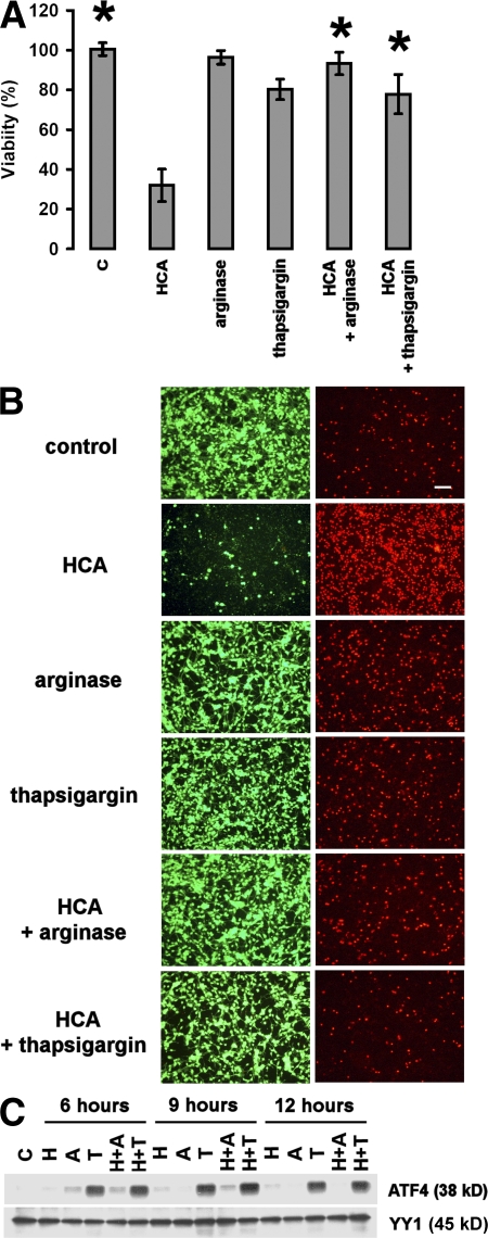
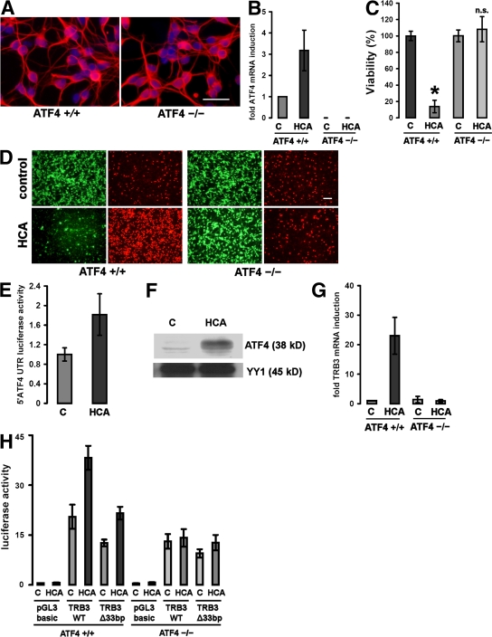
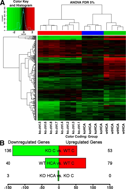
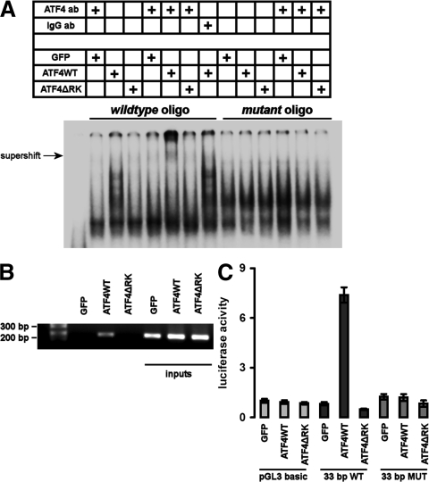
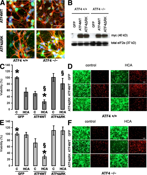
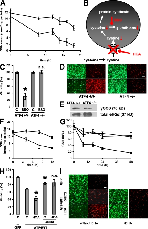
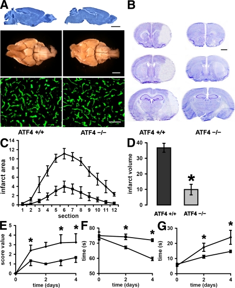
Oxidative stress is pathogenic in neurological diseases, including stroke. The identity of oxidative stress–inducible transcription factors and their role in propagating the death cascade are not well known. In an in vitro model of oxidative stress, the expression of the bZip transcription factor activating transcription factor 4 (ATF4) was induced by glutathione depletion and localized to the promoter of a putative death gene in neurons. Germline deletion of ATF4 resulted in a profound reduction in oxidative stress–induced gene expression and resistance to oxidative death. In neurons, ATF4 modulates an early, upstream event in the death pathway, as resistance to oxidative death by ATF4 deletion was associated with decreased consumption of the antioxidant glutathione. Forced expression of ATF4 was sufficient to promote cell death and loss of glutathione. In ATF4−/− neurons, restoration of ATF4 protein expression reinstated sensitivity to oxidative death. In addition, ATF4−/− mice experienced significantly smaller infarcts and improved behavioral recovery as compared with wild-type mice subjected to the same reductions in blood flow in a rodent model of ischemic stroke. Collectively, these findings establish ATF4 as a redox-regulated, prodeath transcriptional activator in the nervous system that propagates death responses to oxidative stress in vitro and to stroke in vivo.
Overview
The claims and statistics of this paper haven’t been checked yet.
Run the check — free, no account needed. It reads the open-access text and checks the sources it cites.
Integrity signals
Registry
Cites
Statistics
Figures
Verification
Claims the paper makes
The claims and statistics of this paper haven’t been checked yet.
Run the check — free, no account needed. It reads the open-access text and checks the sources it cites.
Claims other papers make about this one
Each is checked against this paper’s own text: does it say what the citing paper says it does?
The source backs part of the claim, not all of it — or backs it for a different population, condition or comparison.
In the citing paper
“We verified that known ATF4 target genes, Tribbles 3 (Trib3), a modulator of AKT, and Chac1, a cyclohydrolase that degrades glutathione,51”
From 2-Deoxyglucose drives plasticity via an adaptive ER stress-ATF4 pathway and elicits stroke recovery and Alzheimer’s resilience (2023) · cites this paper as [51]
Experimental — accuracy not yet measuredThe source backs part of the claim, not all of it — or backs it for a different population, condition or comparison.
In the citing paper
“GSK2606414 significantly inhibited 2-DG-induced eIF2α phosphorylation (Figures 6G and 6H) and transcription of the ATF4 target gene, Trib3, in primary neurons51”
From 2-Deoxyglucose drives plasticity via an adaptive ER stress-ATF4 pathway and elicits stroke recovery and Alzheimer’s resilience (2023) · cites this paper as [51]
Experimental — accuracy not yet measured
Self-check
Percentages, sums and reported differences re-computed from the paper’s own numbers.
The self-check needs the paper’s full text.
Citations
Cites
Mitochondrial Dysfunction and Oxidative Stress in Neurodegenerative Diseases
Lin, M. T., & Beal, M. F. (2006) · Nature · doi:10.1038/nature05292 · Cited by 6,455
limma: Linear Models for Microarray Data
Smyth, G. K. (2005) · Statistics for Biology and Health · doi:10.1007/0-387-29362-0_23 · Cited by 5,796
Vinson, C. R., Hai, T., & Boyd, S. M. (1993) · Genes & Development · doi:10.1101/gad.7.6.1047 · Cited by 336
Research works printed but not linked: 56
- 2002, Physiol. Rev., 82, 47, 10.1152/physrev.00018.2001 · DOI · Paper page
- 2006, Int. J. Biochem. Cell Biol., 38, 317, 10.1016/j.biocel.2005.09.018 · DOI · Paper page
- 2000, Ann. NY Acad. Sci., 924, 164, 10.1111/j.1749-6632.2000.tb05575.x · DOI
- 2007, FEBS Lett., 581, 3634, 10.1016/j.febslet.2007.04.028 · DOI
- 2007, Mol. Cell., 26, 1, 10.1016/j.molcel.2007.03.016 · DOI · Paper page
- 2003, J. Neurosci., 23, 3597, 10.1523/JNEUROSCI.23-09-03597.2003 · DOI
- 1997, J. Biol. Chem., 272, 24088, 10.1074/jbc.272.38.24088 · DOI
- 2001, Gene., 273, 1, 10.1016/S0378-1119(01)00551-0 · DOI
- 2006, Cancer Biol. Ther., 5, 723
- 2003, Mol. Cell., 11, 619, 10.1016/S1097-2765(03)00105-9 · DOI
- 1998, J. Neurosci., 18, 4083, 10.1523/JNEUROSCI.18-11-04083.1998 · DOI
- 2002, Methods Enzymol., 352, 183, 10.1016/S0076-6879(02)52018-8 · DOI
- 1994, J. Neurochem., 62, 376, 10.1046/j.1471-4159.1994.62010376.x · DOI
- 1995, Clin. Exp. Pharmacol. Physiol., 22, 309, 10.1111/j.1440-1681.1995.tb02006.x · DOI
- 1996, Neurochem. Int., 29, 153, 10.1016/0197-0186(95)00115-8 · DOI
- 2004, J. Nutr., 134, 2812S, 10.1093/jn/134.10.2812S · DOI
- 2003, J. Biol. Chem., 278, 37375, 10.1074/jbc.M303619200 · DOI
- 2002, Blood., 99, 736, 10.1182/blood.V99.3.736 · DOI
- 1994, J. Neurosci., 14, 4385, 10.1523/JNEUROSCI.14-07-04385.1994 · DOI
- 2005, J. Cereb. Blood Flow Metab., 25, 41, 10.1038/sj.jcbfm.9600005 · DOI
- 2007, J. Biol. Chem., 282, 4634, 10.1074/jbc.M609388200 · DOI
- 2007, J. Neurosci., 27, 6320, 10.1523/JNEUROSCI.0449-07.2007 · DOI
- 2003, J. Biol. Chem., 278, 17421, 10.1074/jbc.M300761200 · DOI
- 1998, Genes Cells., 3, 801, 10.1046/j.1365-2443.1998.00230.x · DOI
- 2000, Dev. Biol., 222, 110, 10.1006/dbio.2000.9699 · DOI
- 2001, Neuron., 29, 563, 10.1016/S0896-6273(01)00232-X · DOI
- 2000, Mol. Cell., 6, 1099, 10.1016/S1097-2765(00)00108-8 · DOI
- 2003, Dev. Cell., 4, 442, 10.1016/S1534-5807(03)00100-X · DOI
- 2002, J. Biol. Chem., 277, 24120, 10.1074/jbc.M201959200 · DOI
- 2001, J. Cell Biol., 153, 1011, 10.1083/jcb.153.5.1011 · DOI
- 2001, Mol. Cell. Biol., 21, 2192, 10.1128/MCB.21.6.2192-2202.2001 · DOI
- 2006, Ann. NY Acad. Sci., 1068, 131, 10.1196/annals.1346.027 · DOI
- 1992, Proc. Natl. Acad. Sci. USA., 89, 4820, 10.1073/pnas.89.11.4820 · DOI
- 1995, Cell., 83, 979, 10.1016/0092-8674(95)90213-9 · DOI · Paper page
- 2001, J. Biol. Chem., 276, 20858, 10.1074/jbc.M101198200 · DOI
- 2003, Curr. Pharm. Des., 9, 2499, 10.2174/1381612033453730 · DOI
- 2004, Mol. Cell. Biol., 24, 9456, 10.1128/MCB.24.21.9456-9469.2004 · DOI
- 2004, Cell., 117, 387, 10.1016/S0092-8674(04)00344-7 · DOI
- 2004, J. Biol. Chem., 279, 5288, 10.1074/jbc.M311862200 · DOI
- 2005, EMBO J., 24, 1243, 10.1038/sj.emboj.7600596 · DOI
- 2006, Proc. Natl. Acad. Sci. USA., 103, 768, 10.1073/pnas.0505723102 · DOI
- 1999, Biochem. Biophys. Res. Commun., 258, 260, 10.1006/bbrc.1999.0576 · DOI
- 2004, EMBO J., 23, 169, 10.1038/sj.emboj.7600030 · DOI
- 2006, J. Biomed. Sci., 13, 205, 10.1007/s11373-005-9057-3 · DOI
- 2006, Br. J. Haematol., 134, 145, 10.1111/j.1365-2141.2006.06132.x · DOI
- 2005, Curr. Med. Chem. Anticancer Agents., 5, 15, 10.2174/1568011053352587 · DOI
- 2004, J. Natl. Cancer Inst., 96, 1300, 10.1093/jnci/djh243 · DOI
- 2003, Cancer Res., 63, 8592
- 2006, Cell., 125, 971, 10.1016/j.cell.2006.05.012 · DOI
- 2005, J. Biol. Chem., 280, 30689, 10.1074/jbc.M500750200 · DOI
- 2006, J. Cereb. Blood Flow Metab., 26, 1465, 10.1038/sj.jcbfm.9600298 · DOI
- 2005, Nat. Rev. Neurosci., 6, 775
- 2005, J. Biol. Chem., 280, 41732, 10.1074/jbc.M504963200 · DOI
- 2005, J. Biol. Chem., 280, 3996, 10.1074/jbc.M409223200 · DOI
- 2004, Genome Biol., 5, R80, 10.1186/gb-2004-5-10-r80 · DOI
- 2006, J. Neurosci., 26, 9471, 10.1523/JNEUROSCI.2838-06.2006 · DOI
Cited by
Sorted by citations
ER-stress-induced transcriptional regulation increases protein synthesis leading to cell death
Han, J., Back, S. H., Hur, J., Lin, Y.-H., Gildersleeve, R., Shan, J., Yuan, C. L., Krokowski, D., Wang, S., Hatzoglou, M., et al. (2013) · Nature Cell Biology · doi:10.1038/ncb2738 · Cited by 1,691
Selenium Drives a Transcriptional Adaptive Program to Block Ferroptosis and Treat Stroke
Alim, I., Caulfield, J. T., Chen, Y., Swarup, V., Geschwind, D. H., Ivanova, E., Seravalli, J., Ai, Y., Sansing, L. H., Ste.Marie, E. J., et al. (2019) · Cell · doi:10.1016/j.cell.2019.03.032 · Cited by 1,024
“Germline deletion of ATF4 in neurons renders them resistant to homocysteic acid (HCA)-induced ferroptosis in vitro, and sensitivity to cell death can be reinstated by wildtype ATF4, but not ATF4 with the DNA binding domain mutated (Lange et al., 2008).”
Self-citationTargeting the unfolded protein response in disease
Hetz, C. A., Chevet, É., & Harding, H. P. (2013) · Nature Reviews Drug Discovery · doi:10.1038/nrd3976 · Cited by 942
Antioxidant response elements: Discovery, classes, regulation and potential applications
Raghunath, A., Sundarraj, K., Raju, N., Arfuso, F., Bian, J., Kumar, A. P., Sethi, G., & Perumal, E. (2018) · Redox Biology · doi:10.1016/j.redox.2018.05.002 · Cited by 483
ATF4 Regulates MYC-Mediated Neuroblastoma Cell Death upon Glutamine Deprivation
Qing, G., Li, B., Vu, A., Skuli, N., Walton, Z. E., Liu, X., Mayes, P. A., Wise, D. R., Thompson, C. B., Maris, J. M., et al. (2012) · Cancer Cell · doi:10.1016/j.ccr.2012.09.021 · Cited by 375
“In contrast to fibroblasts, ATF4 is a proapoptotic factor in neurons both in vitro and in vivo (Lange et al., 2008).”
Galehdar, Z., Swan, P., Fuerth, B. J., Callaghan, S. M., Park, D. S., & Cregan, S. P. (2010) · Journal of Neuroscience · doi:10.1523/jneurosci.1598-10.2010 · Cited by 374
“Nonetheless, the involvement of p53 in ER stress may be impacted by the optosis in neurons (Lange et al., 2008).”
BackgroundDivergent Effects of PERK and IRE1 Signaling on Cell Viability
Lin, J. H., Li, H., Zhang, Y., Ron, D., & Walter, P. (2009) · PLoS ONE · doi:10.1371/journal.pone.0004170 · Cited by 322
“Deletion of downstream components of PERK signaling, Atf4 and Chop, impaired or enhanced cell survival in response to protein misfolding depending on the cell type studied [3,13,14,15,16,17].”
BackgroundNakka, V. P., Gusain, A., & Raghubir, R. (2010) · Neurotoxicity research · doi:10.1007/s12640-009-9110-5 · Cited by 287
“Indeed, mice lacking ATF-4 have shown significantly smaller infarcts and improved behavioral outcome as compared with wildtype mice subjected to ischemic stroke ( Lange et al. 2008 ).”
BackgroundCellular Mechanisms of Endoplasmic Reticulum Stress Signaling in Health and Disease. 1. An overview
Dufey, E., Sepúlveda, D., Rojas‐Rivera, D., & Hetz, C. A. (2014) · American Journal of Physiology-Cell Physiology · doi:10.1152/ajpcell.00258.2014 · Cited by 186
Molecular landscapes of human hippocampal immature neurons across lifespan
Zhou, Y. F., Su, Y., Li, S., Kennedy, B. C., Zhang, D. Y., Bond, A. M., Sun, Y. Y., Jacob, F., Lu, L., Hu, P., et al. (2022) · Nature · doi:10.1038/s41586-022-04912-w · Cited by 314
Modulating stress responses by the UPRosome: a matter of life and death.
Woehlbier, U., & Hetz, C. (2011) · TIBS -Trends in Biochemical Sciences. Regular ed · doi:10.1016/j.tibs.2011.03.001 · Cited by 269
Wu, L., Jin, Y., Zhao, X., Tang, K., Zhao, Y., Tong, L., Yu, X., Xiong, K., Luo, C., Zhu, J., et al. (2023) · Cell Metabolism · doi:10.1016/j.cmet.2023.07.001 · Cited by 301
BOK Is a Non-canonical BCL-2 Family Effector of Apoptosis Regulated by ER-Associated Degradation
Llambi, F., Wang, Y.-M., Victor, B., Yang, M., Schneider, D. M., Gingras, S., Parsons, M. J., Zheng, J. H., Brown, S. A., Pelletier, S., et al. (2016) · Cell · doi:10.1016/j.cell.2016.02.026 · Cited by 261
“During ER stress, accumulation of unfolded proteins in the ER activates the UPR through IRE1a, PERK, and ATF6 to upregulate ERAD components (Smith et al., 2011).”
InfluentialATF4-dependent induction of heme oxygenase 1 prevents anoikis and promotes metastasis
Dey, S., Sayers, C. M., Verginadis, I. I., Lehman, S. L., Cheng, Y., Cerniglia, G. J., Tuttle, S. W., Feldman, M. D., Zhang, P. J. L., Fuchs, S. Y., et al. (2015) · Journal of Clinical Investigation · doi:10.1172/jci78031 · Cited by 291
“Inter-estingly, ATF4 activation has also been linked to cells’ protection from oxidative stress, which transcriptionally activates multiple antioxidant genes to maintain redox status in the cells (2, 38).”
Lewerenz, J., Ates, G., Methner, A., Conrad, M., & Maher, P. A. (2018) · Frontiers in Neuroscience · doi:10.3389/fnins.2018.00214 · Cited by 276
“Arginase treatment induces ATF4 in primary embryonic neurons and protects against oxytosis brought about by the system x−c inhibitor homocysteate (Lange et al., 2008) although the same authors showed that induction of ATF4 during activation of oxytosis acts as a pro-death regulator (Lange et al.,…”
BackgroundInfluentialProtein folding stress in neurodegenerative diseases: a glimpse into the ER
Matus, S., Glimcher, L. H., & Hetz, C. A. (2011) · Current Opinion in Cell Biology · doi:10.1016/j.ceb.2011.01.003 · Cited by 233
Nakka, V. P., Phanithi, P.-B., & Vemuganti, R. (2014) · Molecular Neurobiology · doi:10.1007/s12035-014-9029-6 · Cited by 246
Endoplasmic Reticulum Stress is implicated in Retinal Inflammation and Diabetic Retinopathy
Li, J., Wang, J., Yu, Q., Wang, M., & Zhang, S. X. (2009) · FEBS Letters · doi:10.1016/j.febslet.2009.04.007 · Cited by 226
“Several studies demonstrated that various oxidants or oxidatively modified lipids induce ER stress in cultured endothelial cells and neurons [29,36,37].”
BackgroundAberrant iron distribution via hepatocyte-stellate cell axis drives liver lipogenesis and fibrosis
Gao, H., Jin, Z., Bandyopadhyay, G., Wang, G., Zhang, D., Rocha, K. C. E., Liu, X., Zhao, H., Kisseleva, T., Brenner, D. A., et al. (2022) · Cell Metabolism · doi:10.1016/j.cmet.2022.07.006 · Cited by 224
Ishimura, R., Nagy, G., Dotu, I., Chuang, J. H., & Ackerman, S. L. (2016) · eLife · doi:10.7554/elife.14295 · Cited by 221
Torrence, M. E., MacArthur, M. R., Hosios, A. M., Valvezan, A. J., Asara, J. M., Mitchell, J. R., & Manning, B. D. (2021) · eLife · doi:10.7554/elife.63326 · Cited by 243
Zhong, Y., Li, J., Chen, Y.-M., Wang, J., Ratan, R., & Zhang, S. X. (2012) · Diabetes · doi:10.2337/db11-0315 · Cited by 184
“To assess the potential role of ATF4 in Müller cells, we manipulated ATF4 expression and/or activity using AdATF4WT or Ad-ATF4DN (13) in rMC-1 cells.”
MethodSelf-citationAhola, S., Rivera-Mejías, P., Hermans, S., Chandragiri, S., Giavalisco, P., Nolte, H., & Langer, T. (2022) · Cell Metabolism · doi:10.1016/j.cmet.2022.08.017 · Cited by 193
CHOP is a critical regulator of acetaminophen-induced hepatotoxicity.
Uzi, D., Barda, L., Scaiewicz, V., Mills, M., Mueller, T., González-Rodríguez, Á., Valverde, Á. M., Iwawaki, T., Nahmias, Y., Xavier, R., et al. (2013) · Journal of Hepatology · doi:10.1016/j.jhep.2013.04.024 · Cited by 180
Lewerenz, J., & Maher, P. A. (2008) · Journal of Biological Chemistry · doi:10.1074/jbc.m807325200 · Cited by 173
Multiple Mechanisms of Unfolded Protein Response–Induced Cell Death
Hiramatsu, N., Chiang, W.-C. J., Kurt, T. D., Sigurdson, C. J., & Lin, J. H. (2015) · American Journal Of Pathology · doi:10.1016/j.ajpath.2015.03.009 · Cited by 181
“0 DTD AJPA2030_pro cell death.(45,47) These findings point to ATF4 as an important determinant in regulating cell fate during ER stress, with too little and too much ATF4 both producing deleterious effects.”
BackgroundSignaling Pathways from the Endoplasmic Reticulum and Their Roles in Disease
Kadowaki, H., & Nishitoh, H. (2013) · Genes · doi:10.3390/genes4030306 · Cited by 211
“homeostasis, and ER stress-induced apoptosis [51 53] (Figure 1).”
Targeting the IRE1α–XBP1 branch of the unfolded protein response in human diseases
Jiang, D., Niwa, M., & Koong, A. C. (2015) · Seminars in Cancer Biology · doi:10.1016/j.semcancer.2015.04.010 · Cited by 185
“ATF4 transactivates target genes involved in redox processes, amino acid metabolism, ER chaperones and foldases [4, 5].”
BackgroundThe Chemical Biology of Ferroptosis in the Central Nervous System
Ratan, R. R. (2020) · Cell chemical biology · doi:10.1016/j.chembiol.2020.03.007 · Cited by 183
“Consistent with this model, germline deletion of ATF4 in mouse cortical neurons has been shown to preserve glutathione levels in response to glutamate or the glutamate analog, HCA, by inhibiting degradation, not synthesis (Lange et al., 2008).”
BackgroundInfluentialSelf-citationRen, P., Yue, M., Xiao, D., Xiu, R., Gan, L., Liu, H., & Qing, G. (2014) · The Journal of Pathology · doi:10.1002/path.4429 · Cited by 157
REGULATION OF ER STRESS-INDUCED CELL DEATH BY ATF4 IN NEUROECTODERMAL TUMOUR CELLS
Armstrong, J., Flockhart, R., Veal, G., Lovat, P., & Redfern, C. (2009) · Journal of Biological Chemistry · doi:10.1074/jbc.m109.014092 · Cited by 156
Ren, J., Li, C., Yan, X., Qu, Y., Yang, Y., & Guo, Z. (2021) · Oxidative Medicine and Cellular Longevity · doi:10.1155/2021/6643382 · Cited by 176
“Increased ATF4 mRNA levels and translation levels after oxidative stress make ATF4 overexpression sufficient to induce cell death [56].”
BackgroundInfluentialAutophagy protects the retina from light-induced degeneration
Chen, Y., Sawada, O., Kohno, H., Le, Y.-Z., Subauste, C., Maeda, T., & Maeda, A. (2013) · Journal of Biological Chemistry · doi:10.1074/jbc.m112.439935 · Cited by 152
“3B), indicating the activation of oxidative stress by light exposure (18).”
BackgroundThe neuropathobiology of multiple sclerosis
Woo, M. S., Engler, J. B., & Friese, M. A. (2024) · Nature reviews. Neuroscience · doi:10.1038/s41583-024-00823-z · Cited by 205
“However, because the UPR is different in postmitotic neurons, PERK-dependent and ATF4-independent signalling pathways in these neurons need further investigation.”
Endoplasmic reticulum stress and the unfolded protein responses in retinal degeneration
Zhang, S. X., Sanders, E. N., Fliesler, S. J., & Wang, J. J. (2014) · Experimental Eye Research · doi:10.1016/j.exer.2014.04.015 · Cited by 152
Endoplasmic reticulum-mediated unfolded protein response and mitochondrial apoptosis in cancer
Bhat, T. A., Chaudhary, A. K., Kumar, S., O’Malley, J., Inigo, J. R., Kumar, R., Yadav, N., & Chandra, D. (2016) · Biochimica et Biophysica Acta (BBA) - Reviews on Cancer · doi:10.1016/j.bbcan.2016.12.002 · Cited by 157
Speer, R. E., Karuppagounder, S. S., Basso, M., Sleiman, S. F., Kumar, A., Brand, D., Smirnova, N., Gazaryan, I. G., Khim, S. J., & Ratan, R. R. (2013) · Free Radical Biology and Medicine · doi:10.1016/j.freeradbiomed.2013.01.026 · Cited by 157
“regulated transcriptional pathways that can compensate for persistent glutathione depletion and oxidative stress in order to foster cell survival [2, 3].”
BackgroundSelf-citationKaruppagounder, S. S., Alim, I., Khim, S. J., Bourassa, M. W., Sleiman, S. F., John, R., Thinnes, C. C., Yeh, T.-L., Demetriades, M., Neitemeier, S., et al. (2016) · Science Translational Medicine · doi:10.1126/scitranslmed.aac6008 · Cited by 141
“Glutamate analog (HCA) treatment in mouse cortical neurons induced robust gene expression changes, significantly overlapping with those we reported previously (27).”
BackgroundMethodResultInfluentialSelf-citationActivation of the unfolded protein response enhances motor recovery after spinal cord injury
Hetz, C., Valenzuela, V., Collyer, E., Armentano, D., Parsons, G., & Court, F. A. (2012) · Cell Death and Disease · doi:10.1038/cddis.2012.8 · Cited by 141
“ATF4 deficiency results in resistance to oxidative stress in models of brain ischemia.(34,35) PERK/eIF2a signaling has protective effects on axonal remyelination in models of experimental autoimmune encephalomyelitis, possibly because of the control of oligodendrocyte viability and function.”
BackgroundIntegrated Stress Response Modulates Cellular Redox State via Induction of Cystathionine γ-Lyase
Dickhout, J. G., Carlisle, R. E., Jerome, D. E., Mohammed-Ali, Z., Jiang, H., Yang, G., Mani, S., Garg, S. K., Banerjee, R., Kaufman, R. J., et al. (2012) · Journal of Biological Chemistry · doi:10.1074/jbc.m111.304576 · Cited by 129
“Conversely, ATF4 / neurons were protected from oxidative stress-induced cell death, stimulated by the glutamate analogHCA (53).”
BackgroundMizuuchi, M., Cindrová‐Davies, T., Olovsson, M. H., Jones, D. S., Burton, G. J., & Yung, H. W. (2015) · The Journal of Pathology · doi:10.1002/path.4678 · Cited by 105
Chen, Y., Wang, J.-J., Li, J., Hosoya, K., Ratan, R., Townes, T., & Zhang, S.-X. (2012) · Diabetologia · doi:10.1007/s00125-012-2594-1 · Cited by 125
“Adenoviral infection of TR-iBRB cells Adenoviral vectors expressing mouse wild-type (WT) Atf4 (Ad-ATF4WT) and dominant negative Atf4 (Ad-ATF4ΔRK) harbouring a mutation in its DNA-binding domain ((292)RYRQKKR(298) to (292)GYLEAAA(298)) were generated as described previously [19].”
MethodSelf-citationATF4 protects against neuronal death in cellular PD models by maintaining levels of parkin
Sun, X., Liu, J., Crary, J. F., Malagelada, C., Sulzer, D., Greene, L., & Levy, O. (2013) · Journal of Neuroscience · doi:10.1523/jneurosci.2292-12.2013 · Cited by 123
“For example, ATF4-null mice show less neuronal loss in stroke models (Lange et al., 2008), and ATF4-deficient neurons are more resistant to ERS (Galehdar et al.”
BackgroundLiao, M., Liao, W., Xu, N., Li, B., Liu, F., Zhang, S., Wang, Y., Wang, S., Zhu, Y., Chen, D., et al. (2019) · EBioMedicine · doi:10.1016/j.ebiom.2019.01.035 · Cited by 152
“Increased ROS contributes to cellular stress, which in turn activated of P-eIF2α/ATF4 pathway [26,27].”
BackgroundOhri, S., Maddie, M. A., Zhao, Y.-M., Qiu, M., Hetman, M., & Whittemore, S. (2011) · Glia · doi:10.1002/glia.21191 · Cited by 119
“Indeed, the contribution of UPR-activated cell death has been reported in ischemic stroke (Lange et al., 2008), multiple sclerosis (Lin et al.”
Molecular pathway of near-infrared laser phototoxicity involves ATF-4 orchestrated ER stress
Khan, I., Tang, E., & Arany, P. (2015) · Scientific Reports · doi:10.1038/srep10581 · Cited by 118
Hiramatsu, N., Messah, C., Han, J., LaVail, M. M., Kaufman, R. J., & Lin, J. H. (2014) · Molecular Biology of the Cell · doi:10.1091/mbc.e13-11-0664 · Cited by 135
“Microarray and RNA sequencing studies predict that ATF4’s transcriptional targets include a number of ubiquitin ligases (Harding et al., 2003; Lange et al., 2008; Han et al., 2013).”
BackgroundResultFelice, F. G. D., & Lourenco, M. V. (2015) · Frontiers in Aging Neuroscience · doi:10.3389/fnagi.2015.00094 · Cited by 122
“…the translation of a small fraction of mRNAs (Buffington et al., 2014), among which is activating transcription factor 4 (ATF4), a protein linked to oxidative stress, enhanced γ-secretase activity and neuronal dysfunction when abnormally elevated (Mitsuda et al., 2007; Lange et al., 2008).”
BackgroundTranscriptional and metabolic adaptation of human neurons to the mitochondrial toxicant MPP+
Krug, A. K., Gutbier, S., Zhao, L., Pöltl, D., Kullmann, C., Ivanova, V., Förster, S., Jagtap, S., Meiser, J., Leparc, G., et al. (2014) · Cell Death and Disease · doi:10.1038/cddis.2014.166 · Cited by 111
Fang, Y., Gao, S., Wang, X., Cao, Y., Lü, J., Chen, S., Lenahan, C., Zhang, J. H., Shao, A., & Zhang, J. (2020) · Frontiers in Cellular Neuroscience · doi:10.3389/fncel.2020.00068 · Cited by 114
Karuppagounder, S. S., & Ratan, R. R. (2012) · Journal of Cerebral Blood Flow & Metabolism · doi:10.1038/jcbfm.2012.28 · Cited by 97
“ATF4 is a leucine zipper transcription factor that has been shown to have prodeath and prosurvival roles in response to oxidative stress or endoplasmic reticulum stress in neurons (Lange et al, 2008).”
BackgroundSelf-citationPerk-dependent repression of miR-106b-25 cluster is required for ER stress-induced apoptosis
Gupta, S., Read, D. E., Deepti, A., Cawley, K., Gupta, A., Oommen, D., Verfaillie, T., Matus, S., Smith, M. A., Mott, J. L., et al. (2012) · Cell Death and Disease · doi:10.1038/cddis.2012.74 · Cited by 101
“Atf4 has been shown to act as a pro-death transcriptional activator in the nervous system that propagates death responses to oxidative stress in vitro and to stroke in vivo.(34) Atf4 also mediates ER stress-induced cell death of neuroectodermal tumor cells in response to fenretinide or bortezomib.”
Bajpai, R., Sharma, A., Achreja, A., Edgar, C. L., Wei, C., Siddiqa, A., Gupta, V. A., Matulis, S. M., McBrayer, S. K., Mittal, A., et al. (2020) · Nature Communications · doi:10.1038/s41467-020-15051-z · Cited by 103
“Complex II is linked to the generation of ROS, accumulation of succinate and the induction of HIF1-α 54 that can lead to induction of ATF4 55 .”
BackgroundMatus, S., López, E., Valenzuela, V., Nassif, M., & Hetz, C. A. (2013) · PLoS ONE · doi:10.1371/journal.pone.0066672 · Cited by 98
“Finally, based on our results in the animal model of ALS and in reports suggesting a protective role of ATF4 deficiency on neuronal survival [21,22], we analyzed the susceptibility of shATF4 motoneuron cells to ER stress-mediated cell death.”
ResultThe transcription factor ATF5: role in neurodevelopment and neural tumors
Greene, L. A., Lee, H. Y., & Angelastro, J. M. (2008) · Journal of Neurochemistry · doi:10.1111/j.1471-4159.2008.05749.x · Cited by 85
“For instance, while ATF4 plays a major role in cellular/neuronal responses to stresses (Ameri and Harris 2008; Lange et al. 2008), there is currently no evidence that ATF5, which can also be induced by stress, has similar actions.”
BackgroundKasetti, R. B., Maddineni, P., Patel, P. D., Searby, C. C., Sheffield, V. C., & Zode, G. S. (2018) · Journal of Biological Chemistry · doi:10.1074/jbc.ra118.002540 · Cited by 99
“We also knocked down ATF4 using adenovirus 5– expressing CRISPR-Cas9 –targeting ATF4 or dominantnegative ATF4 RK (which inhibits endogenous ATF4 activity) as described previously (45) at multiplicity of infection 100 for 48 h.”
BackgroundMethodInfluentialLarhammar, M., Huntwork-Rodriguez, S., Jiang, Z., Solanoy, H., Sengupta Ghosh, A., Wang, B., Kaminker, J. S., Huang, K., Eastham-Anderson, J., Siu, M., et al. (2017) · eLife · doi:10.7554/elife.20725 · Cited by 90
“In the current study, our efforts to understand neuronal stress signaling pathways after acute injury uncovered a functional role for PERK signaling in regulating apoptotic responses downstream of DLK.”
BackgroundMethodResultInfluentialGADD34 induces cell death through inactivation of Akt following traumatic brain injury
Farook, J. M., Shields, J., Tawfik, A., Markand, S., Sen, T., Smith, S. B., Brann, D., Dhandapani, K., & Sen, N. (2013) · Cell Death and Disease · doi:10.1038/cddis.2013.280 · Cited by 87
“Intriguingly, recent work has further revealed that induction of oxidative stress in neurons leads to an increase of ATF4 in cells.(12) Upon selective activation of ATF4, several stress-related genes such as GADD34 are known to be upregulated.”
BackgroundLimbu, S. M., Ma, Q., Zhang, M., & Du, Z. (2019) · The Science of The Total Environment · doi:10.1016/j.scitotenv.2019.05.067 · Cited by 106
ATF4 leads to glaucoma by promoting protein synthesis and ER client protein load
Kasetti, R. B., Patel, P. D., Maddineni, P., Patil, S., Kiehlbauch, C. C., Millar, J. C., Searby, C. C., Raghunathan, V. K., Sheffield, V. C., & Zode, G. S. (2020) · Nature Communications · doi:10.1038/s41467-020-19352-1 · Cited by 93
Lin, Y.-W., Chen, T.-Y., Hung, C.-Y., Tai, S.-H., Huang, S.-Y., Chang, C.-C., Hung, H.-Y., & Lee, J. E. (2018) · International Journal of Molecular Medicine · doi:10.3892/ijmm.2018.3607 · Cited by 81
“It has been reported that ATF4 is an oxidative stress-inducible, prodeath transcription factor for neurons after stroke (50).”
Oxidative stress up-regulates presenilin 1 in lipid rafts in neuronal cells
Oda, A., Tamaoka, A., & Araki, W. (2010) · Journal of Neuroscience Research · doi:10.1002/jnr.22271 · Cited by 76
“…found that activating transcription factor 4 (ATF4) binds to the regulatory region of the PS1 gene and up-regulates PS1 expression during amino acid imbalance (Mitsuda et al., 2007), and that ATF4 is a pro-death transcription factor induced by oxidative stress in neurons (Lange et al., 2008).”
BackgroundZille, M., Kumar, A., Kundu, N., Bourassa, M. W., Wong, V. S. C., Willis, D. E., Karuppagounder, S. S., & Ratan, R. R. (2019) · eNeuro · doi:10.1523/eneuro.0263-18.2019 · Cited by 84
“Glutamate analog (HCA)-induced ferroptosis in neurons requires de novo transcription via the leucine zipper transcription factor ATF4 leading to upregulation of putative pro-death genes such as Trib3, Chop, and Chac1 (Lange et al., 2008; Karuppagounder et al., 2016).”
BackgroundSelf-citationATF4 deficiency protects mice from high-carbohydrate-diet-induced liver steatosis
Li, H., Meng, Q., Xiao, F., Chen, S., Du, Y., Yu, J., Wang, C., & Guo, F. (2011) · Biochemical Journal · doi:10.1042/bj20110263 · Cited by 87
Restoring endoplasmic reticulum homeostasis improves functional recovery after spinal cord injury
Ohri, S., Hetman, M., & Whittemore, S. (2013) · Neurobiology of Disease · doi:10.1016/j.nbd.2013.04.021 · Cited by 79
“Involvement of ERSR-activated cell death has been reported in ischemic stroke (Lange et al., 2008), multiple sclerosis (Lin et al., 2006), and Alzheimer's disease (Milhavet et al., 2002).”
BackgroundEmerging Role of Ferroptosis in the Pathogenesis of Ischemic Stroke: A New Therapeutic Target?
Bu, Z., Yu, H., Wang, J., He, X., Cui, Y., Feng, J., & Feng, J. (2021) · ASN NEURO · doi:10.1177/17590914211037505 · Cited by 91
“And the protective effect of adaptaquin was related to the reduced activity of activating transcription factor 4 (ATF4) (Karuppagounder et al., 2016; Lange et al., 2008).”
BackgroundInfluentialLysosomal cystine governs ferroptosis sensitivity in cancer via cysteine stress response
Swanda, R. V., Ji, Q., Wu, X., Yan, J., Dong, L., Mao, Y., Uematsu, S., Dong, Y., & Qian, S. (2023) · Molecular Cell · doi:10.1016/j.molcel.2023.08.004 · Cited by 84
Wu, C.-X., Liu, R., Gao, M., Zhao, G., Wu, S., Wu, C.-F., & Du, G.-H. (2013) · Neuroscience Letters · doi:10.1016/j.neulet.2013.04.060 · Cited by 74
Lewerenz, J., Sato, H., Albrecht, P., Henke, N., Noack, R., Methner, A., & Maher, P. A. (2011) · Cell Death and Differentiation · doi:10.1038/cdd.2011.165 · Cited by 68
Wang, Y., Wang, M., Liu, Y., Tao, H., Banerjee, S., Srinivasan, S., Nemeth, E., Czaja, M. J., & He, P. (2022) · Redox Biology · doi:10.1016/j.redox.2022.102407 · Cited by 76
Lourenco, M. V., Ferreira, S. T., & Felice, F. G. D. (2015) · Progress in Neurobiology · doi:10.1016/j.pneurobio.2015.03.003 · Cited by 74
Amino Acid Homeostasis in Mammalian Cells with a Focus on Amino Acid Transport
Bröer, S., & Gauthier-Coles, G. (2021) · Journal of Nutrition · doi:10.1093/jn/nxab342 · Cited by 86
PKR Inhibition Rescues Memory Deficit and ATF4 Overexpression in ApoE ε4 Human Replacement Mice
Segev, Y., Barrera, I., Ounallah-Saad, H., Wibrand, K., Sporild, I., Livne, A., Rosenberg, T., David, O., Mints, M., Bramham, C. R., et al. (2015) · Journal of Neuroscience · doi:10.1523/jneurosci.5241-14.2015 · Cited by 81
“Furthermore, ATF4 has been described as playing an important role in normal brain physiology (Frank et al., 2010), as well as in the integrated stress response (Lange et al., 2008; Baleriola et al., 2014).”
BackgroundSalubrinal protects against rotenone-induced SH-SY5Y cell death via ATF4-parkin pathway
Wu, L., Luo, N., Zhao, H.-R., Gao, Q., Lu, J., Pan, Y., Shi, J., Tian, Y., & Zhang, Y. (2014) · Brain Research · doi:10.1016/j.brainres.2014.01.003 · Cited by 61
Halterman, M. W., Gill, M., DeJesus, C., Ogihara, M., Schor, N. F., & Federoff, H. J. (2010) · Journal of Biological Chemistry · doi:10.1074/jbc.m109.095299 · Cited by 56
Kumar, A., Karuppagounder, S. S., Chen, Y., Corona, C., Kawaguchi, R., Cheng, Y., Balkaya, M., Sagdullaev, B. T., Wen, Z., Stuart, C., et al. (2023) · Neuron · doi:10.1016/j.neuron.2023.06.013 · Cited by 64
Self-citationZonated leucine sensing by Sestrin-mTORC1 in the liver controls the response to dietary leucine
Cangelosi, A. L., Puszynska, A. M., Roberts, J. M., Armani, A., Nguyen, T. P., Spinelli, J. B., Kunchok, T., Wang, B., Chan, S. H., Lewis, C. A., et al. (2022) · Science · doi:10.1126/science.abi9547 · Cited by 75
Basso, M., Berlin, J., Xia, L., Sleiman, S. F., Ko, B., Haskew-Layton, R., Kim, E., Antonyak, M. A., Cerione, R., Iismaa, S., et al. (2012) · Journal of Neuroscience · doi:10.1523/jneurosci.3353-11.2012 · Cited by 64
“…factor prolyl hydroxylases (Siddiq et al., 2005), class I and II histone deacetylases (Langley et al., 2008), activating transcription factor-4 (Lange et al., 2008), Sp1 (Ryu et al., 2003; Sleiman et al., 2011), Myc (Sleiman et al., 2011), and Keap (a negative regulator of Nrf2) (Smirnova et…”
BackgroundSelf-citationCurtis, W. M., Seeds, W. A., Mattson, M. P., & Bradshaw, P. C. (2022) · Cells · doi:10.3390/cells11152416 · Cited by 72
“Low‐level or transient activation of ATF4 appears to be cytoprotective [41], while chronic high‐level ATF4 activation can lead to cell death [42].”
BackgroundYang, Y., Ma, F., Liu, Z., Su, Q., Liu, Y., Liu, Z., & Li, Y. (2018) · Journal of Biological Chemistry · doi:10.1074/jbc.ra118.005166 · Cited by 75
“It has been reported that the ATF6 –NF-Y key stress factor ATF4 is known as a chaperone activator (5, 39, 40).”
BackgroundGu, X., Li, K., Laybutt, D. R., He, M., Zhao, H., Chan, J. C. N., & Xu, G. (2010) · Life Sciences · doi:10.1016/j.lfs.2010.10.012 · Cited by 63
Pauneto, C. D. M. A., Riesenberg, B., Gandy, E. J., Kennedy, A. S., Clutton, G., Hem, J. W., Hurst, K. E., Hunt, E. G., Green, J. M., Miller, B. C., et al. (2025) · Immunity · doi:10.1016/j.immuni.2025.09.003 · Cited by 69
The UPR and the Anti-oxidant Response: Relevance to Sleep and Sleep Loss
Brown, M. K., & Naidoo, N. N. (2010) · Molecular Neurobiology · doi:10.1007/s12035-010-8114-8 · Cited by 58
“This same study further describes resistance to neuronal cell death with an ATF4 deletion [62].”
BackgroundAmyotrophic lateral sclerosis pathogenesis: a journey through the secretory pathway
Nassif, M., Matus, S., Castillo, K., & Hetz, C. (2010) · Antioxidants & Redox Signaling · doi:10.1089/ars.2009.2991 · Cited by 60
“…eIF2 a phosphorylation augments the specific translation of the mRNA encoding Activation of Transcription-4 (ATF4), a UPR transcription factor essential for the upregulation of many UPR-associated genes that function in amino acid metabolism, redox homeostasis, and apoptosis (1, 106, 167).”
ATF4 is a novel regulator of MCP-1 in microvascular endothelial cells
Huang, H., Jing, G., Wang, J., Sheibani, N., & Zhang, S. X. (2015) · Journal of Inflammation · doi:10.1186/s12950-015-0076-1 · Cited by 60
“ATF4 is a major stress response gene activated in cells under conditions of ER stress and oxidative stress [4,5,17-20].”
BackgroundGlahn, F., Schmidt-Heck, W., Zellmer, S., Guthke, R., Wiese, J., Golka, K., Hergenröder, R., Degen, G., Lehmann, T., Hermes, M., et al. (2008) · Archives of Toxicology · doi:10.1007/s00204-008-0331-9 · Cited by 60
Trib3 Is Elevated in Parkinson's Disease and Mediates Death in Parkinson's Disease Models
Aimé, P., Sun, X., Zareen, N., Rao, A., Berman, Z., Volpicelli‐Daley, L. A., Bernd, P., Crary, J. F., Levy, O. A., & Greene, L. A. (2015) · Journal of Neuroscience · doi:10.1523/jneurosci.0614-15.2015 · Cited by 59
“, 2009), oxidative stress (Lange et al., 2008), mitochondrial stress (Ishikawa et al.”
BackgroundInfluentialBagheri‐Yarmand, R., Sinha, K. M., Gururaj, A. E., Ahmed, Z., Rizvi, Y. Q., Huang, S., Ladbury, J. E., Bögler, O., Williams, M. D., Cote, G. J., et al. (2015) · Journal of Biological Chemistry · doi:10.1074/jbc.m114.619833 · Cited by 57
“Some studies indicate that ATF4 plays a prosurvival role (23), whereas others indicate a proapoptotic role, suggesting that the function of ATF4 is context-dependent (16, 17, 24, 25).”
BackgroundYeh, K.-Y., Lai, C., Lin, C.-Y., Hsu, C.-C., Lo, C.-P., & Her, G. (2017) · Scientific Reports · doi:10.1038/s41598-017-16587-9 · Cited by 56
Role of the unfolded protein response in organ physiology: Lessons from mouse models
Cornejo, V. H., Pihán, P., Vidal, R. L., & Hetz, C. A. (2013) · IUBMB Life · doi:10.1002/iub.1224 · Cited by 54
“Gene expression profile analysis revealed that ATF4 regulates a subset of genes that are induced in response to oxidative stress and controls antioxidant response (20).”
BackgroundInfluentialChronic oxycodone induces axonal degeneration in rat brain
Fan, R., Schrott, L. M., Arnold, T., Snelling, S., Rao, M., Graham, D. D., Cornelius, A. P., & Korneeva, N. L. (2018) · BMC Neuroscience · doi:10.1186/s12868-018-0417-0 · Cited by 50
“It was suggested that ATF4 serves as a pro-death transcription factor sensitizing neurons to oxidative stress [83].”
Bose, S., & Cho, J. (2016) · Ageing Research Reviews · doi:10.1016/j.arr.2016.09.004 · Cited by 56
Liu, J., Pasini, S., Shelanski, M. L., & Greene, L. A. (2014) · Frontiers in Cellular Neuroscience · doi:10.3389/fncel.2014.00177 · Cited by 56
Demmings, M. D., Tennyson, E. C., Petroff, G. N., Tarnowski-Garner, H. E., & Cregan, S. P. (2020) · Cell Death and Differentiation · doi:10.1038/s41418-020-00688-6 · Cited by 63
Williams, C. H., Neitzel, L. R., Cornell, J. L., Rea, S., Mills, I. G., Silver-Isenstadt, M., Ahmad, J. D., Birukov, K. G., Birukova, A. A., Brem, H., et al. (2024) · Experimental Hematology and Oncology · doi:10.1186/s40164-023-00468-1 · Cited by 50
“ATF4, implicated in ferroptotic neuronal death during stroke, demonstrated a similar induction of ferroptosis in GBM cells [84].”
BackgroundBhootada, Y., Kotla, P., Zolotukhin, S., Gorbatyuk, O. S., Bebok, Z. M., Athar, M., & Gorbatyuk, M. S. (2016) · PLoS ONE · doi:10.1371/journal.pone.0154779 · Cited by 45
“It is known that ATF4 may exert either protective or deleterious effects on cell survival, depending on the paradigm [10, 11].”
BackgroundLeu, J. I.-J., Murphy, M. E., & George, D. L. (2020) · Proceedings of the National Academy of Sciences · doi:10.1073/pnas.2009943117 · Cited by 47
“A number genes implicated in cysteine-depletion-mediated ferroptotic cell death are ATF4 targets, including CHAC1 and SLC7A11/xCT (52), and germline deletion of ATF4 can render primary neurons resistant to glutamate-induced ferroptosis (6, 53).”
BackgroundROS is the major player in regulating altered autophagy and lifespan insin-3mutants ofC. elegans
Sharma, M., Pandey, R., & Saluja, D. (2018) · Autophagy · doi:10.1080/15548627.2018.1474312 · Cited by 50
Yang, B., Xu, Y., Hu, Y., Luo, Y., Lü, X., Tsui, C., Lü, L., & Liang, X. (2016) · Biomedicine & Pharmacotherapy · doi:10.1016/j.biopha.2016.10.015 · Cited by 50
Gully, J. C., Sergeyev, V., Bhootada, Y., Mendez-Gomez, H. R., Meyers, C., Zolotukhin, S., Gorbatyuk, M., & Gorbatyuk, O. (2016) · Neuroscience Letters · doi:10.1016/j.neulet.2016.05.039 · Cited by 47
“With this said, the role of ATF4 in triggering the prosurvival [6,15] or pro-death [10] trend is a source of continuous debates, which suggest that ATF4 is an important element in cell fate decision.”
BackgroundResult
Metadata
- Authors
- Lange, P. S., Chavez, J. C., Pinto, J. T., Coppola, G., Sun, C.-W., Townes, T. M., Geschwind, D. H., & Ratan, R. R.
- Affiliations
- University of Bonn (DE)
- Cornell University (US)
- New York Medical College (US)
- 6Program in Neurogenetics, Department of Neurology,
- University of Alabama at Birmingham (US)
- University of California, Los Angeles (US)
- Venue
- The Journal of Experimental Medicine
- Publisher
- Rockefeller University Press
- Published
- 2008-05-05
- Volume
- 205
- Issue
- 5
- Pages
- 1227-1242
- ISSN
- 0022-1007 (ISSN-L)
- 1540-9538
- PMC
- PMC2373852
- OpenAlex
- W2105818495
- Cited by
- 272
- References
- 62
- Citations by year
- 2026: 15 · 2025: 16 · 2024: 17 · 2023: 19 · 2022: 17 · 2021: 21 · 2020: 12 · 2019: 18 · 2018: 19 · 2017: 9 · 2016: 17 · 2015: 17 · 2014: 20 · 2013: 16 · 2012: 13
- FWCI
- 3.79
- Type
- journal article
- Language
- en
- Open access
- green
- www.ncbi.nlm.nih.gov
- Licence
- cc-by-nc-sa
- Locations
- The Journal of Experimental Medicine (publishedVersion)
- PubMed Central (publishedVersion, cc-by-nc-sa)
- PubMed (publishedVersion)
- eScholarship (California Digital Library) (submittedVersion, cc-by-nc-sa)
- Touro Scholar (Touro College) (submittedVersion, cc-by-nc-sa)
- eScholarship (California Digital Library) (submittedVersion)
- Funders
- American Heart Association
- Dr. Miriam and Sheldon G. Adelson Medical Research Foundation
- Burke Foundation
- Deutsche Forschungsgemeinschaft
- Universität zu Köln
- National Institutes of Health
- National Institute of Neurological Disorders and Stroke (R01NS040591, NS40591)
- Topics
- Life Sciences › Biochemistry, Genetics and Molecular Biology › Cell Biology › Endoplasmic Reticulum Stress and Disease
- Life Sciences › Biochemistry, Genetics and Molecular Biology › Molecular Biology › Redox biology and oxidative stress
- Life Sciences › Biochemistry, Genetics and Molecular Biology › Molecular Biology › Cell death mechanisms and regulation
- Keywords
- ATF4, oxidative stress, neuronal cell death, transcription factors, glutathione depletion, ischemic stroke, infarct size, redox regulation, bZIP transcription factors, neuroprotection, stroke recovery
- MeSH terms
- Animals, Cell Nucleus, Cell Survival, Cells, Cultured, Cerebral Cortex, Chromatin, DNA, Neurons, Plasmids, Polymerase Chain Reaction, Leucine Zippers, Cell Death, Mice, Knockout, Oxidative Stress, Mice, Activating Transcription Factor 4
About these checks
Registry, retraction, Cites and statistics results are computed, not judged, with one exception: a printed reference that no index lists is read once by a model before it counts as a citation error. Claims are extracted by a model and kept only when their quote appears verbatim in the text; each cited source is then checked, with a model’s help, for whether it says what the claim relies on. Those verdicts can be wrong — read the claim’s quote and the cited source before relying on one.
Statistics and arithmetic are recomputed, not judged.
Confidence bands are shown only for fields calibrated on our annotated test set. None is calibrated yet, so no band is shown.
What counts as a claim ›How we measure confidence ›How we check this ›
Are you an author, or do you see a problem with this page? Request a review
