

The DOI matches its Crossref record; 14 of the 14 references it cites were found in a registry or an index.
Progressive replacement of embryo-derived cardiac macrophages with age
Abstract
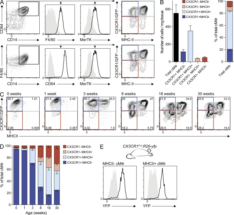
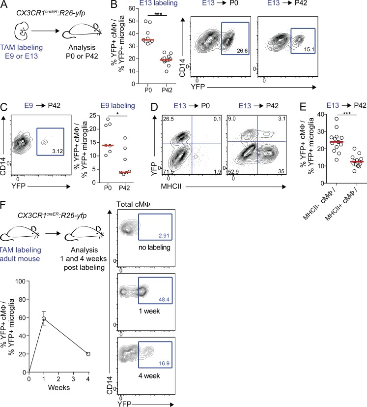
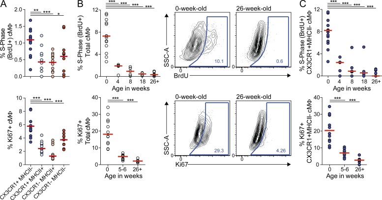
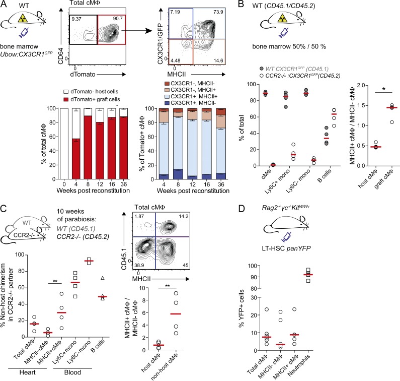
Cardiac macrophages (cMΦ) are critical for early postnatal heart regeneration and fibrotic repair in the adult heart, but their origins and cellular dynamics during postnatal development have not been well characterized. Tissue macrophages can be derived from embryonic progenitors or from monocytes during inflammation. We report that within the first weeks after birth, the embryo-derived population of resident CX3CR1+ cMΦ diversifies into MHCII+ and MHCII− cells. Genetic fate mapping demonstrated that cMΦ derived from CX3CR1+ embryonic progenitors persisted into adulthood but the initially high contribution to resident cMΦ declined after birth. Consistent with this, the early significant proliferation rate of resident cMΦ decreased with age upon diversification into subpopulations. Bone marrow (BM) reconstitution experiments showed monocyte-dependent quantitative replacement of all cMΦ populations. Furthermore, parabiotic mice and BM chimeras of nonirradiated recipient mice revealed a slow but significant donor contribution to cMΦ. Together, our observations indicate that in the heart, embryo-derived cMΦ show declining self-renewal with age and are progressively substituted by monocyte-derived macrophages, even in the absence of inflammation.
Overview
The claims and statistics of this paper haven’t been checked yet.
Run the check — free, no account needed. It reads the open-access text and checks the sources it cites.
Integrity signals
Registry
Cites
Statistics
Figures
Verification
Claims the paper makes
The claims and statistics of this paper haven’t been checked yet.
Run the check — free, no account needed. It reads the open-access text and checks the sources it cites.
Claims other papers make about this one
Each is checked against this paper’s own text: does it say what the citing paper says it does?
In the citing paper
“We now know that, in the steady state, arterial24 and cardiac1, 25–27 macrophages are mostly independent of monocytes”
From Abandoning M1/M2 for a Network Model of Macrophage Function (2016) · cites this paper as [26]
Self-check
Percentages, sums and reported differences re-computed from the paper’s own numbers.
The self-check needs the paper’s full text.
Citations
Cites
Fate Mapping Analysis Reveals That Adult Microglia Derive from Primitive Macrophages
Ginhoux, F., Greter, M., Leboeuf, M., Nandi, S., See, P., Gokhan, S., Mehler, M. F., Conway, S. J., Ng, L. G., Stanley, E. R., et al. (2010) · Science · doi:10.1126/science.1194637 · Cited by 5,175
Fate Mapping Reveals Origins and Dynamics of Monocytes and Tissue Macrophages under Homeostasis
Yona, S., Kim, K.-W., Wolf, Y., Mildner, A., Varol, D., Breker, M., Strauss-Ayali, D., Viukov, S., Guilliams, M., Misharin, A., et al. (2013) · Immunity · doi:10.1016/j.immuni.2012.12.001 · Cited by 3,245
Self-citationA Lineage of Myeloid Cells Independent of Myb and Hematopoietic Stem Cells
Schulz, C., Perdiguero, E. G., Chorro, L., Szabo-Rogers, H., Cagnard, N., Kierdorf, K., Prinz, M., Wu, B., Jacobsen, S. E. W., Pollard, J. W., et al. (2012) · Science · doi:10.1126/science.1219179 · Cited by 2,578
Self-citationTransient Regenerative Potential of the Neonatal Mouse Heart
Porrello, E. R., Mahmoud, A. I., Simpson, E., Hill, J. A., Richardson, J. A., Olson, E. N., & Sadek, H. A. (2011) · Science · doi:10.1126/science.1200708 · Cited by 2,545
Hashimoto, D., Chow, A., Noizat, C., Teo, P., Beasley, M. B., Leboeuf, M., Becker, C. D., See, P., Price, J., Lucas, D., et al. (2013) · Immunity · doi:10.1016/j.immuni.2013.04.004 · Cited by 2,311 · cited 3×
“(Chorro et al., 2009; Ginhoux et al., 2010; Hoeffel et al., 2012; Schulz et al., 2012; Hashimoto et al., 2013; Yona et al., 2013).”
Nahrendorf, M., Swirski, F. K., Aikawa, E., Stangenberg, L., Wurdinger, T., Figueiredo, J.-L., Libby, P., Weissleder, R., & Pittet, M. J. (2007) · The Journal of Experimental Medicine · doi:10.1084/jem.20070885 · Cited by 2,300
Identification of splenic reservoir monocytes and their deployment to inflammatory sites
Swirski, F. K., Nahrendorf, M., Etzrodt, M., Wildgruber, M., Cortez-Retamozo, V., Panizzi, P., Figueiredo, J.-L., Kohler, R. H., Chudnovskiy, A., Waterman, P., et al. (2009) · Science · doi:10.1126/science.1175202 · Cited by 2,221
Davies, L. C., Jenkins, S. J., Allen, J. E., & Taylor, P. R. (2013) · Nature Immunology · doi:10.1038/ni.2705 · Cited by 2,096
the Immunological Genome Consortium, Gautier, E. L., Shay, T., Miller, J., Greter, M., Jakubzick, C., Ivanov, S., Helft, J., Chow, A., Elpek, K. G., et al. (2012) · Nature Immunology · doi:10.1038/ni.2419 · Cited by 1,972 · cited 2×
“Tissue-resident macrophages were identified as positive for the core macrophage signature markers CD14, CD64, and MerTK (Gautier et al., 2012; Tamoutounour et al., 2013) and the classical macrophage marker F4/80 (Fig.”
Epelman, S., Lavine, K. J., Beaudin, A. E., Sojka, D. K., Carrero, J. A., Calderon, B., Brija, T., Gautier, E. L., Ivanov, S., Satpathy, A. T., et al. (2014) · Immunity · doi:10.1016/j.immuni.2013.11.019 · Cited by 1,494
Research works printed but not linked: 15
- Ajami, 2007, Local self-renewal can sustain CNS microglia maintenance and function throughout adult life, Nat. Neurosci., 10, 1538, 10.1038/nn2014 · DOI · Paper page
- Aurora, 2014, Macrophages are required for neonatal heart regeneration, J. Clin. Invest., 124, 1382, 10.1172/JCI72181 · DOI
- Aziz, 2009, MafB/c-Maf deficiency enables self-renewal of differentiated functional macrophages, Science., 326, 867, 10.1126/science.1176056 · DOI · Paper page
- Chorro, 2009, Langerhans cell (LC) proliferation mediates neonatal development, homeostasis, and inflammation-associated expansion of the epidermal LC network, J. Exp. Med., 206, 3089, 10.1084/jem.20091586 · DOI
- Geissmann, 2010, Development of monocytes, macrophages, and dendritic cells, Science., 327, 656, 10.1126/science.1178331 · DOI · Paper page
- Ghigo, 2013, Multicolor fate mapping of Langerhans cell homeostasis, J. Exp. Med., 210, 1657, 10.1084/jem.20130403 · DOI
- Goldmann, 2013, A new type of microglia gene targeting shows TAK1 to be pivotal in CNS autoimmune inflammation, Nat. Neurosci., 16, 1618, 10.1038/nn.3531 · DOI · Paper page
- Hoeffel, 2012, Adult Langerhans cells derive predominantly from embryonic fetal liver monocytes with a minor contribution of yolk sac-derived macrophages, J. Exp. Med., 209, 1167, 10.1084/jem.20120340 · DOI · Paper page
- Jenkins, 2011, Local macrophage proliferation, rather than recruitment from the blood, is a signature of TH2 inflammation, Science., 332, 1284, 10.1126/science.1204351 · DOI
- Jung, 2000, Analysis of fractalkine receptor CX3CR1 function by targeted deletion and green fluorescent protein reporter gene insertion, Mol. Cell. Biol., 20, 4106, 10.1128/MCB.20.11.4106-4114.2000 · DOI · Paper page
- Luche, 2013, In vivo fate mapping identifies pre-TCRα expression as an intra- and extrathymic, but not prethymic, marker of T lymphopoiesis, J. Exp. Med., 210, 699, 10.1084/jem.20122609 · DOI
- Serbina, 2006, Monocyte emigration from bone marrow during bacterial infection requires signals mediated by chemokine receptor CCR2, Nat. Immunol., 7, 311, 10.1038/ni1309 · DOI
- van Furth, 1968, The origin and kinetics of mononuclear phagocytes, J. Exp. Med., 128, 415, 10.1084/jem.128.3.415 · DOI
- Waskow, 2009, Hematopoietic stem cell transplantation without irradiation, Nat. Methods., 6, 267, 10.1038/nmeth.1309 · DOI
- Zigmond, 2012, Ly6C hi monocytes in the inflamed colon give rise to proinflammatory effector cells and migratory antigen-presenting cells, Immunity., 37, 1076, 10.1016/j.immuni.2012.08.026 · DOI
Cited by
Sorted by citations
Tissue-Resident Macrophage Ontogeny and Homeostasis
Ginhoux, F., & Guilliams, M. (2016) · Immunity · doi:10.1016/j.immuni.2016.02.024 · Cited by 1,884
Understanding the Mysterious M2 Macrophage through Activation Markers and Effector Mechanisms
Rőszer, T. (2015) · Mediators of Inflammation · doi:10.1155/2015/816460 · Cited by 1,762
Origin, fate and dynamics of macrophages at central nervous system interfaces
Goldmann, T., Wieghofer, P., Jordão, M. J. C., Prutek, F., Hagemeyer, N., Frenzel, K., Amann, L., Staszewski, O., Kierdorf, K., Krueger, M., et al. (2016) · Nature Immunology · doi:10.1038/ni.3423 · Cited by 1,209
Self-citationMacrophages Facilitate Electrical Conduction in the Heart
Hulsmans, M., Clauss, S., Xiao, L., Aguirre, A. D., King, K. R., Hanley, A., Hucker, W. J., Wülfers, E. M., Seemann, G., Courties, G., et al. (2017) · Cell · doi:10.1016/j.cell.2017.03.050 · Cited by 1,011
Macrophages: Development and Tissue Specialization
Varol, C., Mildner, A., & Jung, S. (2015) · Annual Review of Immunology · doi:10.1146/annurev-immunol-032414-112220 · Cited by 962
Self-citation
Metadata
- Authors
- Molawi, K., Wolf, Y., Kandalla, P. K., Favret, J., Hagemeyer, N., Frenzel, K., Pinto, A. R., Klapproth, K., Henri, S., Malissen, B., Rodewald, H.-R., Rosenthal, N. A., Bajenoff, M., Prinz, M., Jung, S., & Sieweke, M. H.
- Affiliations
- Centre d’Immunologie de Marseille-Luminy (CIML), Aix-Marseille Université, UM2, 13288 Marseille, France 1
- Department of Immunology, The Weizmann Institute of Science, 7610001 Rehovot, Israel 5
- Institute of Neuropathology 6 and 7
- Australian Regenerative Medicine Institute (ARMI), Monash University, Clayton 3800, Victoria, Australia 8
- Division of Cellular Immunology, German Cancer Research Center (DKFZ), D-69120 Heidelberg, Germany 9
- Venue
- Journal of Experimental Medicine
- Publisher
- Rockefeller University Press
- Published
- 2014-09-22
- Volume
- 211
- Issue
- 11
- Pages
- 2151-2158
- ISSN
- 1540-9538
- 0022-1007
- PMC
- PMC4203946
- Cited by
- 513
- References
- 14
- Language
- en
About these checks
Registry, retraction, Cites and statistics results are computed, not judged, with one exception: a printed reference that no index lists is read once by a model before it counts as a citation error. Claims are extracted by a model and kept only when their quote appears verbatim in the text; each cited source is then checked, with a model’s help, for whether it says what the claim relies on. Those verdicts can be wrong — read the claim’s quote and the cited source before relying on one.
Statistics and arithmetic are recomputed, not judged.
Confidence bands are shown only for fields calibrated on our annotated test set. None is calibrated yet, so no band is shown.
What counts as a claim ›How we measure confidence ›How we check this ›
Are you an author, or do you see a problem with this page? Request a review
How to cite
Cite CoChat for the checks on this page, and the paper for its own findings.
- This page
- CoChat. (2026). Citation check: Progressive replacement of embryo-derived cardiac macrophages with age. https://app.cochat.ai/papers/10.1084/jem.20140639
- The paper
- Molawi, K., Wolf, Y., Kandalla, P. K., Favret, J., Hagemeyer, N., Frenzel, K., Pinto, A. R., Klapproth, K., Henri, S., Malissen, B., Rodewald, H.-R., Rosenthal, N. A., Bajenoff, M., Prinz, M., Jung, S., & Sieweke, M. H. (2014). Progressive replacement of embryo-derived cardiac macrophages with age. Journal of Experimental Medicine. https://doi.org/10.1084/jem.20140639
