

The DOI matches its Crossref record; 44 of the 44 references it cites were found in a registry or an index.
A neuroprotective role for microglia in prion diseases
Open accessAbstract
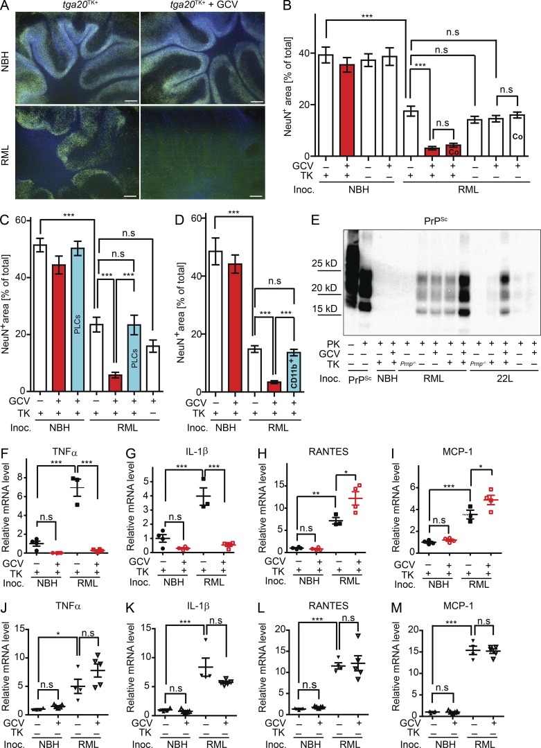
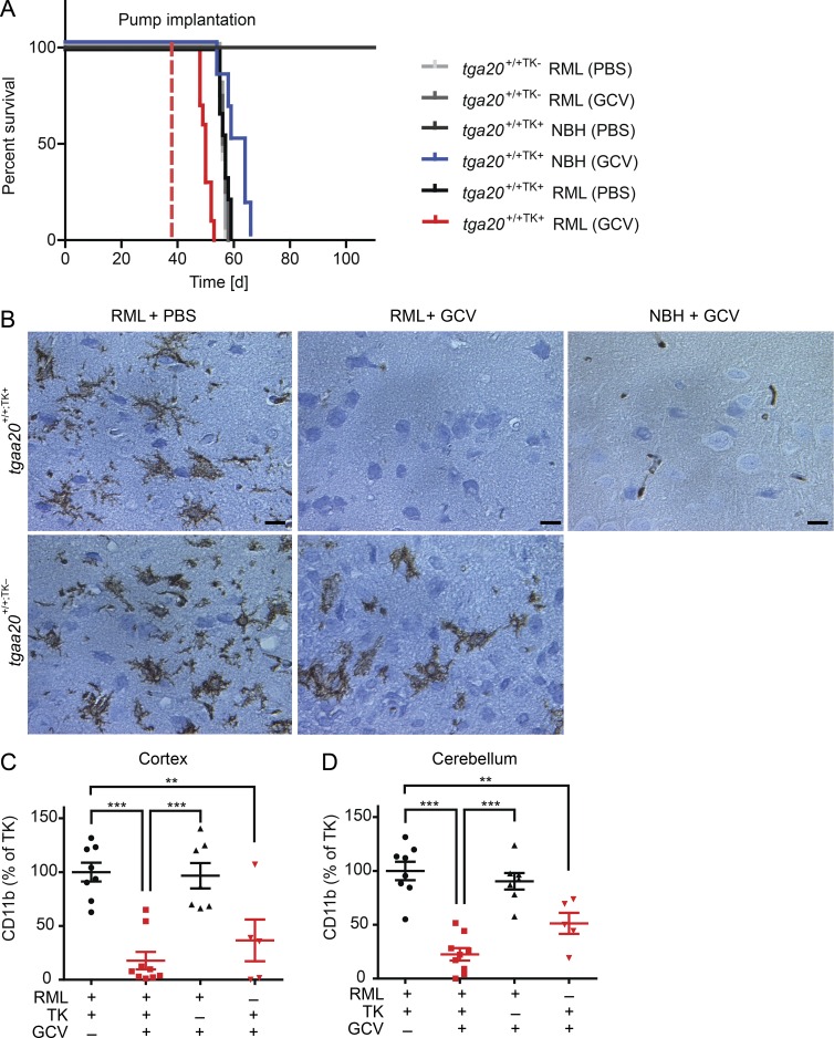
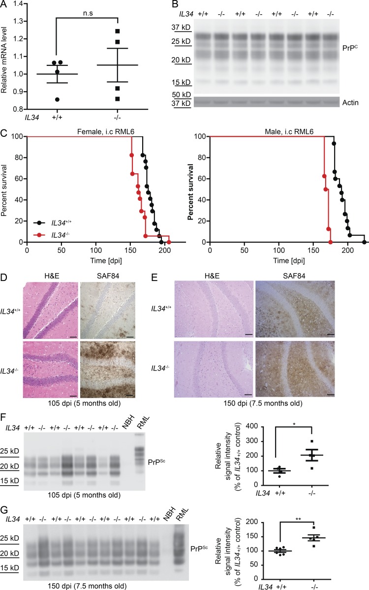
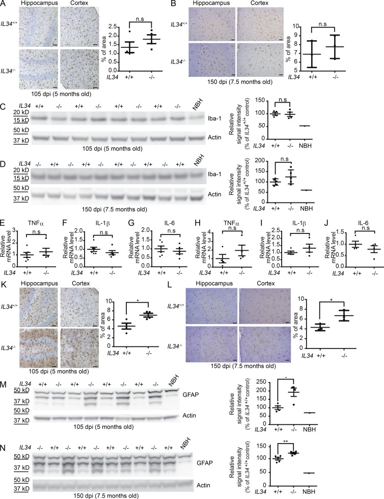
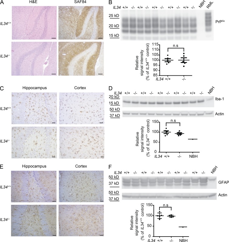
Microglial activation is a hallmark of most neurodegenerative disorders, and is particularly conspicuous in prion diseases. However, the role of microglia, which function as both primary immune effector cells and professional phagocytes in the central nervous system, remains contentious in the context of neurodegeneration. Here, we evaluated the effect of microglial depletion/deficiency on prion pathogenesis. We found that ganciclovir-mediated microglial ablation on tga20/CD11b-thymidine kinase of Herpes simplex virus (HSVTK) cerebellar organotypic cultured slices markedly aggravated prion-induced neurotoxicity. A similar deterioration of disease was recapitulated in in vivo microglial depletion in prion-infected tga20/CD11b-HSVTK mice. Additionally, deficiency of microglia in interleukin 34 knockout (IL34−/−) mice again resulted in significantly augmented proteinase K–resistant prion protein deposition and accelerated prion disease progression. These results provide unambiguous evidence for a general protective role of microglia in prion pathogenesis.
Overview
The claims and statistics of this paper haven’t been checked yet.
Run the check — free, no account needed. It reads the open-access text and checks the sources it cites.
Integrity signals
Registry
Cites
Statistics
Figures
Verification
Claims the paper makes
The claims and statistics of this paper haven’t been checked yet.
Run the check — free, no account needed. It reads the open-access text and checks the sources it cites.
Claims other papers make about this one
Each is checked against this paper’s own text: does it say what the citing paper says it does?
In the citing paper
“Work on prion-infected organotypic brain-slice cultures support the prion clearing and neuroprotective functions of microglia [50].”
From Loss of Homeostatic Microglia Signature in Prion Diseases (2022) · cites this paper as [50]
In the citing paper
“In contrast, reducing microglia numbers by knocking out interleukin 34 (IL-34), an essential signaling molecule, led to augmented PrPSc deposition and shortened survival in a prion mouse model [50,51]”
From Loss of Homeostatic Microglia Signature in Prion Diseases (2022) · cites this paper as [50]
Self-check
Percentages, sums and reported differences re-computed from the paper’s own numbers.
The self-check needs the paper’s full text.
Citations
Cites
Polazzi, E., Mengoni, I., Caprini, M., Peña‐Altamira, E., Kurtys, E., & Monti, B. (2012) · Neurosignals · doi:10.1159/000337115 · Cited by 9,023
Resting microglial cells are highly dynamic surveillants of brain parenchyma in vivo
Nimmerjahn, A., Kirchhoff, F., & Helmchen, F. (2005) · Science · doi:10.1126/science.1110647 · Cited by 5,827
Fate Mapping Analysis Reveals That Adult Microglia Derive from Primitive Macrophages
Ginhoux, F., Greter, M., Leboeuf, M., Nandi, S., See, P., Gokhan, S., Mehler, M. F., Conway, S. J., Ng, L. G., Stanley, E. R., et al. (2010) · Science · doi:10.1126/science.1194637 · Cited by 5,175
Synaptic Pruning by Microglia Is Necessary for Normal Brain Development
Paolicelli, R. C., Bolasco, G., Pagani, F., Maggi, L., Scianni, M., Panzanelli, P., Giustetto, M., Ferreira, T. A., Guiducci, E., Dumas, L., et al. (2011) · Science · doi:10.1126/science.1202529 · Cited by 4,244
Microglia Sculpt Postnatal Neural Circuits in an Activity and Complement-Dependent Manner
Schafer, D. P., Lehrman, E. K., Kautzman, A. G., Koyama, R., Mardinly, A. R., Yamasaki, R., Ransohoff, R. M., Greenberg, M. E., Barres, B. A., & Stevens, B. (2012) · Neuron · doi:10.1016/j.neuron.2012.03.026 · Cited by 4,089 · cited 1×
“In the brain, microglia interact with neurons and exert a wide variety of physiological functions (Chen et al., 2010; Paolicelli et al., 2011; Rogers et al., 2011; Derecki et al., 2012; Schafer et al., 2012; Parkhurst et al., 2013; Zhan et al., 2014).”
BackgroundATP mediates rapid microglial response to local brain injury in vivo
Davalos, D., Grutzendler, J., Yang, G., Kim, J. V., Zuo, Y., Jung, S., Littman, D. R., Dustin, M. L., & Gan, W.-B. (2005) · Nature Neuroscience · doi:10.1038/nn1472 · Cited by 3,913
Microglia: Active sensor and versatile effector cells in the normal and pathologic brain
Hanisch, U.-K., & Kettenmann, H. (2007) · Nature Neuroscience · doi:10.1038/nn1997 · Cited by 3,676
Microglia promote learning-dependent synapse formation through brain-derived neurotrophic factor
Parkhurst, C. N., Yang, G., Ninan, I., Savas, J. N., Yates, J. R., Lafaille, J. J., Hempstead, B. L., Littman, D. R., & Gan, W.-B. (2013) · Cell · doi:10.1016/j.cell.2013.11.030 · Cited by 2,599
A Lineage of Myeloid Cells Independent of Myb and Hematopoietic Stem Cells
Schulz, C., Perdiguero, E. G., Chorro, L., Szabo-Rogers, H., Cagnard, N., Kierdorf, K., Prinz, M., Wu, B., Jacobsen, S. E. W., Pollard, J. W., et al. (2012) · Science · doi:10.1126/science.1219179 · Cited by 2,578
Microglia emerge from erythromyeloid precursors via Pu.1- and Irf8-dependent pathways
Kierdorf, K., Erny, D., Goldmann, T., Sander, V., Schulz, C., Perdiguero, E. G., Wieghofer, P., Heinrich, A., Riemke, P., Hölscher, C., et al. (2013) · Nature Neuroscience · doi:10.1038/nn.3318 · Cited by 1,457
Zhan, Y., Paolicelli, R. C., Sforazzini, F., Weinhard, L., Bolasco, G., Pagani, F., Vyssotski, A. L., Bifone, A., Gozzi, A., Ragozzino, D., et al. (2014) · Nature Neuroscience · doi:10.1038/nn.3641 · Cited by 1,236
Drabkin, D. L., & Austin, J. H. (1935) · Journal of Biological Chemistry · doi:10.1016/s0021-9258(18)74965-x · Cited by 1,018
Wang, Y., Szretter, K. J., Vermi, W., Gilfillan, S., Rossini, C., Cella, M., Barrow, A. D., Diamond, M. S., & Colonna, M. (2012) · Nature Immunology · doi:10.1038/ni.2360 · Cited by 1,011
Microglia derive from progenitors, originating from the yolk sac, and which proliferate in the brain
Alliot, F., Godin, I., & Pessac, B. (1999) · Developmental Brain Research · doi:10.1016/s0165-3806(99)00113-3 · Cited by 884
Fischer, M. B., Rülicke, T., Raeber, A. J., Sailer, A. W., Moser, M., Oesch, B., Brandner, S., Aguzzi, A., & Weissmann, C. (1996) · The EMBO Journal · doi:10.1002/j.1460-2075.1996.tb00467.x · Cited by 877
Self-citationMicroglia: Scapegoat, saboteur, or something else?
Aguzzi, A., Barres, B. A., & Bennett, M. L. (2013) · Science · doi:10.1126/science.1227901 · Cited by 836
Self-citationExperimental autoimmune encephalomyelitis repressed by microglial paralysis
Heppner, F. L., Greter, M., Marino, D., Falsig, J., Raivich, G., Hövelmeyer, N., Waisman, A., Rülicke, T., Prinz, M., Priller, J., et al. (2005) · Nature Medicine · doi:10.1038/nm1177 · Cited by 732 · cited 5×
“…evaluated the role of microglia in COCS prepared from tga20 :CD11b -HSV TK mice (tga20TK+), which were obtained by intercrossing mice overexpressing the cellular prion protein PrPC (Fischer et al., 1996) and mice expressing the HSV TK under the control of the CD11b promoter (Heppner et al., 2005).”
InfluentialSelf-citationGreter, M., Lelios, I., Pelczar, P., Hoeffel, G., Price, J., Leboeuf, M., Kündig, T. M., Frei, K., Ginhoux, F., Merad, M., et al. (2012) · Immunity · doi:10.1016/j.immuni.2012.11.001 · Cited by 630
CX3CR1 deficiency leads to impairment of hippocampal cognitive function and synaptic plasticity
Rogers, J. T., Morganti, J. M., Bachstetter, A. D., Hudson, C. E., Peters, M. M., Grimmig, B. A., Weeber, E. J., Bickford, P. C., & Gemma, C. (2011) · The Journal of Neuroscience · doi:10.1523/jneurosci.3667-11.2011 · Cited by 624
Wild-type microglia arrest pathology in a mouse model of Rett syndrome
Derecki, N. C., Cronk, J. C., Lu, Z., Xu, E., Abbott, S. B. G., Guyenet, P. G., & Kipnis, J. (2012) · Nature · doi:10.1038/nature10907 · Cited by 607
Role of microglia and host prion protein in neurotoxicity of a prion protein fragment
Brown, D. R., Schmidt, B., & Kretzschmar, H. A. (1996) · Nature · doi:10.1038/380345a0 · Cited by 499
Hematopoietic origin of pathological grooming in Hoxb8 mutant mice
Chen, S.-K., Tvrdik, P., Peden, E., Cho, S., Wu, S., Spangrude, G., & Capecchi, M. R. (2010) · Cell · doi:10.1016/j.cell.2010.03.055 · Cited by 450
Regulation of Microglial Proliferation during Chronic Neurodegeneration
Gómez-Nicola, D., Fransen, N. L., Suzzi, S., & Perry, V. H. (2013) · The Journal of Neuroscience · doi:10.1523/jneurosci.4440-12.2013 · Cited by 407
Formation and maintenance of Alzheimer's disease β-amyloid plaques in the absence of microglia
Grathwohl, S. A., Kälin, R. E., Bolmont, T., Prokop, S., Winkelmann, G., Kaeser, S. A., Odenthal, J., Radde, R., Eldh, T., Gandy, S., et al. (2009) · Nature Neuroscience · doi:10.1038/nn.2432 · Cited by 401
Self-citationMildner, A., Schlevogt, B., Kierdorf, K., Böttcher, C., Erny, D., Kummer, M. P., Quinn, M., Brück, W., Bechmann, I., Heneka, M. T., et al. (2011) · The Journal of Neuroscience · doi:10.1523/jneurosci.6209-10.2011 · Cited by 351
Role of microglia in neuronal cell death in prion disease
Giese, A., Brown, D. R., Groschup, M. H., Feldmann, C., Haist, I., & Kretzschmar, H. A. (1998) · Brain Pathology · doi:10.1111/j.1750-3639.1998.tb00167.x · Cited by 239
The toxicity of antiprion antibodies is mediated by the flexible tail of the prion protein
Sonati, T., Reimann, R., Falsig, J., Baral, P. K., O’Connor, T., Hornemann, S., Yaganoglu, S., Li, B., Herrmann, U., Wieland, B., et al. (2013) · Nature · doi:10.1038/nature12402 · Cited by 210
Self-citationPrP Deposition, Microglial Activation, and Neuronal Apoptosis in Murine Scrapie
Williams, A., Lucassen, P. J., Ritchie, D., & Bruce, M. (1997) · Experimental Neurology · doi:10.1006/exnr.1997.6424 · Cited by 204
Follicular dendritic cells: origin, phenotype, and function in health and disease
Aguzzi, A., Kranich, J., & Krautler, N. J. (2014) · Trends in Immunology · doi:10.1016/j.it.2013.11.001 · Cited by 176
Self-citationA versatile prion replication assay in organotypic brain slices
Falsig, J., Julius, C., Margalith, I., Schwarz, P., Heppner, F. L., & Aguzzi, A. (2007) · Nature Neuroscience · doi:10.1038/nn2028 · Cited by 153
Self-citationThe immunobiology of prion diseases
Aguzzi, A., Nuvolone, M., & Zhu, C. (2013) · Nature reviews. Immunology · doi:10.1038/nri3553 · Cited by 150
Self-citationKlein, T. R., Kirsch, D., KAUFMANN, R. L., & Riesner, D. (1998) · Biological Chemistry · doi:10.1515/bchm.1998.379.6.655 · Cited by 143
Baker, C. A., Martin, D., & Manuelidis, L. (2002) · Journal of Virology · doi:10.1128/jvi.76.21.10905-10913.2002 · Cited by 128
Prion propagation, toxicity and degradation
Aguzzi, A., & Falsig, J. (2012) · Nature Neuroscience · doi:10.1038/nn.3120 · Cited by 126
Self-citationKranich, J., Krautler, N. J., Falsig, J., Ballmer, B., Li, S., Hutter, G., Schwarz, P., Moos, R., Julius, C., Miele, G., et al. (2010) · Journal of Experimental Medicine · doi:10.1084/jem.20092401 · Cited by 110
Self-citationPrion Infections and Anti-PrP Antibodies Trigger Converging Neurotoxic Pathways
Herrmann, U., Sonati, T., Falsig, J., Reimann, R., Dametto, P., O’Connor, T., Li, B., Lau, A., Hornemann, S., Sorce, S., et al. (2015) · PLoS Pathogens · doi:10.1371/journal.ppat.1004662 · Cited by 97
Self-citationHughes, M. M., Field, R. H., Perry, V. H., Murray, C. L., & Cunningham, C. (2010) · Glia · doi:10.1002/glia.21070 · Cited by 94
Stereological estimation of total microglia number in mouse hippocampus
Long, J. M., Kalehua, A. N., Muth, N. J., Hengemihle, J. M., Jucker, M., Calhoun, M. E., Ingram, D. K., & Mouton, P. R. (1998) · Journal of Neuroscience Methods · doi:10.1016/s0165-0270(98)00100-9 · Cited by 87
The prion organotypic slice culture assay—POSCA
Falsig, J., & Aguzzi, A. (2008) · Nature Protocols · doi:10.1038/nprot.2008.13 · Cited by 87
Self-citationPrion Pathogenesis Is Faithfully Reproduced in Cerebellar Organotypic Slice Cultures
Falsig, J., Sonati, T., Herrmann, U., Saban, D. V., Li, B., Arroyo, K., Ballmer, B., Liberski, P. P., & Aguzzi, A. (2012) · PLoS Pathogens · doi:10.1371/journal.ppat.1002985 · Cited by 79
Self-citationRouvinski, A., Karniely, S., Kounin, M., Moussa, S., Goldberg, M. D., Warburg, G., Lyakhovetsky, R., Papy-García, D., Kutzsche, J., Korth, C., et al. (2014) · The Journal of Cell Biology · doi:10.1083/jcb.201308028 · Cited by 72
Morphogenesis of amyloid plaques in 87V murine scrapie†
Jeffrey, M., Goodsir, C. M., Bruce, M. E., McBride, P., & Farquhar, C. F. (1994) · Neuropathology and Applied Neurobiology · doi:10.1111/j.1365-2990.1994.tb01007.x · Cited by 68
Gómez‐Nicola, D., Schetters, S. T. T., & Perry, V. H. (2014) · Glia · doi:10.1002/glia.22660 · Cited by 60
ZyFISH: A Simple, Rapid and Reliable Zygosity Assay for Transgenic Mice
McHugh, D., O’Connor, T., Bremer, J., & Aguzzi, A. (2012) · PLoS ONE · doi:10.1371/journal.pone.0037881 · Cited by 7
Self-citation
Cited by
Sorted by citations
Microglia Function in the Central Nervous System During Health and Neurodegeneration
Colonna, M., & Butovsky, O. (2017) · Annual Review of Immunology · doi:10.1146/annurev-immunol-051116-052358 · Cited by 2,894
Prions, prionoids and protein misfolding disorders
Scheckel, C., & Aguzzi, A. (2018) · Nature Reviews Genetics · doi:10.1038/s41576-018-0011-4 · Cited by 311
Self-citationYan, Z., Gibson, S. A., Buckley, J. A., Qin, H., & Benveniste, E. N. (2018) · Clinical Immunology · doi:10.1016/j.clim.2016.09.014 · Cited by 318
Lee, J.-W., Nam, H., Kim, L. E., Jeon, Y., Min, H., Ha, S., Lee, Y., Kim, S.-Y., Lee, S. J., Kim, E.-K., et al. (2019) · Autophagy · doi:10.1080/15548627.2018.1556946 · Cited by 260
Kröller‐Schön, S., Daiber, A., Steven, S., Oelze, M. W., Frenis, K., Kalinovic, S., Heimann, A., Schmidt, F. P., Pinto, A. V., Kvandová, M., et al. (2018) · European Heart Journal · doi:10.1093/eurheartj/ehy333 · Cited by 229
Self-citation
Metadata
- Authors
- Zhu, C., Herrmann, U. S., Falsig, J., Abakumova, I., Nuvolone, M., Schwarz, P., Frauenknecht, K., Rushing, E. J., & Aguzzi, A.
- Affiliations
- University Hospital Zurich (CH)
- Corresponding author
- Zhu, Caihong
- Aguzzi, Adriano
- ORCID
- Venue
- Journal of Experimental Medicine
- Publisher
- Rockefeller University Press
- Published
- 2016-05-16
- Volume
- 213
- Issue
- 6
- Pages
- 1047-1059
- ISSN
- 0022-1007 (ISSN-L)
- 1540-9538
- PMC
- PMC4886355
- OpenAlex
- W2406691442
- Cited by
- 134
- References
- 44
- Citations by year
- 2026: 4 · 2025: 10 · 2024: 9 · 2023: 7 · 2022: 17 · 2021: 11 · 2020: 12 · 2019: 17 · 2018: 17 · 2017: 25 · 2016: 5
- FWCI
- 11.08
- Type
- journal article
- Language
- en
- Open access
- bronze
- doi.org
- Locations
- The Journal of Experimental Medicine (publishedVersion)
- Europe PMC (PubMed Central) (publishedVersion, cc-by-nc-sa)
- PubMed Central (publishedVersion, cc-by-nc-sa)
- PubMed (publishedVersion)
- Zurich Open Repository and Archive (University of Zurich) (acceptedVersion, cc-by-nc)
- Zurich Open Repository and Archive (University of Zurich) (submittedVersion, cc-by-nc)
- Zurich Open Repository and Archive (University of Zurich)
- Funders
- European Research Council (250356)
- Swiss National Foundation
- Novartis Research Foundation
- Stavros Niarchos Foundation
- Topics
- Life Sciences › Biochemistry, Genetics and Molecular Biology › Molecular Biology › Prion Diseases and Protein Misfolding
- Life Sciences › Neuroscience › Neurology › Neuroinflammation and Neurodegeneration Mechanisms
- Life Sciences › Neuroscience › Cellular and Molecular Neuroscience › Alzheimer's disease research and treatments
- Keywords
- microglia, prion disease, neuroprotection, microglial depletion, ganciclovir, IL-34, prion protein deposition, neurodegeneration
- MeSH terms
- Animals, Interleukins, Prions, Prion Diseases, Microglia, Mice, Knockout, Mice
About these checks
Registry, retraction, Cites and statistics results are computed, not judged, with one exception: a printed reference that no index lists is read once by a model before it counts as a citation error. Claims are extracted by a model and kept only when their quote appears verbatim in the text; each cited source is then checked, with a model’s help, for whether it says what the claim relies on. Those verdicts can be wrong — read the claim’s quote and the cited source before relying on one.
Statistics and arithmetic are recomputed, not judged.
Confidence bands are shown only for fields calibrated on our annotated test set. None is calibrated yet, so no band is shown.
What counts as a claim ›How we measure confidence ›How we check this ›
Are you an author, or do you see a problem with this page? Request a review
