

The DOI matches its Crossref record; 16 of the 16 references it cites were found in a registry or an index.
The Role of Reactive Oxygen Species in Mesenchymal Stem Cell Adipogenic and Osteogenic Differentiation: A Review
Abstract
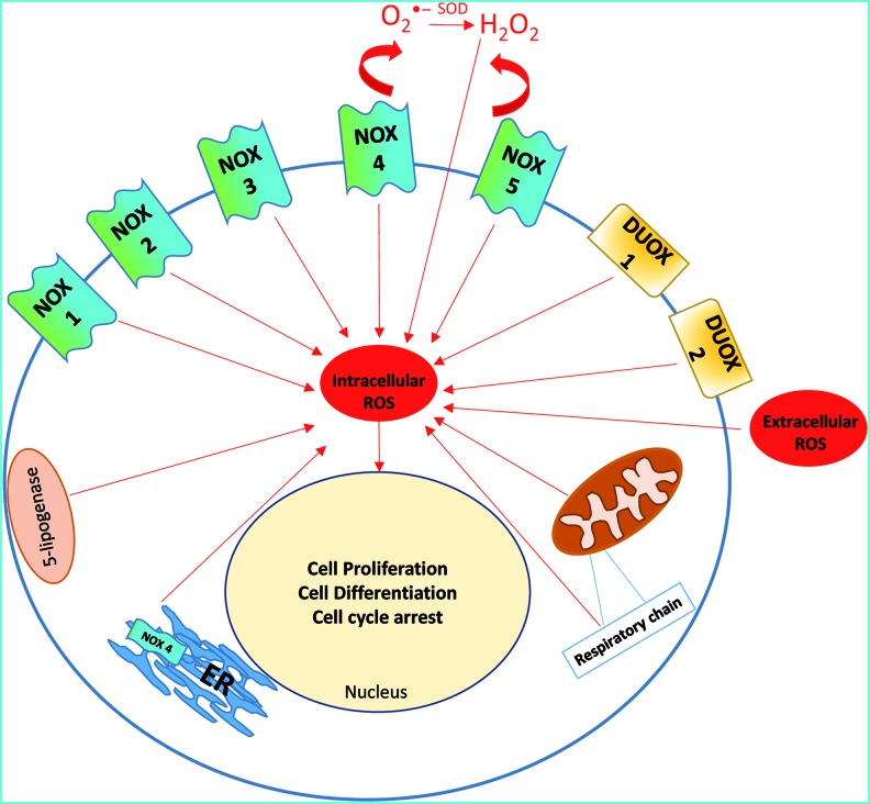
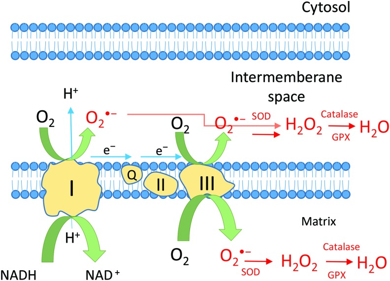
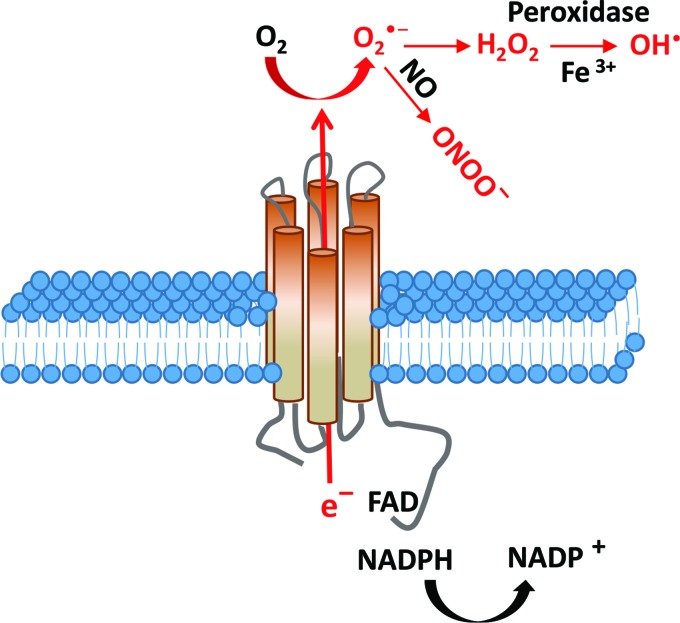
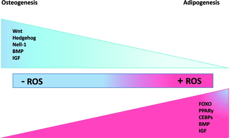
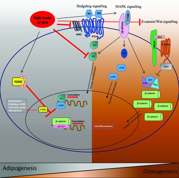
Mesenchymal stromal cells (MSCs) are promising candidates for tissue engineering and regenerative medicine. The multipotent stem cell component of MSC isolates is able to differentiate into derivatives of the mesodermal lineage including adipocytes, osteocytes, chondrocytes, and myocytes. Many common pathways have been described in the regulation of adipogenesis and osteogenesis. However, stimulation of osteogenesis appears to suppress adipogenesis and vice-versa. Increasing evidence implicates a tight regulation of these processes by reactive oxygen species (ROS). ROS are short-lived oxygen-containing molecules that display high chemical reactivity toward DNA, RNA, proteins, and lipids. Mitochondrial complexes I and III, and the NADPH oxidase isoform NOX4 are major sources of ROS production during MSC differentiation. ROS are thought to interact with several pathways that affect the transcription machinery required for MSC differentiation including the Wnt, Hedgehog, and FOXO signaling cascades. On the other hand, elevated levels of ROS, defined as oxidative stress, lead to arrest of the MSC cell cycle and apoptosis. Tightly regulated levels of ROS are therefore critical for MSC terminal differentiation, although the precise sources, localization, levels and the exact species of ROS implicated remain to be determined. This review provides a detailed overview of the influence of ROS on adipogenic and osteogenic differentiation in MSCs.
Overview
The claims and statistics of this paper haven’t been checked yet.
Run the check — free, no account needed. It reads the open-access text and checks the sources it cites.
Integrity signals
Registry
Cites
Statistics
Figures
Verification
Claims the paper makes
The claims and statistics of this paper haven’t been checked yet.
Run the check — free, no account needed. It reads the open-access text and checks the sources it cites.
Claims other papers make about this one
Each is checked against this paper’s own text: does it say what the citing paper says it does?
In the citing paper
“It was reported that high levels of ROS induce adipogenesis through Forkhead box O (FOXO), peroxisome proliferator‐activated receptor γ (PPARγ), and CCAAT‐enhancer‐binding protein signaling, whereas the absence of ROS induces osteogenesis through Wnt/β‐catenin, mitogen‐activated protein kinase (MAPK), and Hedgehog signaling in mesenchymal stem cells.40”
From Phlebotomy as a preventive measure for crocidolite-induced mesothelioma in male rats (2018) · cites this paper as [40]
Self-check
Percentages, sums and reported differences re-computed from the paper’s own numbers.
The self-check needs the paper’s full text.
Citations
Cites
Dominici, M., Le Blanc, K., Mueller, I., Slaper-Cortenbach, I., Marini, F. C., Krause, D. S., Deans, R. J., Keating, A., Prockop, D. J., & Horwitz, E. M. (2006) · Cytotherapy · doi:10.1080/14653240600855905 · Cited by 17,766
Biochemistry and molecular cell biology of diabetic complications
Brownlee, M. (2001) · Nature · doi:10.1038/414813a · Cited by 9,107
How mitochondria produce reactive oxygen species
Murphy, M. P. (2009) · Biochemical Journal · doi:10.1042/bj20081386 · Cited by 8,207
The NOX Family of ROS-Generating NADPH Oxidases: Physiology and Pathophysiology
Bedard, K., & Krause, K.-H. (2007) · Physiological Reviews · doi:10.1152/physrev.00044.2005 · Cited by 6,989
Stress-dependent regulation of FOXO transcription factors by the SIRT1 deacetylase
Brunet, A., Sweeney, L. B., Sturgill, J. F., Chua, K. F., Greer, P. L., Lin, Y., Tran, H., Ross, S. E., Mostoslavsky, R., Cohen, H. Y., et al. (2004) · Science · doi:10.1126/science.1094637 · Cited by 3,239
Signal transduction by reactive oxygen species
Finkel, T. (2011) · Journal of Cell Biology · doi:10.1083/jcb.201102095 · Cited by 2,322
Fat and Beyond: The Diverse Biology of PPARγ
Tontonoz, P., & Spiegelman, B. M. (2008) · Annual Review of Biochemistry · doi:10.1146/annurev.biochem.77.061307.091829 · Cited by 2,085
Bourin, P., Bunnell, B. A., Casteilla, L., Dominici, M., Katz, A. J., March, K. L., Redl, H., Rubin, J. P., Yoshimura, K., & Gimble, J. M. (2013) · Cytotherapy · doi:10.1016/j.jcyt.2013.02.006 · Cited by 1,890
Hypercholesterolemia increases endothelial superoxide anion production.
Ohara, Y., Peterson, T. E., & Harrison, D. G. (1993) · Journal of Clinical Investigation · doi:10.1172/jci116491 · Cited by 1,765
Forkhead transcription factor FOXO3a protects quiescent cells from oxidative stress
Kops, G. J. P. L., Dansen, T. B., Polderman, P. E., Saarloos, I., Wirtz, K. W. A., Coffer, P. J., Huang, T.-T., Bos, J. L., Medema, R. H., & Burgering, B. M. T. (2002) · Nature · doi:10.1038/nature01036 · Cited by 1,580
Research works printed but not linked: 126
- 10.1093/geronj/11.3.298 · DOI · Paper page
- 10.1152/physrev.1998.78.2.547 · DOI · Paper page
- 10.1016/j.drudis.2006.04.003 · DOI
- 10.1089/ars.2011.4489 · DOI
- 10.1089/ars.2010.3405 · DOI
- 10.1186/1755-1536-4-20 · DOI
- 10.1186/ar614 · DOI
- 10.1007/s10517-005-0430-z · DOI
- 10.1182/blood-2003-05-1664 · DOI
- 10.1186/1478-811X-9-12 · DOI
- 10.1007/s12015-012-9365-8 · DOI
- 10.1089/ten.tec.2008.0031 · DOI
- 10.4252/wjsc.v4.i6.53 · DOI
- 10.1016/S1471-4914(01)02016-0 · DOI
- 10.1016/j.stem.2008.03.002 · DOI
- 10.1097/TP.0b013e3181c7dc99 · DOI
- 10.1002/jcb.20886 · DOI · Paper page
- 10.1089/scd.2010.0093 · DOI
- 10.1089/scd.2012.0306 · DOI
- 10.1196/annals.1402.075 · DOI
- 10.1016/j.scr.2013.07.004 · DOI
- 10.1186/scrt190 · DOI
- 10.1172/JCI21311 · DOI
- 10.1242/jcs.102.2.341 · DOI
- 10.1083/jcb.142.1.295 · DOI
- 10.1155/2013/684736 · DOI
- 10.1016/j.bbrc.2011.06.111 · DOI
- 10.1242/jcs.114.11.2085 · DOI
- 10.1038/nrrheum.2009.137 · DOI
- 10.1089/scd.2008.0130 · DOI
- 10.1007/s00467-004-1613-y · DOI
- 10.1016/0026-0495(95)90220-1 · DOI
- 10.1016/S0021-9258(19)76555-7 · DOI
- 10.1073/pnas.88.22.10045 · DOI
- 10.1515/CCLM.2008.302 · DOI
- 10.1016/j.mito.2011.02.001 · DOI
- 10.1161/01.RES.86.5.494 · DOI · Paper page
- 10.2174/138920112798868782 · DOI
- 10.1042/bj1280617 · DOI
- 10.1073/pnas.0506448103 · DOI
- 10.1161/01.RES.59.6.612 · DOI · Paper page
- 10.1016/0005-2760(94)90041-8 · DOI
- 10.1042/bj3180805 · DOI
- 10.1152/ajpheart.00515.2003 · DOI
- 10.1634/stemcells.2007-0509 · DOI
- 10.1016/j.freeradbiomed.2011.06.033 · DOI · Paper page
- 10.1074/jbc.M105395200 · DOI
- 10.1089/ars.2000.2.4-811 · DOI
- 10.1152/physrev.00018.2001 · DOI · Paper page
- 10.1073/pnas.0906805106 · DOI
- 10.1182/blood-2010-02-269894 · DOI
- 10.1038/276515a0 · DOI
- 10.1038/sj.bjp.0701171 · DOI
- 10.1139/y90-241 · DOI
- 10.1038/43459 · DOI · Paper page
- 10.1517/14728222.2011.628315 · DOI
- 10.1097/01.MIB.0000442012.45038.0e · DOI
- 10.1089/thy.2012.0003 · DOI
- 10.1089/ars.2006.1467 · DOI · Paper page
- 10.1002/jcp.20641 · DOI
- 10.1161/01.RES.0000135902.99383.6f · DOI
- 10.3727/096368910X507204 · DOI
- 10.1097/PRS.0b013e31824a2b19 · DOI
- 10.3727/096368912X639035 · DOI
- 10.1371/journal.pone.0077077 · DOI
- 10.1038/cddis.2013.117 · DOI
- 10.1165/rcmb.2006-0165OC · DOI
- 10.1016/S0891-5849(01)00610-4 · DOI
- 10.1006/bbrc.2001.5747 · DOI
- 10.1002/jcb.20497 · DOI
- 10.1182/blood-2004-09-3645 · DOI
- 10.1074/jbc.M702811200 · DOI
- 10.1074/jbc.M702810200 · DOI
- 10.1161/ATVBAHA.108.174219 · DOI
- Turker I, , Zhang YH, , Zhang YM, and Rehman J. (2007). Oxidative stress as a regulator of adipogenesis. Faseb J, 21:830.5.
- 10.1172/JCI21625 · DOI · Paper page
- 10.1007/s11010-014-2047-x · DOI
- 10.1179/135100009X392467 · DOI
- 10.1089/scd.2011.0516 · DOI
- 10.1074/jbc.M808742200 · DOI
- 10.1016/j.lfs.2011.06.007 · DOI
- 10.1194/jlr.M200305-JLR200 · DOI
- 10.1038/nature01657 · DOI
- 10.1038/nrm3198 · DOI · Paper page
- 10.1016/j.abb.2008.02.030 · DOI
- 10.1242/dev.02480 · DOI
- 10.1126/science.289.5481.950 · DOI
- 10.1016/j.bbrc.2003.12.073 · DOI
- 10.1172/JCI68049 · DOI
- 10.1126/science.1109083 · DOI
- Katoh M, and Katoh M. (2004). Human FOX gene family (Review). Int J Oncol, 25:1495–1500.
- 10.1016/j.ceb.2008.02.005 · DOI · Paper page
- 10.1016/S0070-2153(03)53002-2 · DOI · Paper page
- 10.1161/01.RES.0000147315.71699.51 · DOI
- 10.1038/nature03100 · DOI
- 10.1002/jcb.22846 · DOI
- 10.1172/JCI67603 · DOI
- 10.1016/j.tem.2008.09.002 · DOI
- Laudes M. (2011). Role of WNT signalling in the determination of human mesenchymal stem cells into preadipocytes. J Mol Endocrinol, 46:R65–R72.
- 10.1016/j.ceb.2007.09.014 · DOI
- 10.1359/JBMR.0301239 · DOI
- 10.1016/j.bbrc.2005.01.136 · DOI
- 10.1161/CIRCRESAHA.107.156372 · DOI
- 10.1111/j.1745-7254.2007.00671.x · DOI
- 10.1074/jbc.M407050200 · DOI
- 10.1074/jbc.M309069200 · DOI
- 10.1091/mbc.E08-06-0647 · DOI
- 10.1007/s11626-009-9230-5 · DOI
- 10.1016/S1534-5807(02)00401-X · DOI
- 10.1093/jn/134.11.2979 · DOI
- 10.1016/j.biochi.2007.08.012 · DOI
- 10.1016/j.cmet.2005.11.012 · DOI
- 10.1093/oxfordjournals.jhered.a022981 · DOI
- 10.1016/j.bone.2003.07.004 · DOI
- 10.1016/j.chembiol.2004.06.010 · DOI
- 10.1016/j.tem.2008.11.005 · DOI
- 10.1074/jbc.272.34.21473 · DOI
- 10.1242/jcs.079699 · DOI
- 10.3390/cells1040976 · DOI
- 10.1371/journal.pone.0048874 · DOI
- 10.1038/nature02583 · DOI
- 10.1016/j.cmet.2007.07.003 · DOI
- 10.1016/j.cmet.2011.08.007 · DOI
- 10.1016/S0014-5793(03)00862-7 · DOI
- 10.1016/j.bbamcr.2007.03.003 · DOI
- 10.3945/an.112.002626 · DOI
Cited by
Sorted by citations
Brand, M. D. (2016) · Free Radical Biology and Medicine · doi:10.1016/j.freeradbiomed.2016.04.001 · Cited by 1,063
The Signaling Pathways Involved in Chondrocyte Differentiation and Hypertrophic Differentiation
Li, J., & Dong, S. (2016) · Stem Cells International · doi:10.1155/2016/2470351 · Cited by 170
Ali, D., Tencerova, M., Figeac, F., Kassem, M., & Jafari, A. (2022) · Frontiers in Endocrinology · doi:10.3389/fendo.2022.981487 · Cited by 122
“Aging is also accompanied with increased levels of Reactive Oxygen Species (ROS) and oxidative stress in the bone microenvironment , leading to impaired osteoblast differentiation and function, and enhanced adipocyte differentiation (33).”
BackgroundQi, T., Weng, J., Yu, F., Zhang, W., Li, G., Qin, H., Tan, Z., & Zeng, H. (2021) · Biological Trace Element Research · doi:10.1007/s12011-020-02183-y · Cited by 115
Iron, Zinc, Copper, Cadmium, Mercury, and Bone Tissue
Ciosek, Ż., Kot, K., & Rotter, I. (2023) · International Journal of Environmental Research and Public Health · doi:10.3390/ijerph20032197 · Cited by 86
Morozzi, G., Beccafico, S., Bianchi, R., Riuzzi, F., Bellezza, I., Giambanco, I., Arcuri, C., Minelli, A., & Donato, R. (2017) · Cell Death & Differentiation · doi:10.1038/cdd.2017.132 · Cited by 58
Phlebotomy as a preventive measure for crocidolite-induced mesothelioma in male rats
Ohara, Y., Chew, S., Shibata, T., Okazaki, Y., Yamashita, K., & Toyokuni, S. (2018) · Cancer Science · doi:10.1111/cas.13460 · Cited by 28
Savvidou, M. G., Georgiopoulou, I., Antoniou, N., Tzima, S., Kontou, M., Louli, V., Fatouros, C., Magoulas, K., & Kolisis, F. N. (2023) · Plants · doi:10.3390/plants12020361 · Cited by 20
Metadata
- Authors
- Atashi, F., Modarressi, A., & Pepper, M. S.
- Affiliations
- University Hospitals of Geneva
- University of Geneva
- Venue
- Stem Cells and Development
- Publisher
- SAGE Publications
- Published
- 2015-01-20
- Volume
- 24
- Issue
- 10
- Pages
- 1150-1163
- ISSN
- 1547-3287
- 1557-8534
- PMC
- PMC4424969
- Cited by
- 603
- References
- 16
- Language
- en
About these checks
Registry, retraction, Cites and statistics results are computed, not judged, with one exception: a printed reference that no index lists is read once by a model before it counts as a citation error. Claims are extracted by a model and kept only when their quote appears verbatim in the text; each cited source is then checked, with a model’s help, for whether it says what the claim relies on. Those verdicts can be wrong — read the claim’s quote and the cited source before relying on one.
Statistics and arithmetic are recomputed, not judged.
Confidence bands are shown only for fields calibrated on our annotated test set. None is calibrated yet, so no band is shown.
What counts as a claim ›How we measure confidence ›How we check this ›
Are you an author, or do you see a problem with this page? Request a review
